3-D Reconstruction Using a Touch Sensor for Mouse Stereotactic Surgery in Unstructured Environment Sin-Jung Kim The Korea University of Science and Technology (UST) and the Cognitive Robotics Center, Korea Institute of Science and Technology (KIST), Korea
[email protected] Abstract— This paper addresses the three dimensional reconstruction method to identify pose and landmarks (here, bregma and lambda points) of a mouse skull for accurate placement of electrodes at clinically pre-determined brain cells during stereotactic surgery. Though the surgery is a widely used technique to investigate the function of brain of animals alive, the success rate (accuracy of placing electrodes) is still poor (30 to 40 percent). Our goal is to provide an accurate electrode positioning human-robot interface to improve the success rate of the surgery. In this paper, a new three dimensional reconstruction method using a touch sensor in unstructured environment is presented. The accuracy ( ≤ 12µ m) of the proposed method is verified via experiments. Keywords— Stereotactic surgery, surgical robot, 3-D reconstruction, human-robot interface, touch sensor
Keehoon Kim, Sungon Lee, Bum-Jae You and Sang-Rok Oh The Cognitive Robotics Center, Korea Institute of Science and Technology (KIST), Korea
[email protected],
[email protected],
[email protected],
[email protected]
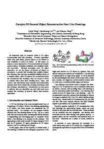
Electrode Stereotactic Device Anesthetized Mouse Fig. 1. Stereotactic surgery using a passive positioning device and anesthetized mouse
1. Introduction Stereotactic surgery is to place electrodes at clinically predetermined brain cells. The first stereotactic surgical method and apparatus were developed by Horsley and Clake in 1908 ([1]). In the field of the animal neuro-science, the surgery is a widely used technique to investigate the function of brain of animals alive. For diagnosis of human Parkinson’s disease, depression, and Tourette syndrome, the stereotactic surgery as the form of deep brain stimulation is recently used. Fig. 1 shows the widely used mouse stereotactic surgery to place an electrode at brain cell using landmarks (bregma and lambda points in Fig. 2) using maps of mice brains (Fig. 3, [2]), and passive positioning device (see Fig. 1). Since the surgery highly depends on the surgeon’s experience and physical condition, the success rate is at most 30 to 40 percents though surgeons spend almost an hour for the surgery. In other words, the data from 60 to 70 percentile mice become useless. Our ultimate goal is to improve the surgical technique using the human guided robotic device in the accuracy and completion time standpoint. This paper introduces a novel three dimensional reconstruction method to identify pose and landmarks(here, bregma and lambda points) of a mouse skull for highly accurate stereotactic surgery. 2. Apparatus This human guided robotic stereotactic surgery interface is consist of four parts; i) a coarse controlled 3-DOF linear stage (Mark-204AM, Sigma KOKI Co.), ii) a fine controlled
Bregma Fig. 2.
Lambda
Landmarks of a mouse skull, bregma and lambda points
Fig. 3.
A mouse brain atlas
5-DOF hybrid type positioning (3-DOF pose and 2-DOF orientation) device (Fig. 5), iii) a touch sensor (will be described in the subsequent section), and iv) haptic input devices (Novint FALCON, Novint technology Co. and Freedom 2.4 Joystick, Logitech Co.). The developed stereotactic
Fig. 4. The developed Human guided robotic stereotactic surgery interface
For the 3-D reconstruction, the electrode mounted on the end of the positioning device (Fig. 4) is connected to a touch sensor (AT42QT1010, ATMEL Co.). It detects electrically conducting objects like metals and body fluid. During the entire process, this touch sensor keep sensing whether it touches the target object or not via the data acquisition device. Prior to applying the proposed method to a living mouse skull, we performed three dimensional reconstruction of a coin texture for accuracy verification of our device. Note that the texture of a coin is within 20 - 50 µ m. Two experiments have been done to verify the accuracy of the proposed method; i) four points repeatability test, ii) 3-D reconstruction of a coin texture. For the four points repeatability tests, the tip scans the depth of four pre-determined points to see the resolution of the proposed method. For the 3-D reconstruction of a coin, the tip scanned texture (height) of a coin with 0.5 mm planar workspace resolution.
5-DOF hybrid type positioning device
3-DOF Linear Stage Controller
Electrode
Fig. 5. Illustration of the Human guided robotic stereotactic surgery interface
surgery interface is operated by a realtime OS (RTX, Ardence Co.) with 1kHz control frequency and programmed by Visual C++ (Microsoft Co.). For control inputs and sensor data acquisition, two PCI boards (PCI 1723, Advantech Co. and PCI 6602, National Instruments Co.) are used.
4. Result For the four points repeatability test, the tip measure the depth of four points, 100 times each points. Fig. 6 shows the mean value and the standard deviation (std.) of measured points. The std. values of each point were 0.0081, 0.0068, 0.0102 and 0.0117 mm, respectively. Thus, we can conclude the resolution of the proposed method is under 12 µ m. For 3-D reconstruction of a coin, 500 Korean won coin was used in this experiment (both side head and tail). Fig. 7 - 10 show the results of the 3-D reconstruction of both sides of coin. The scanned images show successfully reconstructed texture of “bird” and the number “500” that have 20 µ m (at the middle) 50 µ m (at the edges of “500” texture) resolution embossment. Note that the heights of the middle and the edges of the texture is not constant.
3. Methods
0.12 0.1 Value (mm)
In order to identify pose and landmarks (here, bregma and lambda points) of a mouse skull, at the beginning of this research, we considered two widely used three dimensional reconstruction devices; i) stereo vision cameras, and ii) laser scanners. Stereo vision camera is easy to implement and visualize the images directly, however, there are the tradeoffs between the absolute resolution and field of depth. In other words, we tried to use highly magnified camera lens to raise the resolution but it was not able to provide sufficient depth of field. Also, the mouse skull is covered by blood, thus it was difficult to detect features. Laser scanners provide fine three dimensional images of targets (less than 50 µ m), however, it cannot be used as a realtime image source, and it is not applicable to objects covered by fluid like a mouse skull wet by body fluid. Thus, we have developed a novel touch sensor type three dimensional reconstruction method and apparatus that will be merged with coarse level vision camera sensors in the future for unstructured and movable environment.
0.08 0.06 0.04 0.02 0.0
1
Fig. 6.
2 3 Mean value of points
Repeatability test result
4
5. Conclusion
Fig. 7.
In this paper, the three dimensional reconstruction method using a touch sensor to identify pose and landmarks (here, bregma and lambda points) of a mouse skull for accurate stereotactic surgery is presented. In the experiment, we demonstrated three dimensional reconstruction of a coin with 20 µ m to 50 µ m embossment. Thus, it is promising that the proposed method can be implemented for a mouse skull pose and feature estimation of the accurate human guided robotic stereotactic surgery interface. For the future work, in addition to the system in Fig. 4, 2-DOF orientation and 1-DOF linear actuators will be implemented for accurate coarse-fine 5DOF positioning systems as in Fig. 5. In addition, a vision camera system will be used for intuitive and visualizable graphical user interface and three-dimensional reconstruction method as a low resolution but quick 3-D estimation device. Then, we believe that an accurate and convenient human guided robotic surgical interface that covers whole process of mouse stereotactic surgery with high success rate can be developed.
Head of 500 won coin
z (mm) 20
19.5 19 20 10 0
References
10 y (mm) 20
Fig. 8.
5
10
15
0 x (mm)
10
5
15
3-D reconstruction result of coin head
Fig. 9.
Tail of 500 won coin
z (mm) 20.5
20 10 5 0 y (mm) 5 10
Fig. 10.
10
5
0 x (mm)
5
3-D reconstruction result of coin tail
10
[1] Horsley V, Clarke RH, ”The structure and functions of the serebellum examined by a new method”, Brain 31:45-124, 1908. [2] G Paxinos, KBJ Franklin, The Mouse Brain in Stereotaxic Coordinates, Second Edition, Academic Press, 2001.