USO0RE39057E
(19) United States (12) Reissued Patent Hoxie (54)
(10) Patent Number: US (45) Date of Reissued Patent:
ANTIBODIES DIRECTED AGAINST CELLULAR CORECEPTORS FOR HUMAN IMMUNODEFICIENCY VIRUS AND METHODS OF USING THE SAME
RE39,057 E *Apr. 4, 2006
Cohen, 1996, Science vol. 272:809*810.* Collins, “Scientists make AIDS breakthrough”, The Phila
delphia Inquirer (Jun. 20, 1996 ed.).*
(75) Inventor: James A. Hoxie, BerWyn, PA (US)
Crise et al., 1990, J. Virol. 64:5585*5593.* Deng et al., 1996, Nature 381:661*666.* DoranZ et al., 1996, Cell 85:1149*1158.*
(73) Assignee: The Trustees of the University of
Dragic et al., 1996, Nature 381:667*673.* Dragic et al., 1995, J. Virol. 69:1013*1018.*
Pennsylvania, Philadelphia, PA (US) (*)
Notice:
This patent is subject to a terminal dis claimer.
(21) Appl. No.: 10/829,475 (22) Filed: Apr. 22, 2004 Related US. Patent Documents
Reissue of:
(64) Patent No.:
5,994,515 Nov. 30, 1999
12: p. 128.*
Appl. No.:
08/882,435
Filed:
Jun. 25, 1997
HarloW et al. 1988, In: Antibodies, A Laboratory Manual, Cold Spring Harbor, NY.* Harouse et al., 1991, Science 253:320i323.* Haynes et al., 1996, The Finnish Medical Society DUO DECIM, Ann Med’ 28:39*41.*
Provisional application No. 60/020,396, ?led on Jun. 25, 1996, and provisional application No. 60/020,647, ?led on Jun. 27, 1996.
Hesselgesser et al., 1997, “CDi4iindependent association between HlVil gp 120 and CXCR4: functional chemokine
(51) Int. Cl. C07K 16/28
(2006.01)
(52)
US. Cl. ............................ .. 530/388.22; 530/3871;
(58)
Field of Classi?cation Search ............ .. 530/3871,
530/3891; 424/1431; 424/1441
530/3881, 388.15, 388.21, 388.22, 388.3, 530/388.35, 389.1; 424/1311, 137.1, 141.1, 424/1421,1431,1441 See application ?le for complete search history. (56)
Fox 1994, No Winners Against AIDS, Bio/Technology, vol.
Issued:
US. Applications: (60)
D’SouZa et al., 1996, “Chemokines and HlVil second receptors”, Nature Medicine 2:1293*1300.* Earl et al., 1994, J. Virol. 68:3015*3026.* Endres et al., 1996, “CD4ilndependent Infection by HIV*2 Is Mediated by Fusin CXCR4”, Cell 87:745*756.* Fahey et al., 1992, Clin. exp. Immunol., 88: 1*5.* Fauci et al., 1996, Nature 384:529*534.* Feng et al., 1996, “HlVil Entry Cofactor: Functional cDNA Cloning of a SeveniTransmembrance, G Protein£oupled Receptor”, Science 272:872*876.*
References Cited
receptors are expressed in human neurons”, Current Biology 7:112*121.* Hoxie et al., 1988, J. Virol. 62:2557*2568.* Hoxie et al., 1986, Science 234:1123*1127.* Ikeuchi et al., 1990, J. Virol. 64:226*4231.* Jonker et al., 1993, “In vivo treatment With a monoclonal
chimeric antiiCD4 antibody results in prolonged depletion of circulating CD4+ cells in chimpanzees”, Clin. Exp. Immunol. 93:301*307.* Koot et al., 1992, AIDS 6:49*54.* LaBranche et al., 1994, J. Virol. 68:5509*5522.*
PUBLICATIONS
Lapham et al., 1996, Science 274:602*605.* Leung et al., 1994, “ChimeriZation of LL2, a Rapidly
Hoxie, et al. : Biological characterization of a Simian . . . :
InternaliZing Antibody Speci?c for B Cell Lymphoma”, Hybridoma 13:4694176.>X<
J. Vir. : pp. 2557*2568, Aug. 1988.* Feng, et al. : HlVil Entry Cofactor: ?lnctional cDNA. . . :
Science: vol. 272: pp. 872*877, May 1996.* Cohen: Likely HIV cofactor Found: Science: vol. 272: pp.
809*810, May 1996* Alkhatib et al., 1996, Science 272:1955*1958.* Berson et al., 1996, J. Virol. 70:6288*6295.* Bleul et al., 1996, Nature 282:829i833.* Bleul et al., 1997, Proc. Natl. Acad. Sci. USA 94:1925*1930.* Brass et al., 1994, J. Biol. Chem. 269:2943i2952.* Brelot et al., 1997, J. Virol. 71:4744*4751.* Burton et al., 1994, Adv. Immunol. 57:191*280.* Chaudhuri et al. , 1994, J. Biol. Chem. 269:7835*7838.*
Chesebro et al., 1990, J. Virol. 64:215*221.* Choe et al., 1996, Cell 85:1135*1148.* Clackson et al., 1991, Nature 352:624.* Clapham et al., 1992, J. Virol. 66:3531*3537.* Clapham, 1991, Rev. in Med. Virol. 1:51*58.*
Clapham et al., 1991, Virology 181:703*715.* Clapham et al., 1987, Virology 158:44*51.* Cocchi et al., 1995, Science 270:1811*1815.*
Li et al., 1990, J. Virol. 64138341387.>X< LoBuglio et al., 1989, “Mouse/human chimeric monoclonal antibody inman: Kinetics and immune response”. Proc. Natl. Acad. Sci. USA 86:4220*4224.* Lu et al., 1997, Proc. Natl. Acad. Sci. USA, vol. 94:6426*6431.* Maddon et al., 1986, Cell 47:333*348.*
McDougal et al., 1986, Science 231:382*385.*
(Continued) Primary ExamineriJeifrey Stucker (74) Attorney, Agent, or FirmiDrinker, Biddle & Reath, LLP
(57)
ABSTRACT
The invention relates to an anti-immunode?ciency virus
antibody Which binds to a cellular protein and diagnostic and therapuetic methods of using the same.
7 Claims, 14 Drawing Sheets
US RE39,057 E Page 2
McKnight et al., 1997 “Inhibitionof Human Immunode? ciency Virus Fusion by a Monoclonal Antibody to a Core
Samson et al., 1996, Biochemistry 35:3362*3367.* Sattentau, et al., 1986, Epitopes of the CD4 Antigen and HIV Infection, Science, V01. 234: 1 12041 122.*
ceptor (CXCR4) is both Cell Type and Virus Strain Depen dent”, J. Virol. 71:1692*1696.* McKnight et al., 1996, J. Virol. 70:459841606.>X< McKnight et al., 1995, J. Virol. 69:3167*3170.*
SchneideriSchaulies et al., 1995, Proc. Natl. Acad. Sci. USA 92:3943*3947.*
Simmons et al., 1996, Virology 70:8355*8360.* Simmons et al., 1995, Virology 209:696*700.*
McKnight et al., 1994, Virology, 201:8*18.*
Stefano et al., 1993, J. Virol. 67:6707*6715.* StriZiki et al., 1997. J. Virol. p. 5678*5683.* Sung Co et al., 1992, “Chimeric and HumaniZed Antibodies With Speci?city for the CD33 Antigen”, J. Immunol. 148:1149*1154.* Takeuchi et al., 1991, J. Virol. 65:1710*1718.* Tateno et al., 1989, Proc. Natl. Acad. Sci. USA 86:428741290.>X< Weiner et al., 1991, Pathobiol. 59:361*371.* Willett et al., 1997, “Common mechanism of infection by lentiviruses”, Nature 385:587.* Weiss et al., 1996, Nature 381:647*648.*
OTHER PUBLICATIONS
Miyoshi et al., 1981 Nature, 294:77(¥771.* Moore, 1997, Science 276:51.* Nara et al., 1988, Nature 332:469*470.* Nara et al., 1987, AIDS Res. and Hum. Retroviruses 3:283*202.* Neote et al., 1993, Cell 72:415*425.* Oberlin et al., 1996, Nature 382:833*835.* PelcheniMatthews et al., 1989, EMBO J. 8:3641*3649.*
Picard et al., 1997, Virology, 231:105*111.* PoWer et al., 1996, Trends Pharmacol. Sci. 17:209*213.* PoWer et al., 1995, J. Biol. Chem. 270:19495*19500.* Premack et al., 1996, “Chemokine receptors: GatWays to in?ammation and infection”, Nature Medicine 2:1174*1178.* Queen et al., 1989 Poc. Natl. Acad. Sci USA, V01. 86:10029*10033.* Ratner et al., 1987, AIDS Res. and Hum. Retroviruses 3:57*69.*
Sambrook et al., 1989, Molecular Cloning: A Laboratory Manual, Cold Spring Harbor, NY.*
Wright et al., 1992, “Genetically Engineered Antibodies: Progress and Prospects”, Critical Ref. in Immunol. 12(3,
4):125*168.* Wu et al., 1996, Nature 384:179*183.* Zagury et al., 1988, Proc. Natl. Acad. Sci. USA 85:5941*5945.* * cited by examiner
U.S. Patent
Apr. 4, 2006
Sheet 1 0f 14
US RE39,057 E
CELL ‘5.A
CELL
NUMBER
FLUORESCENCE INTENSITY
FLUORESCENCE INTENSITY
FIG. IAi
FIG. lAii
CELL
CELL
NUMBER
NUMBER
FLUORESCENCE INTENSITY
FLUORESCENCE INTENSITY
FIG. lAiii
FIG. lAiv
CELL NUMBER
2L.
FLUORESCENCE INTENSITY
FLUORESCENCE INTENSITY
FIG. lAv
FIG. lAvi
U.S. Patent
Apr. 4, 2006
Sheet 2 0f 14
US RE39,057 E
MOLT4
C8166 MAC#1
I ‘ uavicndrv RDJCD4 MAC#2
FIG. 1Bi
‘
MOLT4 C8166 MAC?! mm uswcm
| RDICD4
FIG. ICi
CXCR4
U.S. Patent
Apr. 4, 2006
Sheet 3 0f 14
US RE39,057 E
--o-- LAI + GUN-lwt
——A— GUN-Ivar
I RESIDUAL
+ ROD/A
0
INFECTIVI'I'Y
ROD/B
pg/ml ANTIBODY
FIG. 2A
1
00f‘
-o—- LAI
80 -
>
.
+
60 -
% RESIDUAL
_
INFECTIVITY
40 _
1
20 -
0
.
0
“U- RF
I
I
10
pg/ml ANTIBODY
FIG. 2B
20
GUN-lwt
U.S. Patent
Apr. 4, 2006
Sheet 4 0f 14
US RE39,057 E
100,000
10,000 RT ACTIVITY, 1,000 (cpm) 100 I0
2
4
6 DAY
8
10
FIG. 3A1 100,000 10,000 RT ACTIVITY, 1,000 (cpm) 100
10,000 1,000 RT ACTIVITY,
(cpm)
100
'2
A
6
i
DAY
FIG. 3Aiii 10.000
1,000 RT ACTIVITY.
(cpm)
100
DAY
FIG. 3Aiv
1'0
U.S. Patent
Apr. 4, 2006
Sheet 5 0f 14
US RE39,057 E
1 ,000,000
100,000 10,000
RT ACTIVITY, 1,000
(0pm)
100 10 .
0
5
1'0
15
DAY
FIG. 3Av 1 000,000
100.000 10,000
RT ACTIVITY, 1,000
(cpm)
100 10 r
:
:
:
0
5
l0
15
DAY
FIG. 3Avi
1,000,000 1-
ANTI-CD4
12 NONE
D 10pg/m1 El 100 pg/ml
100,000 -
RTACTIVITY,
(Cpm) 10,000 ..
i
1,00o--
- ~
100
I \ FIG. 3B
U.S. Patent
FIG. 3Ci
Apr. 4, 2006
FIG. 3C5
Sheet 6 0f 14
FIG. 3Ciii
FIG. 3Civ
US RE39,057 E
FIG. 3Cv
U S. Patent
Apr. 4, 2006
FIG. 4Biii
Sheet 8 0f 14
US RE39,057 E
FIG. 4Biv
U.S. Patent
Apr. 4, 2006
FIG. 5Ci
Sheet 10 0f 14
US RE39,057 E
U.S. Patent
Apr. 4, 2006
Sheet 11 0f 14
US RE39,057 E
PIG. 6A1
FIG. 6Aii
FIG. 6Aiii
FIG. 6Aiv
U.S. Patent
Apr. 4, 2006
Sheet 12 0f 14
US RE39,057 E
2000 1600
RTACTIVITY,
~
1200 -
(cpm)
<> U87 800 -
III U87/FUSIN
0 U87/CD4
400 0
.
.
4
6
:
8 DAY
10
12
U.S. Patent
Apr. 4, 2006
Sheet 13 0f 14
E VCP
WA
m w m, n0m w, w m w. m.
mumvm,
g/ ,wem:/ Hw.
US RE39,057 E
ES] BHS
.R MW NM my... m+ mRo+
FIG. 7
%m.
NO
N0
U.S. Patent
Apr. 4, 2006
Sheet 14 0f 14
///// / \\\\\ \
QB$r3ME5l2.bZ%Dm
\ \\\\\\
US RE39,057 E
E5
40 .E2
.UEmm
nL-
"52 0m.
53N2Hm6u.-%26:
=2Em5.8296 .UE
~62cm.4
US RE39,057 E 1
2
ANTIBODIES DIRECTED AGAINST CELLULAR CORECEPTORS FOR HUMAN IMMUNODEFICIENCY VIRUS AND METHODS OF USING THE SAME
Ikeuchi et al., 1990, J. V1rol. 641422644231) in the absence
of CD4. Although infection of CD4-negative cells generally proceeds slowly and without cytopathic eifects, some iso lates of HIV-2 infect CD4-negative cells rapidly and cause
extensive cell fusion (Clapham et al., 1992, supra). The highly cytopathic nature of these infections has suggested
Matter enclosed in heavy brackets [ ] appears in the original patent but forms no part of this reissue speci?
that these isolates can utilize one or more receptors other
than CD4 with high ef?ciency. HIV-1 strains exhibit distinct tropisms for CD4-positive cells. Macrophage tropic (M-tropic) strains of HIV-1 enter and replicate in macrophages and primary T cells but
cation; matter printed in italics indicates the additions made by reissue. CROSS REFERENCE TO RELATED APPLICATIONS
generally fail to enter T cell lines. These isolates character
istically do not induce multinucleated giant cells when cultured with certain immortalized T cell lines and are
This application claims priority under 35 USC §119(e) (1) to US. Provisional Application Ser. Nos. 60/020,396 and 60/020,647, ?led on Jun. 25 and Jun. 27, 1996, respectively.
tropic strains fail to enter macrophages ef?ciently but readily
GOVERNMENT SUPPORT
cell lines (Fauci et al., 1996, Nature 3845294534). This
The invention was supported in part by a grant from the US. Government (NIH Grant No. AI 33854) and the US. Government may therefore have certain rights in the inven tion.
generally non-syncytium inducing (N SI). In contrast, T cell infect primary T cells and induce syncytia (SI) on some T
20
immunode?ciency are typically T-tropic and SI.
FIELD OF THE INVENTION
The ?eld of the invention is infection by and pathogenesis of Human Immunode?ciency Vlrus.
25
BACKGROUND OF THE INVENTION
The human immunode?ciency viruses HIV-1 and HIV-2
difference in cell tropism has been shown to correlate with disease progression in that HIV strains isolated from indi viduals early in the course of their infection are M-tropic and NSI, while viruses isolated from individuals with advanced
CXCR4 is a cellular protein which in conjunction with CD4, forms a functional cellular receptor for entry of certain strains of HIV into cells. This protein is a member of a family of molecules that bind chemokines which are involved in the traf?cking of T cells and phagocytic cells to areas of in?ammation (Power and Wells, 1996, Trends
Pharmacol. Sci. 17:209*213). The chemokines MIP-alpha 30
and the closely related simian immunode?ciency viruses
and MIP-beta and RANTES all bind to CCR5 while stromal
cell derived factor (SDF-l) binds to CXCR4 (Bluel, et al.,
(SIV), all use the CD4 molecule as a receptor during
1996, Nature 382:829i832; Oberlin et al., 1996, Nature
infection. Other cellular molecules have long been suspected
382z833i835). Recent reports have indicated that the viral
to form an essential component of the cellular HIV receptor; however, the nature of such cellular molecules was not
envelope glycoprotein gp120 interacts directly with 35
known until the discovery of fusin (Feng et al., 1996, Science 272z872i876; Maddon et al., 1986, Cell 47z333i348; Dragic et al., 1995, J. Virol. 691101341018; Clapham et al., 1992, J. Virol. 661353143537; Chesebro et al., 1990, J. Virol. 64:215*221; Stefano et al., 1993, J. V1rol 671670746715; Hoxie et al., 1988, J. Virol. 621255742568).
binding.
Recently, two molecules, fusin, which is now known as
CXCR4 (also known as Lestr, LCR-1, and HUMSTR) and CCR5, which are members of the chemokine receptor fam ily of proteins, have been shown to function with CD4 as coreceptors for HIV-1 isolates that are tropic for T-cell lines
or macrophages, respectively (Feng et al., 1996, Science 272z872i876; Alkhatib et al., 1996, Science 272:1955*1958; Deng et al., 1996, Nature 38116614666; Dragic et al., 1996, Nature 381:667*673). Other molecules in this family including CCR3 and CCR2b, also appear to
chemokine receptors (Lapham et al., 1996, Science 274z602i605; Moore, 1997, Science 276:51; Wu et al., 1996, Nature 384:179*183; Hesselgesser et al., 1997, Cur rent Biology 7:112*121), generally at a step following CD4
45
CXCR4 ful?lls the requirements of an HIV receptor co-factor. It renders a number of murine, feline, simian, quail, and hamster cell lines, as well as human cell lines, which cells are normally resistant to HIV-1 entry, fully permissive for HIV-1 env mediated syncytia formation. In addition, the T cell tropic HIV strain HIV-1 IIIB, is capable of infecting both murine and feline cells which co-express
human CD4 and CXCR4. However, the macrophage tropic strain Ba-L, is not capable of infecting cells which co-express both CXCR4 and CD4. These results suggest that 50 CXCR4 can serve as a co-factor for T-tropic, but not
M-tropic, HIV-1 strains (Feng et al., 1996, supra). Moreover,
CXCR4 as coreceptors for isolates of simian immunode?
the ?nding that change from M to T-tropic viruses over time in infected individuals correlates with disease progression suggests that the ability of the viral envelope to interact with CXCR4 represents an important feature in the pathogenesis of immunode?ciency and the development of full blown
ciency viruses (SIV) and HIV-2, respectively. This, indicates
AIDS.
that the use of chemokine receptors is a general property of all human and nonhuman lentiviruses. In addition to studies on CD4-dependent infection, sev
Current anti-HIV therapy includes the use of compounds which inhibit various aspects of HIV replication in a cell such as inhibition of replication and/or transcription of viral
function as cofactors for some HIV-1 isolates (DoranZ et al.,
1996, Cell 85:1149*1158; Berson et al., 1997, J. Virol. 71:
169241696; Choe et al., 1996, Cell 851113541148). Moreover, recent studies have also implicated CCR5 and
55
60
nucleic acid and inhibition of protein processing. While these therapies, particularly when used in combination with
eral reports have demonstrated that some HIV isolates are
capable of infecting lymphoid cells (Clapham et al., 1992, supra; Clapham, 1991, Rev. in Med. V1rol. 1151458; McK night et al., 1994, V1rology 201 18418) or non-lymphoid cells (Clapham et al., 1992 supra; Harouse et al., 1991, Science 25313204323; Tateno et al., 1989, Proc. Natl. Acad. Sci. USA86142874290; Li et al., 1990, J. Virol. 641138341387;
one another, are effective, they are frequently short-lived in 65
that viral strains rapidly develop that are resistant to one or more of the compounds used. There therefore remains an
acute need to develop additional therapies and strategies for preventing HIV infection in humans.
US RE39,057 E 3
4
SUMMARY OF THE INVENTION
method comprises isolating a test compound capable of binding to an anti-immunode?ciency virus antibody, Which antibody binds to a cellular protein, and assessing the ability
The invention relates to an antibody capable of binding to
a cellular protein, Which antibody has antiviral activity by
of the test compound to inhibit infection of a cell by an immunode?ciency virus in an antiviral assay, Wherein inhi
virtue of the fact that the cellular protein to Which the
antibody binds is a protein Which is required for entry of virus into a cell expressing that protein.
bition of infection of the cell by the immunode?ciency virus in the presence of the test compound is an indication that the test compound is an anti-immunode?ciency virus com
One aspect of the invention relates to an anti
immunode?ciency virus antibody capable of binding to a cellular protein. In another aspect, the immunode?ciency virus is selected from the group consisting of HIV-1, HIV-2
pound.
and SIV.
to the cell an antibody Which binds to the CXCR4 and
In yet another aspect of the invention, the protein to Which the antibody of the invention binds is a chemokine receptor protein, preferably, an HIV receptor protein and/or a cellular cofactor for a cellular HIV receptor protein. More preferably, the protein to Which the antibody of the invention binds is selected from the group consisting of CXCR4 and CCR5; and most preferably, the protein to Which the antibody binds is CXCR4. In another aspect of the invention, the antibody is selected
The invention also includes a method of measuring the level of expression of CXCR4 on a cell comprising adding
assessing the amount of antibody bound to the cell, Wherein the amount of the antibody bound to the cell is a measure of the level of expression of the CXCR4 on the cell.
BRIEF DESCRIPTION OF THE DRAWINGS
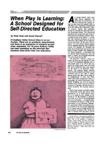
FIG. 1A is a series of graphs graph depicting detection of
CXCR4 cell surface glycoprotein expression folloWing 20
from the group consisting of a monoclonal antibody and a
FIG. 1B is an image of Northern blot analysis of the
synthetic antibody. Preferably, the antibody is a monoclonal antibody, and more preferably, the antibody is MAb 12G5. The invention also relates to an isolated DNA encoding an
staining of MOLT-4, C8166, U87/CD4 and RD/CD4 cells and primary human macrophages (MAC) cultured for 1 and 5 days. indicating cells using CXCR4 or GAPDH nucleic acid as a
probe. 25
anti-immunode?ciency virus antibody capable of binding to
FIG. 1C is an image of reverse transcriptase (RT) PCR analysis on the indicated cells using CXCR4 or GAPDH
a cellular protein. In one aspect, the immunode?ciency virus is selected
nucleic acid as a probe.
from the group consisting of HIV-1, HIV-2 and SIV. In another aspect, the protein to Which the antibody of the invention binds is a chemokine receptor protein. Preferably, the protein is an HIV receptor protein and/or a cellular cofactor for a cellular HIV receptor protein. More
preferably, the protein is selected from the group consisting of CXCR4 and CCR5; and most preferably, the protein is CXCR4 and the antibody is the monoclonal antibody (MAb) 12G5. The invention also relates to a method of inhibiting infection of a cell by HIV comprising adding to the cell an
anti-immunode?ciency virus antibody capable of binding to
30
35
for Sup-T1, all cell lines Were CD4-negative as determined 40
antibody to the cellular protein infection of the cell by HIV 45
an anti-immunode?ciency virus antibody capable of binding
by FACS analysis using a panel of anti-CD4 MAbs and/or by Western blot using an anti-CD4 serum. FIG. 3B is a graph depicting the failure of an anti-CD4 MAb to inhibit HIV-2/vcp infection of a CD4-negative cell line. BC7 or Sup-T1 cells Were inoculated With either HIV-2/vcp or HIV-1I/LAI, respectively, in the presence or absence of anti-CD4 MAb #19. Input virus Was removed
after 24 hours by Washing and cells Were maintained in the presence of anti-CD4 MAb for 8 days at Which time RT
to a cellular protein on a cell, Wherein upon binding of the
antibody to the cellular protein, infection of the cell by HIV is inhibited, thereby treating the HIV infection in the human.
panels Were inoculated With cell-free HIV-2/vcp or HIV-1/ LAI and Were monitored for reverse transcriptase (RT)
activity in culture supernatant at the indicated time points. Input virus Was removed after 24 hours by Washing. Except
a cellular protein on the cell, Wherein upon binding of the is inhibited. Also included in the invention is a method of treating HIV infection in a human comprising administering to the human
FIG. 2A is a graph depicting 12G5-mediated inhibition of cell-free infectivity of RD/CD4 cells. FIG. 2B is a graph depicting 12G5-mediated inhibition of cell-free infectivity of HeLa/CD4 cells. FIG. 3A is a series of graphs depicting infection of CD4 negative cells With HIV-2/vcp. The cell lines indicated in the
activity Was determined. Similar results Were seen using
anti-immunode?ciency virus antibody capable of binding to
OKT4A, another anti-CD4 MAb. FIG. 3C is a series of images of syncytium induction assays Which Were performed on the indicated target cells by
a cellular protein on a cell, the method comprising gener ating a panel of antibodies directed against HIV infected or
cocultivation With HIV-2/vcp-infected BC7 cells or HIV-1/ LAI-infected Hut-78 cells in the presence or absence of 10
50
The invention further includes a method of obtaining an
SIV infected cell proteins, and screening the antibodies for anti-immunode?ciency virus activity to obtain an antibody capable of binding to a cellular protein, Which antibody has
55
anti-immunode?ciency virus activity. Also included in the invention is a method of identifying a target cell for immunode?ciency virus infection, the method comprising adding to a population of cells an
60
anti-immunode?ciency virus antibody capable of binding to a cellular protein on a cell, Wherein binding of the antibody to a cell in the population is an indication that the cell is an
immunode?ciency virus target cell. In addition, there is provided a method of identifying a
candidate anti-immunode?ciency virus compound. This
65
ug/ml of anti-CD4 MAb #19. Cultures Were photographed after either 24 or 48 hours. As shoWn, extensive syncytia
formation is induced by HIV-2/vcp on CD4-negative BC7 cells Which is unaffected by the addition of anti-CD4 MAb. FIG. 4A is a series of graphs depicting inhibition of CP-MAC and HIV-2/vcp infection by the 12G5 MAb. Sup-T1 cells or CD4-negative BC7, Nalm6 and Daudi cells Were preincubated With the indicated concentrations of 12G5 MAb and Were then inoculated With the viruses shoWn. RT activity in infected cell supernatants Was assessed at the indicated times. Dose-dependent inhibition
of CP-MAC and HIV-2/vcp infection by 12G5 MAb is shoWn.
US RE39,057 E 6
5 FIG. 4B is a series of images depicting inhibition of
IL8R-B or the PCR3.1 expression vector alone, and a
CP-MAC and HIV-2/vcp syncytia induction by the 12G5
plasmid containing the luciferase gene drive by a T7 pro moter (Promega Biotech). Where indicated, target cells Were also infected With pT4, Which constitutively expresses CD4
MAb, Sup-T1 or BC7 cells Were cultured With either
CP-MAC-infected Sup-T1 cells, or HIV-2/vcp-infected BC7 cells in the presence or absence of 12G5 (10 ug/ml). Cells Were photographed after 48 hours of culture. Inhibition of syncytium formation by 12G5 is evident in cells infected With either virus. FIG. 5A is a series of graphs depicting reactivity of 1 2G5 MAb With CXCR4. U87 cells stably expressing either CXCR4, CCR1, or the control vector pBABe-puro (Deng et al., 1996, Nature 381:661*666), Were evaluated for reactiv
Nalm6 or Sup-T1 cells that Were either uninfected or infected With the indicated viruses Were evaluated for sur
ity With 12G5 (10 ug/ml) by FACS. The region for positivity,
face reactivity by FACS either With an isotope-matched
designated M1, Was de?ned using a non-reactive control MAb, and the percent of cells falling Within this WindoW for each sample is indicated. As shoWn, a marked shift in reactivity Was observed in the entire population of CXCR4
control MAb or MAb 12G5 (10 ug/ml) and the mean
from the CMV promoter. Luciferase activity as an indication of cell fusion is indicated in terms of relative light units
(RLU) as described (DoranZ et al., 1996, Cell
85:1149*1158). FIG. 8 is a series of graphs depicting doWnregulation of
CXCR4 expression of HIV-2/vcp infection. BC7, Daudi,
channel ?uorescence intensity (MCF) is shoWn for each cell type. Loss of MAb 12G5 reactivity is seen on HIV-2/vcp infected but not CP-MAC-infected cells. No reduction Was
expressing cells. FIG. 5B is a graph depicting reactivity of 12G5 MAb With CXCR4. Control CHO cells or cells stably expressing HA-tagged CXCR4 or the other chemokine receptors indi cated Were evaluated for reactivity to 125I-12G5, using protocols described in Pelchen-MattheWs et al. (1989,
EMBO J. 8z364li3649). Scatchard type analysis indicated that the Kd for 12G5 binding to CHO-CXCR4 cells Was 15 nM; 125I-12G5 binding Was competed to close to back ground levels by 100 nM of unlabeled 12G5 but Was not
seen in expression of HLA class-I using MAb W6/32. The data are representative of three experiments from tWo sepa 20
DETAILED DESCRIPTION OF THE INVENTION 25
FIG. 5C is a series of photographs depicting immunof
CHO-K1 cells expressing HA-tagged CXCR4 or the human ILSR-B receptor Were stained With 12G5, the anti-human CD4 MAb Q4120, or an antibody against the HA-tag
(Pelchen-MattheWs et al. (1989, supra). Only cells With
The antibody of the invention is an antiviral antibody in that it is an antibody Which binds to a speci?c cellular 30
35
The virus against Which the antiviral antibody is directed an immunode?ciency disease. Thus, the antibody of the invention is termed an anti-immunode?ciency virus anti 40
immunode?ciency virus belonging to the group of either HIV type 1 or HIV type 2. By “SIV” as used herein is meant 45
expressing either CXCR4 or CD4, or untransduced cells
any of ?ve recogniZed strains of SIV (SIVmac, SIVsmm, SIVagm, SIVmnd and SIVcpZ) Which are knoWn to infect
(control) Were cocultured With HIV-2/vcp infected BC7 cells
non-human primates. 50
The antibody of the invention is an antibody Which is capable of binding to a cellular protein required to form a functional cellular receptor for entry of HIV into a cell. The
antibody of the invention may be monoclonal antibody (MAb) or may be antibody Which is derived from a phage
CXCR4 is suf?cient to render U87 cells susceptible to HIV-2/vcp infection. Cells Were inoculated With cell-free
HIV-2/vcp (1000 TCID5O units, determined on BC7 cells)
body. Such immunode?ciency virus should be construed to include any strain of HIV or SIV. By “HIV” as used herein, is meant any strain of a human
recombinant CXCR4 is suf?cient to render U87 cells sus
and syncytium formation Was assessed by photography. Large syncytia are evident only in U87 cells Which express CXCR4. Induction of syncytia Was completely inhibited in cells Which Were preincubated With 12G5 (10 ug/ml). FIG. 6B is a graph depicting the fact that recombinant
of the virus into the cell and is therefore termed an antiviral antibody despite the fact that it does not bind to a viral protein, but rather, binds to a cellular protein.
is an immunode?ciency virus, that is, a virus Which causes
expressing CCR5. Scale bar=20 um. FIG. 6A is a series of photographs depicting the fact that
ceptible to HIV-2/vcp syncytium induction. U87 cells stably
protein Which is essential for virus entry into the cell in
Which the cellular protein is expressed. By binding to the cellular protein, the antibody of the invention inhibits entry
HA-tagged CXCR4 stained With MAb 12G5, While both samples stained brightly With the anti-HA MAb. None of the cells exhibited staining With Q4120. Similarly, no binding of MAb 12G5 Was seen in CHO cells stably expressing IL8R A, or CCR1, CCR2b, CCR3 or CCR4, or in cells transiently
The invention relates to an antiviral antibody Which binds to a cellular protein essential for entry of a virus into a cell
expressing that protein.
in?uenced by the anti-HA antibody 12G5. luorescence confocal microscopy of CHO cells stably expressing CXCR4 or chemokine receptors using 12G5.
rate infections.
library or a humanized or a synthetic antibody, or an 55
antibody fragment expressed intracellularly.
and the amount of RT in the culture supematants Was
The antibody of the invention is an antibody Which binds
monitored at the indicated time points. Only CXCR4 expressing cells Were infected With virus. Extensive syncytia
to a cellular co-factor required for entry of HIV into a cell.
formation and cell killing Were also observed in the infected CXCR4-expressing cells Which correlated With the time of
protein Which is required, in association With a cellular receptor for HIV; for entry of HIV into cells.
A “cellular co-factor” as used herein, is de?ned as a 60
production of RT. FIG. 7 is a graph depicting induction of cell fusion by the HIV-2/vcp envelope glycoprotein in a gene reporter fusion assay. HeLa effector cells Were transfected With pCR3.1 expressing either HIV-2/vcp env or BH8 HIV-1 env clones and Were then infected With vaccinia virus. QT6 target cells Were transfected With constructs expressing CXCR4,
Since the preferred antibody of the invention, MAb 12G5, binds to a protein Which is both a cellular cofactor and an
HIV-receptor protein, depending upon the cell type to be infected, it Will be appreciated that the antibody of the 65
invention should be construed to be one Which binds to a
cellular protein Which may be a cellular cofactor and may also be a receptor protein in its oWn right. In other Words, the
US RE39,057 E 7
8
antibody of the invention is one Which binds to a cellular
cell lines folloWing transient or stable cellular expression of a recombinant CXCR4 protein. In addition, in assays designed to evaluate the antiviral effects of CXCR4 12G5, this antibody is capable of inhibiting both infection as Well as syncytia formation by a number of HIV-1, HIV-2 and SIV isolates. The extent of inhibition of virus replication is dependent upon the test virus and also the target cell. By “antiviral activity” as used herein, is meant an anti
protein Which is necessary for virus entry into cells. Since binding of the antibody to the cellular protein serves to block virus entry into cells, the antibody is an antiviral antibody. Preferably, the antibody of the invention is directed
against the cellular protein CXCR4; more preferably, the antibody is a monoclonal antibody and even more
preferably, the antibody of the invention is MAb 12G5. According to the invention, the antibody of the invention
body Which When added to an immunode?ciency virus or to a cell to be infected With such a virus, mediates a reduction
is useful in a method of inhibiting infection of a cell by HIV as described herein. The antibody is further useful for the generation of antibody derivatives Which are useful for inhibiting infection of a cell by HIV also as described herein. Moreover, the antibody of the invention is useful in a
in the ability of the virus to infect and/or replicate in the cell compared With the ability of virus to infect and/or replicate in the cell in the absence of the antibody. Examples of assays for antiviral activity are described in detail in the experi
method of screening compounds for anti-HIV activity as described herein. Additional uses for the antibody of the invention include the identi?cation of cells in the body Which are potential targets for viral infection. The antibody is thus also useful for the isolation of such cells using ?oW cytometry technology or other cellular isolation techniques Which are common in the art. The invention also relates to
methods of use of the antibody of the invention, Which methods include diagnostic and therapeutic uses. The antibody or derivatives thereof may be expressed intracellularly, reducing CXCR4 expression on the cell surface and rendering these cells resistant to HIV infection. In addition, the antibody of the invention inhibits SDF-l
mental detail section and include, but are not limited to, reverse transcriptase assays, immuno?orescence assays,
assays for formation of syncytia, antigen capture assays and the like. 20
To generate the antibody of the invention, Wherein the antibody is a monoclonal antibody, cells Which are suspected
25
to encode a cellular protein essential for virus entry are ?rst infected With HIV or SIV. Extracts are prepared from the cells and are used to generate a panel of monoclonal antibodies directed against the infected cells. Antibodies are screened for antiviral activity in a functional antiviral assay as described herein, and for the ability to bind cellular rather than viral proteins, also as described herein. Antiviral anti bodies Which bind cellular proteins are selected and are
binding and signaling by CXCR4. Thus, the antibody or derivatives thereof, may be used to antagonize SDF-l func tion in vivo.
30
The antibody of the invention, exempli?ed herein by the MAb 12G5, Was generated in a hybridoma screening pro tocol in Which mice Were immunized With an SIV-infected
human cell line in order to generate MAbs reactive With the
SIV envelope glycoproteins. Eight MAbs Were generated Which exhibited potent antiviral properties. Of these eight MAbs, seven reacted speci?cally With the viral envelope glycoproteins, While one antibody, MAb 12G5, reacted With
35
40
MAbs speci?c for viral proteins and the screening assay Was an antiviral assay. Thus, the MAbs Which Were generated
folloWing immunization of mice With SIV-infected human cells Were screened for antiviral activity, i.e., a functional antiviral assay Was used to select the MAbs, rather than the more common antigen-antibody binding assay. Since this assay Was expected to reveal the presence of MAbs Which bind viral antigens, the generation of an antiviral MAb
speci?c for a cellular protein Was unexpected. The preferred antibody of the invention, MAb 12G5, Was discovered to bind to a cell protein termed CXCR4, Which protein is essential for entry of an immunode?ciency virus into cells. Since MAb 12G5 possesses antiviral activity, this antibody is speci?c for epitopes on CXCR4 Which are essential for virus infection. The functional assay by Which the antibody is generated is unique in that it facilitates the
45
teristics described herein. It should be appreciated that the present invention also
occurring proteins by conservative amino acid sequence differences or by modi?cations Which do not affect sequence, or by both.
50
For example, conservative amino acid changes may be made, Which although they alter the primary sequence of the peptide, do not normally alter its function. Conservative amino acid substitutions typically include substitutions Within the folloWing groups:
55
glycine, alanine; valine, isoleucine, leucine; aspar‘tic acid, glutamic acid;
asparagine, glutamine;
identi?cation of an antiviral antibody Which binds a cellular
into cells using CD4 as the cellular receptor molecule. In other instances, CXCR4 functions as a cellular receptor for HIV in the absence of CD4. Thus, CXCR4 is both a cellular co-factor, as de?ned herein, and is a cellular virus receptor protein in its oWn right for the entry of HIV into certain cells. As described in detail herein, MAb 12G5 is speci?c for CXCR4. MAb 12G5 binds to both human and nonhuman
provided they give rise to an antibody having the charac provides for analogs of CXCR4 obtained according to the methods of the invention. Analogs may differ from naturally
serine, threonine;
protein rather than a viral protein. As the data presented herein establish, in some instances, CXCR4 functions as a cellular co-factor for entry of HIV
anti-CXCR4 antibodies since the entire molecule is in the correct conformation for the generation of anti-CXCR4 antibodies Which block virus entry into cells. HoWever, the invention should not be construed to be limited solely to the use of the entire CXCR4 molecule for the generation of
antibodies, it being understood that peptides, Which can be made according to Well knoWn procedures, may also be used
both infected and uninfected cells. This result Was unex
pected since the protocol Was originally designed to identify
further characterized With respect to the proteins to Which they bind and to their antiviral capabilities. Preferably, the entire CXCR4 molecule is used to generate
60
lysine, arginine; phenylalanine, tyrosine. Modi?cations (Which do not normally alter primary
65
sequence) include in vivo, or in vitro chemical derivatization of proteins, e.g., acetylation, or carboxylation. Also included are modi?cations of glycosylation, e.g., those made by modifying the glycosylation patterns of a protein during its synthesis and processing or in further processing steps; e.g., by exposing the protein to enzymes which affect