articles JNK1 as a Molecular Target To Limit Cellular Mortality under Hypoxia Seema Betigeri, Refika I. Pakunlu, Yang Wang, Jayant J. Khandare, and Tamara Minko* Department of Pharmaceutics, Rutgers, The State UniVersity of New Jersey, Piscataway, New Jersey 08854-8020 Received February 12, 2006
Abstract: Many pathological conditions and environmental impacts lead to a decrease in tissue oxygen supply and severe cellular hypoxia. This secondary hypoxia can disturb cellular homeostasis, limiting the efficacy of the prescribed treatment for the primary lesion, eventually leading to cellular and organismal death. Jun N-terminal kinase 1 (JNK1) plays a major role in the hypoxic cellular damage. Therefore, we hypothesized that suppression of JNK1 activity will decrease cellular mortality under hypoxia and might increase the efficacy of traditional treatment of many pathological conditions. These investigations are aimed at studying the influence of the suppression of JNK1 activity on the development of cellular hypoxic damage. We used antisense oligonucleotides (ASO) and small interfering RNA (siRNA) targeted to JNK1 mRNA to inhibit the protein synthesis. Experiments were carried out on a cell culture under normoxia and hypoxic conditions that led to the death of ∼50% of cells. ASO or siRNA was delivered by neutral or cationic liposomes. Intracellular localization of ASO and liposomes and mechanisms of apoptosis were studied. We found that the suppression of JNK1 activity by liposomal antisense oligonucleotides or siRNA limits the caspase-dependent apoptosis signaling pathway and decreases cellular mortality after severe hypoxia. JNK1 protein might be an attractive target for antihypoxic therapy in increasing resistance to many pathological conditions and diseases, leading to the oxygen deficit. Keywords: Liposomal delivery; antisense oligonucleotides; siRNA; intracellular localization; hypoxia; Jun N-terminal kinase 1; apoptosis
Introduction Many known pathological conditions lead to a decrease in systemic oxygen supply, leading to secondary cellular hypoxia. When this hypoxia becomes severe, it causes additional cellular damage, aggravating the primary disorder and leading to cell death. Therefore, remediation of secondary hypoxic damage should significantly increase the efficacy of the treatment of the primary lesion and prevent extensive cellular damage. Moreover, the prevention of cellular death * To whom correspondence should be addressed: Department of Pharmaceutics, Ernest Mario School of Pharmacy, Rutgers, The State University of New Jersey, 160 Frelinghuysen Rd., Piscataway, NJ 08854-8020. Phone: (732) 445-3831, ext. 214. Fax: (732) 445-3134. E-mail:
[email protected]. 424
MOLECULAR PHARMACEUTICS VOL. 3, NO. 4, 424-430
under environmental conditions which cause a reduction in body oxygen supply (high altitude, deep mining, diving, etc.) might increase the working capacity and prevent severe adverse side effects of tissue hypoxia. A cell death signal leading to the activation of caspases and finally causing cell death plays an important role in initiating and promoting of cellular hypoxic damage.1,2 Hypoxia inducible factor R (HIF1A) is a key initiator of such (1) Minko, T.; Wang, Y.; Pozharov, V. Remediation of cellular hypoxic damage by pharmacological agents. Curr. Pharm. Des. 2005, 11, 3185-3199. (2) Minko, T.; Stefanov, A.; Pozharov, V. Lung hypoxia: Antioxidant and antiapoptotic effects of liposomal R-tocopherol. J. Appl. Physiol. 2002, 93, 1550-1560;. 10.1021/mp060014x CCC: $33.50 © 2006 American Chemical Society Published on Web 06/29/2006
articles
JNK1, Molecular Target To Limit Cell Death a signal under hypoxic conditions.3 Therefore, we recently investigated the role of this protein as a possible target for the remediation of hypoxic cellular damage. However, we found that HIF1A plays a bimodal role during hypoxia.4 On one hand, the activation of HIF1A during hypoxia initiates the cell death signal inducing apoptosis (programmed cell death or cellular suicide) and necrosis (pathological cell death). On the other hand, overexpression of HIF1A boosts the power of antihypoxic systems that increase cellular resistance to hypoxia. Moreover, we found that suppression of HIF1A activity predominantly led to the activation of apoptosis and promoted cell death. Therefore, we used this approach to enhance the efficacy of cancer chemotherapy.5 The next protein family which attracts attention as a possible target for the remediation of cellular hypoxic damage is mitogen-activated protein kinases (MAPKs), a family of serine-threonine protein kinases that participate in a major signaling system by which cells transduce extracellular stimuli into intracellular responses.6 Within this group, two subfamilies are usually distinguished: the extracellular regulated kinases (ERKs) and the stress-activated protein kinases (SAPK). The major player in the activation of apoptosis signaling pathways and hypoxic damage is Jun N-terminal kinase (JNK), a stress-activated protein kinase that can be induced by inflammatory cytokines, bacterial endotoxin, osmotic shock, UV radiation, and hypoxia.7-9 Analysis of literature data7,10,11 showed that the activation (3) Hellwig-Burgel, T.; Stiehl, D. P.; Wagner, A. E.; Metzen, E.; Jelkmann, W. Hypoxia-inducible factor-1 (HIF-1): A novel transcription factor in immune reactions. J. Interferon Cytokine Res. 2005, 25, 297-310. (4) Wang, Y.; Pakunlu, R. I.; Tsao, W.; Pozharov, V.; Minko, T. Bimodal effect of hypoxia in cancer: The role of hypoxia inducible factor in apoptosis. Mol. Pharm. 2004, 1, 156-165. (5) Wang, Y.; Minko, T. A novel cancer therapy: Combined liposomal hypoxia inducible factor 1R antisense oligonucleotides and an anticancer drug. Biochem. Pharmacol. 2004, 68, 20312042. (6) Johnson, G. L.; Dohlman, H. G.; Graves, L. M. MAPK kinase kinases (MKKKs) as a target class for small-molecule inhibition to modulate signaling networks and gene expression. Curr. Opin. Chem. Biol. 2005, 9, 325-331. (7) Bennett, B. L.; Sasaki, D. T.; Murray, B. W.; O’Leary, E. C.; Sakata, S. T.; Xu, W.; Leisten, J. C.; Motiwala, A.; Pierce, S.; Satoh, Y.; Bhagwat, S. S.; Manning, A. M.; Anderson, D. W. SP600125, an anthrapyrazolone inhibitor of Jun N-terminal kinase. Proc. Natl. Acad. Sci. U.S.A. 2001, 98, 13681-13686. (8) Chihab, R.; Ferry, C.; Koziel, V.; Monin, P.; Daval, J. L. Sequential activation of activator protein-1-related transcription factors and JNK protein kinases may contribute to apoptotic death induced by transient hypoxia in developing brain neurons. Brain Res. Mol. Brain Res. 1998, 63, 105-120. (9) Zhou, G.; Golden, T.; Aragon, I. V.; Honkanen, R. E. Ser/Thr protein phosphatase 5 inactivates hypoxia-induced activation of an apoptosis signal-regulating kinase 1/MKK-4/JNK signaling cascade. J. Biol. Chem. 2004, 279, 46595-46605. (10) Derijard, B.; Hibi, M.; Wu, I. H.; Barrett, T.; Su, B.; Deng, T.; Karin, M.; Davis, R. J. JNK1: A protein kinase stimulated by UV light and Ha-Ras that binds and phosphorylates the c-Jun activation domain. Cell 1994, 76, 1025-1037.
of cell death under hypoxia includes the following major steps. Activation of JNK by hypoxia mediates phosphorylation of activating transcriptional factor-2 (ATF2) and c-Jun bound to the c-jun promoter and stimulates their transcriptional activities leading to c-jun induction. The newly synthesized c-Jun protein combines with the c-Fos protein to form stable transcriptional factor activator protein-1 (AP1) heterodimers. The formation of AP1 is a key step in the following induction of the central cell death signal leading to the activation of the caspase-dependent apoptosis signal and finally causing cell death. It was found that c-Jun N-terminal kinase 1 (JNK1, SAPK1, and MAPK8) plays a central role in the development of cellular damage under hypoxia, hypoxia/reoxygenation, and ischemia/reperfusion conditions.12-14 Therefore, we selected the JNK1 protein as a molecular target to limit cellular damage and death during hypoxia. Here we report our recent experimental data which clearly support our hypothesis and demonstrate that inhibition of JNK1 by antisense oligonucleotides (ASO) or small interfering RNA (siRNA) targeted to JNK1 mRNA substantially suppresses JNK1 protein expression and effectively limits induction of apoptosis and cellular death under severe hypoxic conditions.
Experimental Section Cell Line. Human embryonic kidney 293 cells were obtained form American Type of Tissue Culture (Manassas, VA). Cells were cultured in Dulbecco’s modified Eagle’s medium (Gibco Inc., Cincinnati, OH) supplemented with 10% fetal bovine serum (Fisher Chemicals, Fairlawn, NJ). All experiments were performed on cells in the exponential growth phase. Hypoxia Model. Cells were maintained at 37 °C in a humidified incubator containing 21% O2 and 5% CO2 in air (termed normoxic conditions). Hypoxia was produced by placing cell culture plates in a modular incubator chamber (Billupus-Rottemberg Inc., Del Mar, CA), and then the chamber was flushed with a mixture of 1% O2, 5% CO2, and 94% N2 at a flow rate of 3 L/min for 15 min.15 The chamber was (11) Karin, M. The regulation of AP-1 activity by mitogen-activated protein kinases. J. Biol. Chem. 1995, 270, 16483-16486. (12) Crenesse, D.; Laurens, M.; Heurteaux, C.; Cursio, R.; Saint-Paul, M. C.; Schmid-Alliana, A.; Gugenheim, J. Rat liver ischemiareperfusion-induced apoptosis and necrosis are decreased by FK506 pretreatment. Eur. J. Pharmacol. 2003, 473, 177-184. (13) Garay, M.; Gaarde, W.; Monia, B. P.; Nero, P.; Cioffi, C. L. Inhibition of hypoxia/reoxygenation-induced apoptosis by an antisense oligonucleotide targeted to JNK1 in human kidney cells. Biochem. Pharmacol. 2000, 59, 1033-1043. (14) Hreniuk, D.; Garay, M.; Gaarde, W.; Monia, B. P.; McKay, R. A.; Cioffi, C. L. Inhibition of c-Jun N-terminal kinase 1, but not c-Jun N-terminal kinase 2, suppresses apoptosis induced by ischemia/reoxygenation in rat cardiac myocytes. Mol. Pharmacol. 2001, 59, 867-874. (15) Rapisarda, A.; Uranchimeg, B.; Scudiero, D. A.; Selby, M.; Sausville, E. A.; Shoemaker, R. H.; Melillo, G. Identification of small molecule inhibitors of hypoxia-inducible factor 1 transcriptional activation pathway. Cancer Res. 2002, 62, 4316-4324. VOL. 3, NO. 4 MOLECULAR PHARMACEUTICS
425
articles sealed and kept at 37 °C for 4, 8, 12, 24, 30, 36, and 48 h. The flushing procedure was repeated every 12 h. Liposomal Delivery of ASO and siRNA. The sequence of used antisense oligonucleotides targeted to JNK1 mRNA was 5′-CTC TCT GTA GGC CCG CTT GG-3′.13 The DNA backbone of all bases in oligonucleotides was P-ethoxymodified to enhance nuclease resistance and increase the efficacy of incorporation into liposomes. Antisense oligonucleotides (ASO) were synthesized by Oligos Etc. (Wilsonville, OR). P-Ethoxy modification of oligonucleotides neutralizes the negative charge of their DNA backbone, making whole ASO neutral. Consequently, neutral liposomes were used to deliver neutral antisense oligonucleotides to cells. Liposomes were prepared using a previously described lipid film rehydration method.2,4,16,17 Briefly, lipids (Avanti Polar Lipids, Alabaster, AL) were dissolved in chloroform, evaporated to a thin film in a rotary evaporator, and rehydrated with citrate buffer. The lipid ratio was 7:3:10 (egg phosphatidylcholine:1,2-dipalmitoyl-sn-glycero-3-phosphatidylcholine:cholesterol). Oligonucleotides were loaded into the liposomes by dissolving the oligonucleotides in the rehydration buffer at concentrations of 1.4 mM. The encapsulation efficacy ranged from 50 to 60% in different series of experiments. The mean liposome diameter was ∼100200 nm. 1,2-siRNA possesses negative charge, requiring positively charged (cationic) liposomes for formation of stable complexes. Dioleoyl-2-trimethylammonium propane (DOTAP, Biontex Laboratories, Munich, Germany) was used to prepare cationic liposomes for the delivery of siRNA. The siRNA (7.5 µg, 300 µL) was dissolved in phosphate-buffered saline (pH 7.4) at room temperature. To this solution was added 50 µL of DOTAP (1 mg/mL), and the solution was mixed by pipet and incubated for 15 min at room temperature. Resulting siRNA-cationic liposome complexes were added to the tissue culture flask containing 5-10 mL of medium and 1.5-2.5 × 10⁶ cells. Intracellular Localization of ASO and Liposomes. To analyze the intracellular localization of ASO released from liposomes, a portion of the oligonucleotides were labeled with fluorescein isothiocyanate (FITC) prior to the incorporation into the liposomes. The labeling was performed by Oligos Etc. (Wilsonville, OR). These labeled ASO were used only in ASO release and localization experiments. Intracellular localization of ASO was studied by fluorescence and confocal microscopy. In these experiments, cell nuclei were additionally stained with Hoechst 33258 nuclear dye (Sigma, St. Louis, MO). Some of the liposomes were labeled with (16) Pakunlu, R. I.; Cook, T. J.; Minko, T. Simultaneous modulation of multidrug resistance and antiapoptotic cellular defense by MDR1 and BCL-2 targeted antisense oligonucleotides enhances the anticancer efficacy of doxorubicin. Pharm. Res. 2003, 20, 351-359. (17) Pakunlu, R. I.; Wang, Y.; Tsao, W.; Pozharov, V.; Cook, T. J.; Minko, T. Enhancement of the efficacy of chemotherapy for lung cancer by simultaneous suppression of multidrug resistance and antiapoptotic cellular defense: Novel multicomponent delivery system. Cancer Res. 2004, 64, 6214-6224. 426
MOLECULAR PHARMACEUTICS VOL. 3, NO. 4
Betigeri et al. rhodamine or osmium tetroxide and visualized by confocal or electron transmission microscopy, respectively. Rhodamine red succinimidyl ester (Invitrogen, Molecular Probes, Carlsbad, CA) was covalently conjugated with the DSPE-PEG lipid. Rhodamine red succinimidyl ester (RRSE, 2 mg, 0.0026 mM) was dissolved in 1.0 mL of anhydrous dimethylformamide (DMF, Fisher Chemicals), and 4.0 mL of N,Ndiisopropylethylamine (Fisher Chemicals) was added to adjust the alkaline pH and maintain the amine group in the DSPE-PEG lipid in nonprotonated form. The DSPE-PEG lipid (14.5 mg, 0.0075 mM) was dissolved separately in 2.0 mL of dimethylformamide and mixed with the RRSE solution. The reaction mixture was stirred for 2 h under subdued light. The DSPE-PEG-RRSE conjugate was purified to remove free RRSE using a dialysis membrane (MW cutoff of ∼2000 Da) in DMF as a solvent. The conjugate was further purified by size exclusion G10 Sephadex column chromatography, and the solution was dried under vacuum. The DSPE-PEG lipid labeled with rhodamine red was used to prepare rhodamine-labeled liposomes as described earlier. Labeling of liposomes for electron transmission microscopy was done by adding osmium tetroxide (0.5%) to rehydratation buffer. Cells were fixed prior to electron microscopy using standard techniques as previously described.18 Briefly, cells were primary fixed for 2 h in Trump’s EM Fixative [combination of a low concentration of both formaldehyde and glutaraldehyde in 0.1 M Milloning’s phosphate buffer (pH 7.3)]. Postfixation was carried out in 1% osmium tetroxide in buffer for 1 h followed by dehydration in graded ethanol series and embedded in Spurr’s low-viscosity resin. Sections were prepared using a diamond knife with an LKB-2088 Ultramicrotome (LKB-Produkter, Bromma, Sweden). Observations and micrographs were made with a JEM-100CXII electron microscope (JEOL Ltd., Tokyo, Japan). Lactic Acid and Protein Concentration. To confirm the existence of cellular hypoxia, the concentration of lactic acid in cell lysates was measured with enzymatic assay kit 75510 (Sigma) and was expressed per gram of protein determined using the BCA protein assay kit (Pierce, Rockford, IL). Gene Expression. Quantitative RT-PCR was used for the analysis of genes encoding JNK1, caspase 9, and β2microglobulin (β2-m) as previously described.2,4,16,17,19 RNA was isolated using an RNeasy kit (Qiagen, Valencia, CA). The following pairs of primers were used: JNK1, 5′-TTG GAA CAC CAT GTC CTG AA-3′ (sense) and 5′-ATG TAC GGG TGT TGG AGA GC-3′ (antisense); caspase 9, 5′-TGA CTG CCA AGA AAA TGG TG-3′ (sense) and 5′-CAG CTG (18) Pakunlu, R. I.; Wang, Y.; Saad, M.; Khandare, J. J.; Minko, T. In vitro and in vivo intracellular liposomal delivery of antisense oligonucleotides and anticancer drug. J. Controlled Release 2006, in press. (19) Dharap, S. S.; Wang, Y.; Chandna, P.; Khandare, J. J.; Qiu, B.; Gunaseelan, S.; Stein, S.; Farmanfarmaian, A.; Minko, T. Tumorspecific targeting of an anticancer drug delivery system by LHRH peptide. Proc. Natl. Acad. Sci. U.S.A. 2005, 102, 12962-12967.
articles
JNK1, Molecular Target To Limit Cell Death GTC CCA TTG AAG AT-3′ (antisense); and β2-m, 5′-ACC CCC ACT GAA AAA GAT GA-3′ (sense) and 5′-ATC TTC AAA CCT CCA TGA TG-3′ (antisense). The level of gene expression was calculated as the ratio of analyzed RT-PCR product to internal standard (β2-m). Protein Expression. To confirm RT-PCR data, the expression of JNK1 protein and caspase 9 was assessed. The identification of the proteins mentioned above was achieved by Western immunoblotting analysis, and they were processed using scanning densitometry to quantify the expressed protein. To this end, harvested cells were lysed in Ripa buffer (Santa Cruz Biotechnologies, Inc., Santa Cruz, CA) using a needle and syringe. Following incubation on ice for 45 min, the cells were centrifuged at 10000g for 10 min. The protein content in the supernatant was determined using the BCA protein assay kit (Pierce), and 50 µg of protein was run on a 15% sodium dodecyl sulfate (SDS)-polyacrylamide gel immersed in Tris/glycine/SDS buffer (Bio-Rad, Hercules, CA) for 90 min at 70 V. Proteins were transferred to an Immobilon-P nitrocellulose membrane (Millipore, Bedford, MA) in Tris/glycine buffer (Bio-Rad) for 90 min at 100 V. The membrane was blocked in nonfat milk for 2 h at room temperature on a rotating shaker to prevent nonspecific binding, washed, and incubated overnight with anti-JNK1 mouse primary antibody (1:2000 dilution, Cell Signaling Technology, Inc., Beverly, MA), anti-caspase 9 rabbit primary antibody (1:2000 dilution, Stress Gen Biotechnologies, Victoria State, BC), and anti-β-actin mice primary antibody (1:2000 dilution, Oncogene Research, San Diego, CA) at 4 °C. Following further washing, the membrane was immersed in goat anti-rabbit and goat anti-mouse IgG biotinylated antibody (1:3000 and 1:1000 dilutions, respectively; Bio-Rad) at room temperature for 1.5 h on a rotating shaker. Bands were visualized using an alkaline phosphatase color development reagent (Bio-Rad). The bands were digitally photographed and scanned using Gel Documentation System 920 (NucleoTech, San Mateo, CA). β-Actin was used as an internal standard to normalize protein expression. The level of protein expression was calculated as the ratio of the mean band density of the analyzed protein to that of the internal standard (β-actin). Apoptosis. Apoptosis was analyzed by measuring the enrichment of histone-associated DNA fragments (mono- and oligo-nucleosomes) in the cell cytoplasm using anti-histone and anti-DNA antibodies by a cell death detection ELISA Plus kit (Roche, Nutley, NJ) as previously described.2,4,16,17 Cellular Viability. Cellular viability after hypoxic exposure and/or JNK1 suppression was assessed using a modified MTT [3-(4,5-dimethylthiazol-2-yl)-2,5-diphenyltetrazolium bromide] assay as previously described.20 Statistical Analysis. Data were analyzed using descriptive statistics, single-factor analysis of variance (ANOVA), and (20) Minko, T.; Kopeckova, P.; Pozharov, V.; Kopecek, J. HPMA copolymer bound adriamycin overcomes MDR1 gene encoded resistance in a human ovarian carcinoma cell line. J. Controlled Release 1998, 54, 223-233.
presented as mean values ( the standard deviation from four to eight independent measurements. The difference between variants is considered significant if P < 0.05.
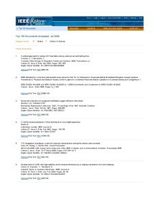
Results Hypoxia. Intracellular Localization of Liposomes and Antisense Oligonucleotides. The use of ASO or siRNA to suppress the expression of a targeted mRNA is limited by their weak ability to penetrate the cellular plasma membrane and interfere with cell genetic material. Therefore, a special delivery system is required to enhance the cell penetration. We have successfully used liposomes to deliver genetic material.4,5,17 To show that liposomes themselves and ASO incorporated into liposomes can penetrate into the cellular cytoplasm and nuclei, we labeled liposomes with rhodamine or osmium tetroxide and ASO with fluorescein isothiocyanate (FITC) and incubated with human embryonic kidney 293 cells within 48 h. In addition, cell nuclei were stained with Hoechst 33258 nuclear dye. The labels were visualized by confocal microscopy (rhodamine), electron transmission microscopy (osmium tetroxide), or fluorescence microscopy (FITC and Hoechst 33258). Typical images obtained in these experiments are presented in Figure 1. Confocal microscope images show that rhodamine-labeled liposomes (red fluorescence) penetrated and accumulated inside cells (Figure 1A,B). Osmium-labeled liposomes were found in cellular perinuclear regions (predominantly in membrane-limited organelles) on electron microscopy images as black spheres with an average diameter of 100-200 nm (identified by arrows in Figure 1C). These data show that liposomes were most probably internalized intact by endocytosis (not by the fusion with the plasma membrane) and after destruction release their payload in the perinuclear region. Further fluorescence microscopy analysis showed that FITC-labeled antisense oligonucleotides delivered by liposomes did penetrate cells and accumulated in the cytoplasm and nuclei (Figure 1D,F). The nuclei can be seen in Figure 1E as bluecolored structures. Therefore, we showed that liposomes can penetrate human cells and deliver genetic material into the cytoplasm and nuclei. Selection of Antisense Oligonucleotides and siRNA Sequences. The sequence of ASO targeted to JNK1 mRNA was selected on the basis of the data reported by Garay et al.13 The DNA backbone of all bases in oligonucleotides was P-ethoxy-modified to enhance nuclease resistance, eliminate electric charge, and increase the level of incorporation into liposomes. Neutral liposomes were used for intracellular delivery of antisense oligonucleotides. Three siRNA sequences targeted to exon 1, exons 1 and 2, and exon 8 of JNK1 protein (locus 5599) were selected as possible candidates for JNK1 suppression. Cationic liposomes were used for intracellular delivery of siRNA. Selected liposomal ASO and siRNA were tested on human embryonic kidney 293 cells. Untreated cells and cells treated with empty neutral and cationic liposomes, free (not incorporated into liposomes) ASO, and siRNA were used as controls. The expression of the gene encoding JNK1 protein was assessed by RT-PCR VOL. 3, NO. 4 MOLECULAR PHARMACEUTICS
427
articles
Betigeri et al.
Figure 3. Lactate concentration in human embryonic kidney 293 cells under normoxia and hypoxia. Means ( the standard deviation are shown: lane 1, control (fresh medium); lane 2, empty neutral liposomes; lane 3, empty cationic liposomes; lane 4, free (nonliposomal) ASO targeted to JNK1; lane 5, free (nonliposomal) siRNA targeted to JNK1 (exon 1); lane 6, liposomal ASO targeted to JNK1; and lane 7, liposomal siRNA targeted to JNK1 (exon 1). Asterisks denote a P of <0.05 when compared with control (normoxia).
Figure 1. Internalization of liposomes and liposomal antisense oligonucleotides in human embryonic kidney 293 cells studied by confocal (A and B), transmission electron (C), and fluorescence (D-F) microscopy. (A and D) Visible light images. Liposomes were labeled with rhodamine (B, red fluorescence) or osmium tetroxide (C, arrows). Cellular nuclei were stained with Hoechst 33258 nuclear dye (E, blue fluorescence). Antisense oligonucleotides were labeled with fluorescein isothiocyanate (F, green fluorescence).
Figure 2. Typical image of RT-PCR products of RNA isolated from human embryonic kidney 293 cells incubated with (1) fresh medium (control), (2) empty neutral liposomes, (3) empty cationic liposomes, (4) free (nonliposomal) ASO targeted to JNK1, (5) liposomal ASO targeted to JNK1, (6) free (nonliposomal) siRNA targeted to JNK1 (exon 1), (7) liposomal siRNA targeted to JNK1 (exon 1), (8) free (nonliposomal) siRNA targeted to JNK1 (exons 1 and 2), (9) liposomal siRNA targeted to JNK1 (exons 1 and 2), (10) free (nonliposomal) siRNA targeted to JNK1 (exon 8), and (11) liposomal siRNA targeted to JNK1 (exon 8).
in cells incubated for 48 h with substances mentioned above (Figure 2). As expected, empty liposomes did not influence the level of targeted mRNA (please compare bands 1 and 3 in Figure 2). Free nonliposomal ASO and all nonliposomal siRNA samples showed a limited or no impact on the expression of JNK1 mRNA. In contrast, liposomal formula428
MOLECULAR PHARMACEUTICS VOL. 3, NO. 4
tions of ASO (band 5) and siRNA targeted to exon 1 (band 7) and exons 1 and 2 (band 9) substantially decreased the level of expression of JNK1 mRNA. On the basis of the results of these experiments, we selected siRNA targeted to exon 1 of JNK1 mRNA (sense sequence GGA GCU CAA GGA AUA GUA UTT) for future experiments. Taken together, these data show that the delivery of ASO and siRNA by liposomes substantially enhanced their specific activity and in turn resulted in a decrease in the level of expression of targeted mRNA. Suppression of JNK1 Activity Attenuates Apoptosis and Mortality under Hypoxia. Hypoxia was induced in cell culture by incubation of human embryonic kidney 293 cells within 48 h in a humidified atmosphere of 1% O2, 5% CO2, and 94% N2 at 37 °C. The incubation led to the statistically significant (P < 0.05) lactate accumulation in cell homogenates, indicating that cellular hypoxia was severe and was accompanied by the substantial activation of anaerobic glycolysis (Figure 3). As expected, suppression of JNK1 did not influence the severity of cellular hypoxia and did not change the concentration of lactic acid under normoxic and hypoxic conditions. Analysis of JNK1 mRNA (Table 1) and protein (Figures 4 and 5) showed that hypoxia significantly (P < 0.05) increased the level of expression of JNK1 mRNA and protein. The level of the expression of JNK1 protein increased with the duration of hypoxic exposure and reached a plateau after 36-48 h of hypoxia (Figure 4). The upregulation of this protein led to the overexpression of procaspase 9 and, more importantly, the active form of caspase 9 (Figure 5), initiating the caspase-dependent signaling pathway of apoptosis. The activation of caspases finally led to the significant (P < 0.05) apoptosis induction (Figure 6A) and the decrease in cellular viability (Figure 6B). Approximately 50% of cells died under hypoxia. Empty liposomes and free (nonliposomal) ASO did not significantly influence the expression of the JNK1 gene and protein, caspase 9 expression, apoptosis induction, and cell death
articles
JNK1, Molecular Target To Limit Cell Death Table 1. Expression of JNK1 mRNA (RT-PCR) in Human Embryonic Kidney 293 Cells under Normoxia and Hypoxiaa series
normoxia
hypoxia
control (fresh medium) empty neutral liposomes empty cationic liposomes free (nonliposomal) ASO targeted to JNK1 free (nonliposomal) siRNA targeted to JNK1 (exon 1) liposomal ASO targeted to JNK1 liposomal siRNA targeted to JNK1 (exon 1)
147.5 ( 5.1 150.0 ( 4.2 151.6 ( 7.5 149.6 ( 0.9
197.7 ( 0.6b 202.2 ( 9.0b 203.9 ( 7.7b 204.7 ( 6.8b
144.4 ( 1.5
169.4 ( 2.3b,c
94.1 ( 3.0b
109.0 ( 0.6b,c
63.7 ( 1.3b
62.8 ( 1.3b,c
a Means ( the standard deviation are shown. b P < 0.05 when compared with control (normoxia). c P < 0.05 when compared with hypoxia, without treatment.
Figure 4. Influence of hypoxia on the expression of JNK1 protein (Western blotting) in human embryonic kidney 293 cells.
under both normoxia and hypoxia. Free siRNA slightly, but significantly (P < 0.05), limited this overexpression under hypoxia. However, such limitation did not change the expression of pro- and active caspase 9 (Figure 5), apoptosis, and cellular viability (Figure 6). In contrast, liposomal ASO and siRNA significantly (P < 0.05) decreased the level of expression of JNK1 mRNA and protein under both normoxia and hypoxia and prevented the overexpression of this JNK1 under hypoxic conditions. The level of expression of JNK1 mRNA and protein under hypoxia after the treatment with liposomal ASO and siRNA was significantly (P < 0.05) lower when compared with the untreated control under normoxic conditions. The suppression of JNK1 protein activity led to the statistically significant decrease in the level of expression of pro- and active caspase 9. Consequently, the level of apoptosis under hypoxic conditions after the treatment with liposomal ASO and siRNA was decreased to
Figure 5. Expression of JNK1 and pro- and active caspase 9 proteins (Western blotting) in human embryonic kidney 293 cells under normoxia and hypoxia. Means ( the standard deviation are shown: lane 1, control (fresh medium); lane 2, empty neutral liposomes; lane 3, empty cationic liposomes; lane 4, free (nonliposomal) ASO targeted to JNK1; lane 5, free (nonliposomal) siRNA targeted to JNK1 (exon 1); lane 6, liposomal ASO targeted to JNK1; and lane 7, liposomal siRNA targeted to JNK1 (exon 1). Asterisks denote a P of <0.05 when compared with control (normoxia). Daggers denote a P of <0.05 when compared with hypoxia, without treatment.
the control levels (Figure 6A), and cellular viability after hypoxia was significantly (P < 0.05) higher (Figure 6B).
Discussion Tissue hypoxia is often a concomitant manifestation of the primary disorders caused by environmental impacts, stress, and diseases of different etiology. This secondary tissue hypoxia and accompanied cellular metabolic disorders complicate the progress of the primary disease and/or the normal physiological adaptive responses to the damaging environmental impacts. Therefore, along with measures VOL. 3, NO. 4 MOLECULAR PHARMACEUTICS
429
articles
Figure 6. Influence of the suppression of JNK1 activity on apoptosis (A) and cellular viability (B) in human embryonic kidney 293 cells under normoxia and hypoxia. Means ( the standard deviation are shown: lane 1, control (fresh medium); lane 2, empty neutral liposomes; lane 3, empty cationic liposomes; lane 4, free (nonliposomal) ASO targeted to JNK1; lane 5, free (nonliposomal) siRNA targeted to JNK1 (exon 1); lane 6, liposomal ASO targeted to JNK1; and lane 7, liposomal siRNA targeted to JNK1 (exon 1). Asterisks denote a P of <0.05 when compared with control (normoxia). Daggers denote a P of <0.05 when compared with hypoxia, without treatment.
directed toward correcting the main cause of secondary tissue hypoxia, it would be beneficial to use preparations that increase the cellular resistance to oxygen deficiency. This pharmacological remediation of secondary hypoxic damage should significantly increase the efficacy of the treatment of primary disease, increase the effectiveness of the adaptive responses, and prevent extensive cellular damage and death. However, despite many investigations aimed at the study of antihypoxic action of different physiologically active substances, antihypoxants are not widely used in clinical practice and environmental physiology. Therefore, the search for an effective antihypoxic therapy is an important task of modern pharmacology. On the basis of our previous research and literature search,1,2 we concluded that the most effective strategy which can lead to the substantial limitation of tissue damage under the conditions of continuous severe secondary hypoxia is the increase in cellular resistance against hypoxia. Because
430
MOLECULAR PHARMACEUTICS VOL. 3, NO. 4
Betigeri et al. apoptosis plays a significant role in the development of tissue damage under severe tissue hypoxia,1,2,4,5 our attention was focused on the apoptosis signaling cascade under severe hypoxia. The study of the main regulator of cellular response to oxygen deficit, hypoxia inducible factor, showed that its activation under hypoxic conditions induces a bimodal effect: it triggers apoptosis and simultaneously increases the power of antiapoptotic defense.4 Further investigations showed that the latter prevails under hypoxic conditions, and the suppression of this protein activity finally leads to an increase in apoptotic cellular damage and death.5 The second protein, Jun N-terminal kinase, attracted our attention as a key regulator of cellular damage under different stresses, including hypoxia.7-9 In this research, we tested antisense oligonucleotides and siRNA targeted to JNK1 mRA as a tool for breaking cell death signal induced by severe hypoxia and therefore preventing apoptosis activation and cell death under the conditions of a severe oxygen deficit. We also developed and tested an effective liposomal delivery system which is capable of delivering active ingredients into the intracellular perinuclear region. The selected ASO and siRNA delivered by our liposomal system exhibited high efficiency in suppressing JNK1 protein activity. Such suppression prevented activation of the caspase-dependent signaling pathway of apoptosis under both continuous hypoxia and accompanied cellular metabolic disturbances. Finally, it decreased the level of hypoxic activation of apoptosis almost to normal levels and consequently significantly limited cellular mortality induced by severe cellular hypoxia. Although the ASO and siRNA targeted to JNK1 decreased the level of expression of this protein to its control levels and prevented caspase-dependent activation of apoptosis under hypoxia, cellular viability was not restored to control levels. This fact shows that in addition to apoptosis, hypoxia induced additional JNK1-independent pathways of cellular mortality. Previously, we found that severe hypoxia limits cellular respiration, induces acidosis, activates lipid peroxidation, stimulates production of prostaglandins and leukotrienes, and substantially disturbs the phospholipid composition of cellular membranes, finally leading to the initiation of both apoptosis and necrosis.2 It is logical to hypothesize that JNK1-independent necrosis was responsible for the additional cell death induction by hypoxia on the background of suppressed JNK1 protein. On the basis of data that have been obtained, one can conclude that JNK1 protein might be an attractive target for antihypoxic therapy to increase resistance to many pathological conditions and diseases leading to the oxygen deficit. MP060014X