150
IEEE TRANSACTIONS ON PLASMA SCIENCE, VOL. 28, NO. 1, FEBRUARY 2000
Cell Deformation and Increase of Cytotoxicity of Anticancer Drugs Due to Low-Intensity Transient Electromagnetic Pulses Changjun Liu, Baoyi Wang, Zishu Wang, and Hong Zhang
Abstract—In this paper, cell deformation induced by low-intensity electromagnetic pulses (EMP’s) is presented. A broad-band transverse electromagnetic wave cell (BTEM cell) was used in the experimental system to simulate the free space transmission condition. The biological samples were exposed to the EMP field in the BTEM cell. After the chick's erythrocytes were exposed to EMP field, pores on their membranes were observed by a scanning electron microscope. Cell fusion was also found between the chick's erythrocytes as well as between the rabbit's. In other experiments, it is found that the EMP field can increase the cytotoxicity of some anticancer drugs. The results suggest that the membrane deformation is a secondary effect of electromagnetic fields. Index Terms—Bioeffect, electromagnetic pulse (EMP), electroporation, membrane.
I. INTRODUCTION
I
NTENSE electric pulses can cause a temporary loss of the semipermeability of cell membranes, thus leading to pores in the membranes. Recently, interest in electroporation has been increased all over the world. A widely applicable method for electroporation is to expose cells to electric pulses with electric fields of kilovolts per centimeter and of microseconds to milliseconds duration. Mechanisms and applications of electroporation have been discussed by Weaver et al. [1]–[3]. Apart from using high-intensity electric field, a few investigators, such as Tsong, have studied some bioeffects related to electroporation by using low-intensity electric field. Tsong applied low-frequency electric field of intensity from 50 to 200 V/cm to E. coli (JM105) cells, and successfully observed the gene transfection while the DNA/cell ratio was 50–75 [4]. In those experiments, different waveforms such as sine, square, and triangle were used, respectively, with frequencies from 0.1 Hz to 1 MHz. However, the gene transfection observed by Tsong cannot prove that electropores have appeared in the membranes. In a study on bioeffects of transient electromagnetic fields, some nonthermal bioeffects, such as dyskaryosis and broken nucleus of human lymph cells, were found in these experiments [5]. In our follow-up study, the same experimental system was used. After animal erythrocytes were exposed to electric pulses of Manuscript received May 11, 1999; revised October 11, 1999. This work was supported by the Natural Science Foundation of China. C. Liu, B. Wang, and H. Zhang are with the Department of Radio-Electronics, Sichuan University, Chengdu 610064, China (e-mail:
[email protected]). Z. Wang is with the Biology Department, Sichuan University, Chengdu 610064, China. Publisher Item Identifier S 0093-3813(00)01166-8.
peak intensity of 20 V/cm for 20–100 min, pores were found in the membranes by using a scanning electron microscope. Moreover, the Sulforhodamine B (SRB) dyeing method was used to determine the cytotoxicity of some anticancer drugs. Two kinds of effective anticancer drugs were examined with five kinds of cancer cells. It was found that the EMP field was able to increase the cytotoxicity of some anticancer drugs. II. MATERIAL AND METHOD The experimental system is shown as Fig. 1. The broad-band transverse electromagnetic wave cell (BTEM cell) consists of an inner and outer conductor. They are isolated from each other. At both ends they are connected to coaxial cables. The frequency band of the BTEM cell is from 0 up to 18 GHz, so the ultrashort electric pulses can be transferred without waveform distortion. The free space radiation condition can also be simulated. The biological samples are placed in the center of the inner conductor where the electric field is uniform. The biological samples are also shielded from outside electromagnetic interference. The detailed design BTEM cell and its applications can be found in [5] and [6]. A temperature control device is available in the system to keep the BTEM cell at 37 C. The electromagnetic pulse (EMP) source is an MFD-1 nanosecond EMP generator, which is able to generate pulses of up to 200 V amplitude and of duration from 2 to 100 ns. The repetition frequency ranges from 1 to 300 Hz. The waveform of each pulse is similar to a square, and the rise time is 1.2 ns. In all experiments, a EMP amplitude of 187 V is used. The electric field intensity on the center of the inner conductor is 20 V/cm. The duration of EMP is 100 ns, and its repetition frequency is 300 Hz. A. Cell Deformation Chick erythrocytes were used in the experiment. Chick blood was mixed with Alsever's solution (containing glucose 2.05 g, sodium citrate 0.676 g, citric acid 0.055 g, and NaCl 0.42 g in 100-mL solution). Ten milliliters of the mixture was rinsed three times by D-hanks and once by isosmotic low-conductivity solution (3 mM/l Tris, 1 mM/l MgCl , and 10% sucrose, with S/cm and pH ). After chick eryconductivity throcytes were detached, they were diluted to 5 mL with isosmotic low-conductivity solution, and then were divided into six groups. Each group was 0.1 mL. Five groups were placed into the BTEM cell and exposed to EMP field for 20, 40, 60, 80,
0093–3813/00$10.00 © 2000 IEEE
LIU et al.: CELL DEFORMATION AND INCREASE OF CYTOTOXICITY OF ANTICANCER DRUGS
Fig. 1.
151
Experimental system and structure of BTEM cell.
and 100 min, respectively. The other group was a control group, which was placed in the incubator. After EMP field exposure, 0.5-mL, 1% glutaraldhyde, was added to each group, and they were supported by Formvar. Then glutaraldhyde-osmic acid was used for cell fixing, and the specimens were dehydrated through four graded acetone-isoamyl acetates (50%, 70%, 90%, and 100%) and critical-point dried from liquid carbon dioxide. A scanning electron microscope was used to observe the chick erythrocyte membranes. B. Field-Induced Cell Fusion The rabbit blood and chick blood were mixed with Alsever's solution, and the procedure followed the same steps described above. After EMP field exposure, RPMI 1640 medium was added to culture the cells for 120 min. Then they were dyed with Giemsa for 10 min. A light microscope was used to take photos for the cells. The chick erythrocyte is larger than the rabbit's, and the chick erythrocyte has a nucleus while the rabbit erythrocyte has none. Therefore, it is easy to distinguish the chick erythrocyte from the rabbit's. The detailed steps and reagent batch formula are described in [7].
80 min, respectively. After 48-h incubation, the anticancer drugs were rinsed out of all groups. b) Test groups are placed into the BTEM cell and exposed to EMP field for 50 and 100 min, respectively. At 120 min, the anticancer drugs, in all groups, were washed out and culture medium was added. They were then incubated for 48 h. All cancer cells attached to the plastic substrata during growth, so they were fixed by gently layering 50 L of 50% trichloroacetic acid (TCA) (4 C) for 1 h. Then they were washed five times with deionized water to remove TCA and other media. TCA-fixed cells were stained with SRB solution for 30 min. Then SRB was removed, and the cultures were quickly rinsed four times with 1% acetic acid to remove the unbound dyes. SRB purchased from Sigma was dissolved in 1% acetic acid for cell staining. Bound dye was solubilized with 10-mM unbuffered Tris base (pH 10.5) for 5 min. The optical density (OD) was read in a Uvmax microtiter plate reader at wavelength of 564 nm (a suboptimal wavelength). The OD value has a linear relation to the number of cells. III. RESULTS AND DISCUSSION
C. Cytotoxicity of Anticancer Drugs
A. Pore Creation and Field-Induced Cell Fusion
Five kinds of cancer cells, Lu-06, Re-07, Bre-04, Col-02, and Col-05, were used in the experiment. Two kinds of anticancer drugs were C7 and hydroxycamptothecine (CPT). They were liposoluble and hydrotropic, respectively. Moreover, they were proven effective in the disease-oriented drug screening test. Cells were cultured in RPMI-1640 medium with glutamine, bicarbonate, and 5% FCS. The culture medium was changed at two-day intervals. Cells were dissociated in NKT buffer (137 mM NaCl, 5.4 mM KCl, and 10 mM Tris, with pH = 7.4). Screening cultures were plated in microtiter plates containing 0.2 mL of medium per well. After 24-h culture, anticancer drugs were added to the plated cells with three concentrations. The concentrations were 0.3, 0.03, and 0 g/mL for the C7 anticancer drug; and 10, 1, and 0 g/mL for the CPT anticancer drug. The following methods were used. a) All groups, except the control group, are placed into the BTEM cell and exposed to EMP field for 20, 40, 60, and
Pores on the erythrocyte membranes can be observed by scanning electron microscopy. The ratio of membrane openings to membrane openings is greater than 2%. The diameter of pores ranges from 20 to 500 nm. For different exposing duration from 20 to 100 min, pores can be found in these groups. However, there is no obvious dosage effect. In the control group, no pores were found. Fig. 2(a) and (b) shows cell membrane deformation. Compared to membrane openings, the yield of cell fusion is low: less than 5%. The erythrocyte cells fusion can be found in chick–chick, rabbit–rabbit and chick–rabbit. Fig. 2(c) illustrates the cell fusion between the chick erythrocyte and the rabbit's. B. Increase of the Cytotoxicity of Anticancer Drugs The results are shown in Tables I and II. In method (a), anticancer drugs stay with cancer cells for 48 h. In both C7 and CPT test groups, there is no obvious difference of the OD value.
152
IEEE TRANSACTIONS ON PLASMA SCIENCE, VOL. 28, NO. 1, FEBRUARY 2000
TABLE I OD VALUES OF C7 DRUG
TABLE II OD VALUES OF CPT DRUG
(a)
the control group for the same anticancer drug concentration. By has been obtained. Moreusing statistical methods, over, effects of EMP fields are stronger in 3.0- g/mL anticancer groups than in 0.3- g/mL groups. For different cancer cells, the sensitivity to drug dosage and EMP field exposure is different. In the CPT groups, there is no obvious difference between the test groups and the control group. C. Discussion (b)
(c) Fig. 2.
Membrane openings and cell fusion.
It means the EMP field has not affected the cytotoxicity of anticancer drugs. In method (b), the anticancer drug stays with cancer cells for only 120 min. For the C7 groups, except the zero concentration group, the OD values of test groups are obviously different from
In the BTEM cell, the maximum electric field intensity in the center of the inner conductor (where the biological sample is located) is 20 V/cm. The maximum induced transmembrane potential of the erythrocytes suspended in low-conductivity solution can be estimated to less than 10 mV. The total transmembrane potential is far less than 500 mV, which is the threshold of transmembrane potential for membrane electric breakdown. Therefore, the mechanism may be different from the high-intensity field-induced electroporation. The induced transmembrane potential can affect the orientation of some voltage sensitive proteins or the function of hemoglobin. Especially, the effective dipole interaction pressure may play an important role in the membrane deformation under low-conductivity condition [8], [9]. In each group, there is a dependence on age. The mechanical elastic ratio of some aged cells may be decreased, which can lead to the transient micropores on the membrane’s being larger when exposed to EMP fields. This result is in agreement with Weaver's and Mintzer's model of interaction between the pores and membrane and the transmembrane potential [10]. If there are some larger pores on membrane, the osmotic equilibrium will cease to exist. Some small ions or molecules may enter the cell by crossing the pores in the membrane. Based on our calculation, pores in the membrane can reduce the membrane energy barrier for ions considerably [11]. Since macromolecules inside the membrane cannot
LIU et al.: CELL DEFORMATION AND INCREASE OF CYTOTOXICITY OF ANTICANCER DRUGS
(a)
(b)
153
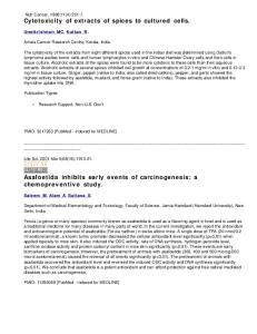
cell increases the membrane surface, which in turn increases the size of pores. Therefore, under repetitive action of EMP fields, a large pore will finally be formed in the membrane. Therefore, the membrane deformation is a secondary effect of electric field, and this pore opening in membranes seems to be secondary electroporation [12]. Fig. 3 shows some photos of cell membrane changes. In (a), the bulge formed by cytoplasm before a pore appears is shown; in (b), the cytoplasm is emitted from a pore on the membrane; in (c), the depression is shown, which occurs when the membrane is healing. These photos are similar to Chang's observations by rapid-freezing electron microscopy [12]. The increase of the cytotoxicity of anticancer drugs may be related to electroporation [13]. If electroporation occurs, anticancer drug molecules can cross the membrane through pores. More drugs can enter the cancer cells, which leads to the increase of cytotoxicity. However, recent research results show that this effect is an electrically stimulated process [14]. Molecules of the drugs are absorbed at the cell surface. Then, by stimulation due to EMP fields, molecules can be transferred into the cell. In further experiments, we plan to study the mechanism for low-intensity EMP stimulation. The experimental results show that EMP field exposure is not effective for all anticancer drugs. The difference between methods (a) and (b) is the duration of anticancer drugs staying with cancer cells. Due to the very long duration of 48 h, the effects of drugs entering the cell after exposure to EMP fields may be inhibited. So only in method (b), some electromagnetic bioeffects can be found. Also, the molecular weight of C7 and CPT is 320 and 677, and the dissolvability of them is different. It is not surprising that the effects of the EMP field on C7 and CPT are different. In the ongoing study, we are trying to perform gene transfection under the same conditions as described, or use some other methods, such as Trypan blue dying, to further the experiment. These may be useful to understand the membrane deformation. Research on membrane deformation caused by low-intensity EMP is in an early phase, and the mechanism is still not understood. Further experimental study and analysis are required. REFERENCES
(c) Fig. 3.
Membrane structure changes.
cross membrane through the pores, the osmotic pressure difference will not vanish until a large pore is present. Swelling of the
[1] J. C. Weaver, “Electroporation in cells and tissues: A biophysical phenomenon due to electromagnetic fields.,” Radio Sci., vol. 30, no. 1, p. 205, 1995. [2] U. Zimmermann, “Electric field-mediated fusion and related electrical phenomena.,” Biochem. Biophys. Acta, vol. 694, p. 227, 1982. [3] T. Y. Tsong, “Electroporation of cell membrane,” Biophys. J., vol. 60, no. 8, p. 297, 1991. [4] , “Study of mechanisms of electric field-induced DNA transfection II,” Biophys. J., vol. 58, no. 10, p. 897, 1990. [5] B. Wang, J. Yang, Q. Guo, X. Runmin, C. Liu, H. Zhang, F. Zou, and Z. Wang, “Experimental and mechanism analysis on bioeffects by nanosecond electromagnetic pulses.,” Sci. China C, vol. 40, no. 3, p. 301, 1997. [6] K. Huang and Y. Liu, “A simple method for calculating electric and magnetic field in GTEM cell.,” IEEE Trans. Electromag. Compat., vol. 36, no. 4, p. 335, 1994. [7] Z. Wang, Experimental Technique in Human and Animal Cytogenetics. Sichuan, China: Sichuan Univ. Press, 1987, pp. 332–337. [8] D. A. Stenger, K. V. Kaler, and S. W. Hui, “Dipole interactions in electrofusion: Contributions of membrane potential and effective dipole interaction pressures,” Biophys. J., vol. 59, no. 5, p. 1074, 1991.
154
IEEE TRANSACTIONS ON PLASMA SCIENCE, VOL. 28, NO. 1, FEBRUARY 2000
[9] M. Krueger and F. Thom, “Deformation and stability of erythrocytes in high-frequency electric fields down to subzero temperatures,” Biophys. J., no. 11, p. 2653, 1997. [10] J. C. Weaver and R. A. Mintzer, “Decreased bilayer stability due to transmembrane potentials,” Phys. Lett., vol. 86A, p. 57, 1981. [11] C. Liu, B. Wang, and H. Zhang, “Energy calculation and analysis of an ion crossing membrane,” J. Sichuan Univ., vol. 35, no. 1, p. 55, 1998. [12] D. C. Chang and T. S. Reese, “Changes in membrane structure induced by electroporation as revealed by rapid-freezing electron microscopy,” Biophys. J., vol. 58, no. 7, p. 1, 1990. [13] O. Stephane, B. Jean, P. Claude, and M. Lluis, “Transient electropermeabilizaion of cells in culture: Increase of the cytotoxicity of anticancer drugs,” Biochem. Pharmacol., vol. 37, no. 24, p. 4727, 1988. [14] V. Ganeva, B. Galutzo, and J. Teissie, “Fast kinetic studies of plasmid DNA transfer in intact yeast cells mediated by electropulsation,” Biochem. Biophys. Res. Commun., vol. 214, no. 3, p. 825, 1995.
Changjun Liu was born in Xingtai, Hebei, China, on April 18, 1973. He received the B.S. degree from Hebei University, China, and the M.S. degree from Sichuan University, China, where he currently is pursuing the Ph.D. degree in biomedical engineering from the Department of Radio-Electronics. His current interests include bioeffects due to weak electromagnetic field, numerical techniques in FDTD method, and microwave chemistry.
Baoyi Wang graduated from Sichuan University, China, in 1962. He was a Visiting Scholar from 1981 to 1982 in the Department of Electronics Engineering, Michigan State University, Ann Arbor. He has been Director of the Department of Radio-Electronics, Sichuan University, for more than ten years, and is a Senior Member of the Chinese Institute of Electronics. His current areas of research include bioelectromagnetics, biological effects due to transient field, and electromagnetic target discrimination.
Zishu Wang graduated from the Department of Biology, Sichuan University, China, in 1958. She became a Professor at Sichuan University in 1990. Her main field is cytogenetics and cytobiology.
Hong Zhang graduated from the Department of Radio-Electronics, Sichuan University, China. She is now a Teacher with Sichuan University. Her field of interest is bioelectromagnetics.