Proc. Natl. Acad. Sci. USA Vol. 93, pp. 4316-4319, April 1996
Biochemistry
Didemnin binds to the protein palmitoyl thioesterase responsible for infantile neuronal ceroid lipofuscinosis CRAIG M. CREWS*t§, WILLIAM S. LANEt,
AND
STUART L. SCHREIBER*
*Howard Hughes Medical Institute, Department of Chemistry and Chemical Biology, and *Microchemistry Facility, Harvard University, 12 Oxford Street, Cambridge, MA 02138
Contributed by Stuart L. Schreiber, January 5, 1996
ABSTRACT The marine natural product didemnin B, currently in clinical trials as an antitumor agent, has several potent biological activities apparently mediated by distinct mechanisms. Our initial investigation of didemnin B resulted in the discovery of its GTP-dependent binding of the translation elongation factor EFla. This finding is consistent with the protein synthesis inhibitory activity of didemnin B observed at intermediate concentrations. To begin to dissect the mechanisms involved in the cytostatic and immunosuppressive activities of didemnin B, observed at low concentrations, additional didemnin-binding proteins were sought. Here we report the purification of a 36-kDa glycosylated didemninbinding protein from bovine brain lysate. Cloning of the human cDNA encoding this protein revealed a strong sequence similarity with palmitoyl protein thioesterase (PPT), an enzyme that removes palmitate from H-Ras and the Gas subunits of heterotrimeric GTP-binding proteins in vitro. Mutations in PPT have recently been shown to be responsible for infantile neuronal ceroid lipofuscinosis, which is a severe brain disorder characterized by progressive loss of brain function and early death.
required for ribosomal recognition of EF-la. This interaction is likely the source of didemnin's reported protein synthesis inhibition. Although it has been proposed that didemnin B's other activities are a consequence of its ability to inhibit protein synthesis (15), the effective concentrations of didemnin B's cytostatic and immunosuppressive activities are one and three orders of magnitude lower than that required for inhibition of protein synthesis, suggesting that distinct mech-
anisms may be involved. To study didemnin's antiproliferative and immunosuppressive activities, affinity chromatography was used to identify additional binding proteins. In this report we describe the purification and cloning of a new didemnin-binding protein with sequence similarity to palmitoyl protein thioesterase (PPT), mutant forms of which are responsible for infantile neuronal ceroid lipofuscinosis (INCL). In light of known characteristics of this disease, models for didemnin's effect on PPT are discussed.1
MATERIALS AND METHODS
Reagents. Didemnin A was a generous gift of K. Rinehart (University of Illinois). Affi-Gel-10 was purchased from BioRad, and e-aminocaproic acid, leupeptin, pepstatin A, phenylmethylsulfonyl fluoride, and 5'-guanylyl imidodiphosphate (GMP-PNP) were obtained from Sigma. Synthesis of Didemnin Affinity Matrix. Didemnin A (1 mg)
Cell-permeant molecules have been used to investigate many
aspects of cellular function. The study of natural products that
bind intracellular proteins has extended this approach into new areas of biology. For example, natural products have facilitated the elucidation of several signal transduction pathways (e.g., brefeldin A, wortmannin, rapamycin, and FK506) (1-4). Various natural products have also been used or are being developed as therapeutic agents. The first natural product derived from marine sources to enter clinical trials, didemnin B (ref. 5; see Fig. 1 for structure), is currently in phase II trials as an antitumor agent for treatment of breast, ovarian, cervical, myeloma, and lung cancers (6-10). Preliminary results of these clinical trials also show a partial response against nonHodgkin lymphoma (11). The potency, specificity, and cell permeability of didemnin B led us to investigate the molecular basis for its actions on cycling cells. Three biological activities have been reported for didemnin B that were of interest to us: its ability to inhibit protein synthesis, induce G, cell cycle arrest, and suppress a mixed lymphocyte reaction (12, 13). These activities range in effective concentrations over five orders of magnitude, suggesting they are mediated by different mechanisms. To investigate these mechanisms, we have have been characterizing proteins that bind didemnin B. Using a didemnin affinity matrix, we have previously shown that a major intracellular didemninbinding protein is the translation elongation factor la (EF-la) (14). It was shown that didemnin B binds uniquely to the activated GTP-bound form of EF-la, albeit with modest affinity. Didemnin B's specificity for GTP-bound EF-la suggests that it recognizes the same structural determinants
in 200 jul of dichloromethane was incubated with 1.2 eq of bromotris (pyrrolidino) phosphonium hexafluorophosphate, >6 eq of triethylamine and 3 eq of fluorenylmethoxycarbonylprotected s-aminocaproic acid at 0°C for 2 min and then warmed to room temperature. After 2 h, the crude reaction mixture was purified by preparative thin-layer chromatography in dichloromethane/methanol (95:5) to provide fluorenylmethoxycarbonyl-aminocaproic acid-tethered didemnin A in a 79% yield. Proton NMR spectra were consistent with the expected product. The protecting group was removed by incubation in 20% piperidine in dimethylformamide for 20 min followed by evaporation under vacuum. Deprotected aminocaproic acid-tethered didemnin A was coupled to 6 ml of Affi-Gel-10 in dimethyl sulfoxide for 14 h at room temperature.
Purification of Didemnin-Binding Proteins.Analytical scale. Bovine brain (4 g) was homogenized on ice in lysis buffer (10 mM K2HPO4, pH 7.1/1 mM EDTA/5 mM EGTA/10 mM MgCl2/50 mM 13-glycerophosphate, pH 7.1/2 mM Na3VO4/2 mM dithiothreitol/5 mM NaF) plus protease inhibitors (10 ,tg/ml pepstatin A, 10 ,tg/ml leupeptin, and 1 mM phenylmethylsulfonyl fluoride) using a Tekmar tissue homogenizer Abbreviations: EF-la, elongation factor la; PPT, palmitoyl protein thioesterase; INCL, infantile neuronal ceroid lipofuscinosis; GMP-
PNP, 5'-guanylyl imidodiphosphate. tPresent address: Yale Biology Department, 219 Prospect Avenue, New Haven, CT 06520-8103; e-mail address:
[email protected]. §To whom reprint requests should be addressed. 1The sequence reported in this paper has been deposited in the GenBank database (accession no. U44772).
publication costs of this article were defrayed in part by page charge payment. This article must therefore be hereby marked "advertisement" in accordance with 18 U.S.C. §1734 solely to indicate this fact.
The
4316
Proc. Natl. Acad. Sci. USA 93 (1996)
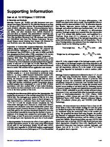
Biochemistry: Crews et al. Me2Tyr
Pro
(ToNH
f*
,H
Me
Didemnin A
R
Didemnin B
R=
Didemnin
Hips
/"P
1n
.
,
Structurf
Compound
OMe MoLeu
e m'M M
Ir
Ist
affigel R
N-Acetyl-
R=
Didemnin A
H
£YknOH x~) 0 S/-NI N-AffoigeIO
R=
,
o
YCH3 0
FIG. 1. Structures of didemnins and of the didemnin-based reagent used for affinity chromatography.
propionic acid. before centrifugation at 100,000 x g for 30 min at 4°C. The high-speed supernatant was batch adsorbed onto 0.4 ml of Sepharose SP to remove the majority of EF-la. The Sepharose SP eluate was incubated with 20 ,ul of didemnin affinity matrix for 4 h in the presence or absence of 100 ,tM GMP-PNP and in the presence or absence of 5 ,tM N-acetyldidemnin A (14). The matrix was washed four times with 1 ml of lysis buffer containing 0.15 M NaCI and 10 mM EDTA. Bound proteins were visualized by SDS/12.5% PAGE (16) and silver staining. Preparative scale. To obtain enough protein for micropeptide sequence analysis, 200 g of bovine brain was homogenized, centrifuged, and adsorbed as described above before incubation with 1 ml of didemnin Affigel 10 for 90 min at 4°C. Bound proteins were washed four times with 10 ml of lysis buffer plus 0.6 M NaCl and transferred to poly(vinylidene difluoride) membrane after SDS/PAGE as described (17). Internal tryptic peptide sequence analysis was performed as described (18). Degenerate PCR and cDNA Cloning. Fragments of the cDNA encoding the p36 didemnin B-binding protein were obtained using PCR and the degenerate oligonucleotides 1
[GGAATTCGT(G/C/T)CA(G/A/)GC(A/T/C)GA(G/A)TA(T/C)TGGCA(T/C)GA(T/C)] and 2 [GCGGATCCTC(T/ C)TG(A/C/G/T)A(G/A)(A/C/G/T)GG(T/G/A)AT(A/ C/G/T)GT(T/C)TC] with 2 ,g of peripheral blood leukocyte plasmid cDNA library as template (19). Reaction conditions were 2 mM MgCl2, 1 ,tM oligonucleotide primer, lx Taq buffer, and 1 unit of Taq polymerase for 3 min at 94°C (template denaturation) followed by two initial cycles of 1-min denaturation at 94°C, 1-min annealing at 45°C, and 1.5-min extension at 72°C. Thirty more cycles were performed under the same conditions as the two initial cycles but at an annealing temperature of 55°C. These cycles were followed by a final extension at 72°C for 5 min. The resulting 249-bp PCR fragment was radioactively labeled using PCR and used to screen a human Jurkat T-cell cDNA library (20). One clone, 29B, was selected for dideoxy-nucleotide DNA sequencing (21). mRNA Tissue Expression. mRNA blots of various human tissues (Clontech) were probed with radiolabeled clone 29B as described (20) and exposed to x-ray film (Kodak, X-Omat)
4317
1st, isostatine; Hip, hydroxyisovaleryl-
didemnin B Affi-Gel. However, unlike the 50-kDa didemnin
B-binding proten (residual EF-la) seen in lane 4, these new didemnin B-binding proteins do not require GTP for didemnin B binding. Nevertheless, binding of all three was blocked by excess free N-acetyldidemnin A (lane 5). Cloning and Sequence Analysis of Human p36 cDNA. Purification of these new didemnin B-binding proteins was scaled up to obtain tryptic peptide sequence information. Amino-terminal sequence analysis of purified p34 and p36 showed that both proteins start at the same internal sequence, DPPAPL, suggesting they are differentially modified forms of the same gene product (data not shown). Based on internal tryptic peptide sequence information obtained from the 36kDa protein (underlined in Fig. 3), degenerate oligonucleotide primers were synthesized and used to amplify by PCR a cDNA
97.4
-
67
low
44
43
11 31
using an intensifing screen at -80°C for 3 days.
RESULTS AND DISCUSSION Purification of a 36-kDa Didemnin-Binding Protein. Be-
the previously identified didemnin-binding protein, EFla, is abundant within cells, the identification of additional didemnin B-binding proteins required the selective removal of this protein. Taking advantage of the high isoelectric point of EF-la, the majority of EF-la was removed by using cationexchange chromatography at pH 7.1. This precleared lysate was then analyzed for additional didemnin B-binding activities in the presence and absence of the nonhydrolyzable GTP analog GMP-PNP. As shown in Fig. 2, a major protein (36 kDa) and a minor protein (34 kDa) bind specifically to cause
21.5 1
2
3
4
5
FIG. 2. Affinity chromatography of didemnin-binding proteins from bovine brain lysate. Lanes: 1, control matrix; 2, didemnin-AffiGel matrix; 3, control matrix with 200 ,/M GMP-PNP; 4, didemninAffi-Gel matrix with 200 ,uM GMP-PNP; 5, didemnin-Affi-Gel matrix with 200 A/M GMP-PNP using lysate preincubated for 60 min with 5 ,uM N-acetyldidemnin A.
Bohmsr:Cese l
4318
1 MASPGCLCVL Bovine MASSSCLWLL Rat MASPGYRRLL Consensus MAS------L Human
Proc. Natl. Acad. Sci. USA 93
~
(1996) 102
AVALLPWTCA ALAFLLGSCA AAALLPWCCA A-A-L---CA
SRALQHLDPP APLPLVIWHG MGDSCCNPLS SLALGHLDPP APLPLVIWHG MGDSCCNPLS AWALGHLDPP SPPPLVIWHG MGDSCCNPMS --AL-HLDPP -P-PLVIWHG MGDSCCNP-S
MGAIKKMVEK MGAIKKMVEK MGSIKKMVEK MG-IKKMVEK
KIPGIYVLSL KIPGIHVLSL EIPGIYVLSL -IPGI-VLSL
EIGKTLMEDV EIGKTLREDV EIGKNMVEDV EIGK---EDV
GFSQGGQFLR GFSQGGQFLR GFSQGGQFLR GFSQGGQFLR
AVAQRCPSPP AVAQRCPSPP AVAQRCPTPP AVAQRCP-PP
PGESSHICDF PGESSHICDF PGESSHICDF PGESSHICDF
IRKTLNAGAY IRKTLNAGAY IRKSLNAGAY IRK-LNAGAY
SKVVQERLVQ AEYWHDPIKE DVYRNHSIFL AD
103
ENSFFLNVNS ENSFFLNVNS ENSFFLNVNL ENSFFLNVN-
QVTTVCQALA QVTTVCQILA QVGMACQILE QV---CQ-L-
KD KD KD KD
204
PKLQQGYNAM Bovine PKLQQGYNAM Rat PKLQHGYNAI Consensus PKLQ-GYNAHuman
MINLISVGGQ MVNLISVGGQ MMTLISVGGQ M--LISVGGQ
HQGVFGLPRC HQGVFGLPRC HQGVFGLPRC HQGVFGLPRC
NKAIQERLVO AEYWHDPIRE DIYRNHSIFL AD SKVVQERLVQ AQYWHDPIKE EVYRNCSIFL AD -K--QERLVQ A-YWHDPI-E --YRN-SIFL AD
205 INQERGINES YKKNLMALKK FVMVKFLNDS IVDPVDSEWF GFYRSGQAKE TIPLQETSLY TQDRLGLKEM DNAGQLVFLA Bovine INOERGVNES YKKNLMALKK FVMVKFLNDT IVDPVDSEWF GFYRSGQAKE TIPLOESTLY TODRLGLKAM DKAGQLVFLA Rat INQERHINES YKENLMALKK FVMVKFFNDS IVDPVDSEWF GFYRSGQAKE TIPLQETTLY TEDRLGLKKM DKAGKLVFLA Consensus INQER--NES YK-NLMALKK FVMVKF-ND- IVDPVDSEWF GFYRSGQAKE TIPLQE--LY T-DRLGLK-M D-AG-LVFLA Human
306 TEGDHLQLSE EWFYAHIIPF LG
LEGDHLQLSE EWFYAHIIPF LE KEGDHLQISK EWFTAHIIPF LK -EGDHLQ-S- EWF-AHIIPF L-
FIG. 3. Amino acid sequence alignment of human, bovine, and rat PPTs. Arrow indicates the amino terminus of the mature protein as determined by amino-terminal sequencing. The consensus sequences that comprise the catalytic site of thioesterases are in boldface type and the tryptic peptide sequences identified from the purified didemnin-binding protein are underlined.
fragment from a human peripheral blood lymphocyte library (19). Using this PCR fragment as a probe, we isolated cDNA clones from a library made from the human Jurkat T-cell line (3). However, DNA sequence analyses of the initial 15 clones revealed that all but 1 began with a G+C-rich region and lacked a starting methionine codon. These incomplete clones are most likely truncated due to the difficulty of reverse transcription through regions of potential RNA secondary structure. The first methionine codon of the longest clone (29B), which is a favorable initiation codon based on the Kozak rule (22), is at nucleotide 41. The open reading frame starting at this methionine encodes a protein of 306 amino acids. This clone has three potential N-glycosylation sites (N197, N212, and N232) as well as a 27-amino acid secretory signal sequence. Edman degradation and mass spectrometry of purified p36 tryptic peptides confirmed the likely glycosylation of at least one asparagine (N232) (data not shown). The results of mRNA tissue distribution analysis of clone 29B using a human mRNA tissue blot are shown in Fig. 4. A 2.4-kb transcript was detected in all tissues examined. Longer exposures revealed an additional 4.5-kb transcript in lower abundance, which is most strongly expressed in heart and skeletal muscle. Because the longest clone isolated (2.0 kb) lacks a polyadenylate tail and the message seen in various tissues is 2.4 kb, this clone is not full length. Nevertheless, this clone probably contains the entire open reading frame of the gene, because in vitro translation of its RNA yields 34- and 36-kDa proteins when synthesized in the absence and presence of microsomal membrane fractions, respectively (data not shown). A
2
3
4
5
6
7
~.' .....~' ..'{'"j . ,'
Kb
8
10 11
9
-
7.5
-
-
4.4
-
12 13 14 15 16
-o2.4 - ..tlo t
-1.35
B
FIG. 4. Tissue distribution of human PPT message. (A) Lanes: 1, spleen; 2, thymus; 3, prostate; 4, testis; 5, ovary; 6, small intestine; 7, colon; 8, peripheral blood leukocytes; 9, heart; 10, brain; 11, placenta; 12, lung; 13, liver; 14, skeletal muscle; 15, kidney; 16, pancreas. (B)
Actin control message levels. Two actin messages are present in cardiac and skeletal muscle.
Human PPT. Comparison of the predicted amino acid sequence of clone 29B with sequences deposited in GenBank revealed high degrees of similarity with rat and bovine PPT (23). Camp and Hofman (24) purified bovine PPT on the basis of its ability to remove palmitate from recombinant H-Ras and the a subunit of heterotrimeric GTP-binding proteins (24). However, these GTP-binding proteins may not be physiological substrates of this didemnin binding protein, because the protein contains a secretory signal sequence and because we have observed that the recombinant protein is at least partially secreted following its expression in insect Sf9 cells (data not shown). In Fig. 3, an alignment of the human, rat, and bovine sequences of PPT is shown. The human sequence reported here is 90 and 94% identical at the amino acid level with the rat and bovine sequences, respectively. In addition, the human sequence contains the conserved amino terminal Gly-XaaSer-Xaa-Gly and COOH-terminal Gly-Asp-His motifs previously suggested to be characteristic of thioesterases (25). Role of PPT in INCL. INCL is an autosomal recessive progressive encephalopathy that is characterized by the accumulation of the autofluorescent pigment lipofuscin in the lysosomal compartments of cells. This disease, which leads to mental deterioration and early death, has recently been shown to be due to mutations in the human PPT gene (26). PPT normally has both secreted and cellular expression patterns (ref. 23 and unpublished data). However, an inactivating mutation near the putative lipase catalytic domain (tryptophan replacing arginine-122) results in the absence of PPT secretion. In addition, mutant PPT is found by immunofluorescence to be associated with the endoplasmic reticulum, whereas wild-type PPT revealed strong staining of the Golgi apparatus and secretory vesicles (26). These PPT staining patterns may reflect the overall perturbation of glycoprotein synthesis observed in INCL cases. Studies of cells and tissues of INCL patients have shown a significant decrease in glycoprotein and oligoglycosphingolipid production (27, 28). In addition, increased dolichol levels in storage cytosomes of INCL patients have been observed, indicating abnormal protein glycosylation
(29-31).
Protease inhibition has been suggested to be the underlying of ceroid lipofuscinosis (32, 33). This theory has been supported by studies showing that infusion of protease inhibitors into rat brain and liver results in the formation of electron-dense lysosomal aggregrates reminiscent of ceroid lipofuscin (34, 35). In addition, endogenous levels of the serine protease inhibitor al-antichymotrypsin are significantly increased in brain tissues from patients with INCL (36, 37). The catalytic mechanisms of serine proteases and the family of serine esterases to which PPT belongs are highly similar. These similarities are underscored by the observations that the serine cause
Biochemistry: Crews et al. proteases have been reported to have esterase activity (38, 39)
and that certain thioesterases have been reported to have protease activity (40). Thus, the observed correlation of lipofuscin induction and increases in endogenous or artificial serine protease inhibitors may be due to the inhibition of PPT, and it is possible that PPT also has protease activity. This hypothesis is in agreement with the results indicating that the loss of PPT correlates with INCL (26). Inconsistent with this model, however, is the finding that PPT is not inhibited by the serine protease inhibitor phenylmethylsulfonyl fluoride (24). Although we have not yet investigated the effects of didemnin on recombinant PPT activity, its structure suggests that it may prove to be an inhibitor of that activity. Statine residues inhibit (aspartyl) proteases by structurally mimicking the tetrahedral intermediate of amide bond hydrolysis (41-44). It is possible that didemnin's structurally similar isostatine residue (Fig. 1) is playing an analogous role in the inhibition of PPT activity. In support of this hypothesis, studies of structureactivity relationships have shown that didemnin's isostatine residue is essential for its biological activity (45). Conclusion. Didemnin B is one of the most potent cytostatic and immunosuppressive marine natural products identified to date. Although didemnin's physiological effects have been examined in detail, the molecular and cellular mechanisms by which this cyclic depsipeptide mediates its actions are unknown. As a step towards the elucidation of these mechanisms, we now report the purification from bovine brain and the molecular cloning from a human T-cell cDNA library of a specific didemnin-binding PPT. Inactivating mutations in this thioesterase have been shown to be responsible for the progressive encephalopathy INCL. Experiments are now underway to investigate the nature of didemnin interactions with human recombinant PPT expressed in Sf9 cells, including the possible effect of didemnin B on the enyzmatic activity of PPT. The cell-permeant didemnin B molecule may prove to be an effective probe of PPT's cellular function and of its role in INCL. We thank members of the Harvard Microchemistry Facility for their
expert assistance and Dr. Jon Collins for his technical advice. This work was supported by a postdoctoral fellowship from the Cancer Research Institute
(C.M.C.). S.L.S. is an Investigator at the Howard
Hughes Medical Institute.
1. Schreiber, S. L. (1992) Chem. Eng. News 70, 22-32. 2. Chung, J. K., Grammer, T. C., Lemon, K. P., Kazlauskas, A. & Blenis, J. (1994) Nature (London) 370, 71-75. 3. Brown, E. J., Albers, M. W., Shin, T. B., Ichikawa, K., Keith, C. T., Lane, W. S. & Schreiber, S. L. (1994) Nature (London) 369, 756-758. 4. Liu, J., Farmer, J. D. J., Lane, W. S., Friedman, J., Weissman, I. & Schreiber, S. L. (1991) Cell 66, 807-815. 5. Chun, H. G., Davies, B., Hoth, D., Suffness, M., Plowman, J., Flora, K., Grieshaber, C. & Leyland, J. B. (1986) Invest. New Drugs 4, 279-284. 6. Benvenuto, J. A., Newman, R. A., Bignami, G. S., Raybould, T. J., Raber, M. N., Esparza, L. & Walter, R. S. (1992) Invest. New Drugs 10, 113-117. 7. Malfetano, J. H., Blessing, J. A. & Jacobs, A. J. (1993) Am. J. Clin. Oncol. 16, 47-49. 8. Jacobs, A. J., Blessing, J. A. & Munoz, A. (1992) Gynecol. Oncol. 44, 268-270. 9. Weiss, R. B., Peterson, B. L., Allen, S. L., Browning, S. M., Duggan, D. B. & Schiffer, C. A. (1994) Invest. New Drugs 12, 41-43.
Proc. Natl. Acad. Sci. USA 93 (1996)
4319
10. Shin, D. M., Holoye, P. Y., Forman, A., Winn, R., Perezsoler, R., Dakhil, S., Rosenthal, J., Raber, M. N. & Hong, W. K. (1994) Invest. New Drugs 12, 243-249. 11. Division of Cancer Treatment (1994) Annual Report to the Food and Drug Administration: Didemnin B, National Service Center report 325319 (National Cancer Institute, Bethesda, MD). 12. Crampton, S. L., Adams, E. G., Kuentzel, S. L., Li, L. H., Badiner, G. & Bhuyan, B. K. (1984) Cancer Res 44, 1796-1801. 13. Montgomery, D. W. & Zukoski, C. F. (1985) Transplantation 40, 49-56. 14. Crews, C. M., Collins, J. L., Lane, W. S., Snapper, M. L. & Schreiber, S. L. (1994) J. Biol. Chem. 269, 15411-15414. 15. Li, L. H., Timmins, L. G., Wallace, T. L., Krueger, W. C., Prairie, M. D. & Im, W. B. (1984) Cancer Lett. 23, 279-288. 16. Laemmli, U. K. (1970) Nature (London) 227, 680-685. 17. Aebersold, R. H., Leavitt, J., Saavedra, R. A., Hood, L. E. & Kent, S. B. H. (1987) Proc. Natl. Acad. Sci. USA 84, 6970-6974. 18. Lane, W. S., Galat, A., Harding, M. W. & Schreiber, S. L. (1991) J. Protein Chem. 10, 151-160. 19. Aruffo, A. & Seed, B. (1987) Proc. Natl. Acad. Sci. USA 84, 8573-8577. 20. Maniatis, T., Fritsch, E. F. & Sambrook, J. (1982) Molecular Cloning: A Laboratory Manual (Cold Spring Harbor Lab. Press,
Plainview, NY).
21. Sanger, F., Nicklen, S. & Coulson, A. R. (1977) Proc. Natl. Acad. Sci. USA 74, 5463-5467. 22. Kozak, M. (1989) J. Cell Biol. 108, 229-241. 23. Camp, L. A., Verkruyse, L. A., Afendis, S. J., Slaughter, C. A. & Hofman, S. L. (1994) J. Biol. Chem. 269, 23212-23219. 24. Camp, L. A. & Hofman, S. L. (1993) J. Biol. Chem. 268, 2256622574. 25. Derewenda, Z. S. & Sharp, A. M. (1993) Trends Biochem. Sci. 18, 20-25. 26. Vesa, J., Hellsten, E., Verkruyse, L. A., Camp, L. A., Rapola, J., Santavuori, P., Hofmann, S. L. & Peltonen, L. (1995) Nature (London) 376, 584-587. 27. Svennerholm, L., Fredman, P., Jungbjer, B., Mansson, J. E., Rynmark, B. M., Bostrom, K., Hagberg, B., Noren, L. & Santavuori, P. (1987) J. Neurochem. 49, 1772-1783. 28. Heaney, K. J., Kieras, F. J. & Wisniewski, K. E. (1992) Biochem. Med. Metab. Biol. 48, 137-142. 29. Wolfe, L. S., Gauthier, S., Haltia, M. & Palo, J. (1988) Am. J. Med. Genet. Suppl. 5, 233-242. 30. Ng, Y. K. N., Palo, J., Haltia, M. & Wolfe, L. S. (1983) J. Neurochem. 40, 1465-1473. 31. Hall, N. A., Thomas, 0. J., Dell, A., Haltia, M., Lake, B. D. & Patrick, A. D. (1992) Am. J. Med. Genet. 42, 580-585. 32. Ivy, G. O., Wenzel, J., Baudry, M. & Lynch, G. (1984) Science 226, 985-987. 33. Ivy, G. 0. (1992) Am. J. Med. Genet. 42, 555-560. 34. Ivy, G. O., Kanai, S., Ohta, M., Sato, Y., Otsubo, K. & Kitani, K. (1991) Mech. Ageing Dev. 57, 213-231. 36. Kitani, K., Ohta, M., Kanai, S., Nokubo, M., Sato, Y., Otsubo, K. & Ivy, G. 0. (1989) Adv. Exp. Med. Biol. 266, 75-92. 37. Wisniewski, K. E. & Kida, E. (1990) Exp. Neurol. 110, 121-126. 38. el Shirbiny, A. (1994) Adv. Clin. Chem. 31, 99-133. 39. Whittaker, R. G., Manthey, M. K., Le, B. D. & Hayes, P. J. (1994) Anal. Biochem. 220, 238-243. 40. Cho, H. & Cronan, J. E. (1994) J. Bacteriol. 176, 1793-1795. 41. Marciniszyn, J., Jr., Hartsuck, J. A. & Tang, J. (1976) J. Biol. Chem. 251, 7088-7094. 42. Bott, R., Subramanian, E. & Davies, D. R. (1982) Biochemistry 21, 6956-6962. 43. James, M. N. G., Sielecki, A., Salituro, F., Rich, D. H. & Hofman, T. (1982) Proc. Natl. Acad. Sci. USA 79, 6137-6141. 44. Rich, D. H., Bernatowicz, M. S. & Schmidt, P. G. (1982) J. Am. Chem. Soc. 104, 3635-3636. 45. Sakai, R., Kishore, V., Kundu, B., Faircloth, G., Gloer, J. B., Carney, J. R., Namikoshi, M., Hughes, R. G., Garcia Gravalos, D., Garcia de Quesada, T., Wilson, G. R., Heid, R. M. & Rinehart, K. L. (1996) J. Med. Chem., in press.