Journal of General Virology (1999), 80, 1119–1126. Printed in Great Britain ...................................................................................................................................................................................................................................................................................
Identification of the RNA-binding sites of the triple gene block protein 1 of bamboo mosaic potexvirus Chiung-Hua Wung,1 Yau-Heiu Hsu,2 Dann-Ying Liou,2 Wei-Cheng Huang,3 Na-Sheng Lin4 and Ban-Yang Chang3 1, 2, 3
Agricultural Biotechnology Laboratories1, Institute of Agricultural Biotechnology2, and Institute of Biochemistry3, National Chung-Hsing University, Taichung, Taiwan 402, Republic of China 4 Institute of Botany, Academia Sinica, Taipei, Taiwan 115, Republic of China
The triple gene block protein 1 (TGBp1) encoded by open reading frame 2 of bamboo mosaic potexvirus (BaMV) was overexpressed in Escherichia coli and purified in order to test its RNAbinding activity. UV crosslinking assays revealed that the RNA-binding activity was present mainly in the soluble fraction of the refolded TGBp1. The binding activity was nonspecific and salt concentration-dependent : activity was present at 0–50 mM NaCl but was almost abolished at 200 mM. The RNA-binding domain was located by deletion mutagenesis to the N-terminal 3–24 amino acids of TGBp1. Sequence alignment analysis of the N-terminal 25 amino acids of the TGBp1 homologues of potexviruses identified three arginine residues. Arg-to-Ala substitution at any one of the three arginines eliminated most of the RNA-binding activity, indicating that they were all critical to the RNA-binding activity of the TGBp1 of BaMV.
Introduction The flexuous rod-shaped particles of potexviruses have single-strand positive-sense RNA genomes that encode five well-conserved open reading frames (ORFs) (Bancroft et al., 1991 ; Zuidema et al., 1989). Mutational analysis of the genome of white clover mosaic potexvirus (WClMV) has revealed that the central three ORFs (ORFs 2, 3 and 4), the ‘ triple gene block ’ (TGB), are required for the systemic spread of virus in host plants (Beck et al., 1991). The TGB counterparts are also found in the genomes of other viruses such as carla-, beny-, pomo-, peclu- and hordeiviruses, the products of which are also essential for virus movement (Gilmer et al., 1992 ; Petty & Jackson, 1990). The movement of plant viruses from cell to cell is assumed to occur by passage through plasmodesmata. Virus-encoded movement proteins have been shown to be involved in this process (Maule, 1991 ; Deom et al., 1992). The P30 protein of tobacco mosaic virus (TMV) induces an increase in the sizeexclusion limit of plasmodesmata (Wolf et al., 1989) and possesses nonspecific single-stranded RNA-binding activity to Author for correspondence : Ban-Yang Chang. Fax j886 4 286 1905. e-mail bychang!mail.nchu.edu.tw
0001-6086 # 1999 SGM
shape viral nucleic acids into a transferable form (Citovsky et al., 1990, 1992). The movement proteins of comoviruses (Shanks et al., 1989 ; van Lent et al., 1990), caulimoviruses (Linstead et al., 1988) and nepoviruses (Wieczorek & Sanfaçon, 1993) participate in the formation of tubular structures extended from plasmodesmata in which virus-like particles are detected. Different from the above-mentioned, the TGBp1 homologues of potexviruses are predominantly associated with cytoplasmic inclusions (Davies et al., 1993 ; Rouleau et al., 1994 ; Chang et al., 1997). None of them was detected in plasmodesmata or in tubular structures. Recently, the TGBp1 homologue of white clove mosaic virus (WClMV) was shown to facilitate the transport of the infectious transcripts in company with capsid protein, and movement of this ribonucleoprotein complex absolutely requires the presence of the other two TGB proteins (Lough et al., 1998). The TGBp1 encoded by ORF2 of foxtail mosaic potexvirus (FMV) is predominantly fractionated in the soluble fraction (S30) of infected tissue homogenate (Rouleau et al., 1994) ; however, the TGBp1 homologue of potato virus X (PVX) is mainly in the insoluble P1 and P30 fractions (Davies et al., 1993), and the TGBp1 homologue of bamboo mosaic potexvirus (BaMV) is associated mainly with the cell wall and P30 fractions (Chang et al., 1997). Despite the difference in BBBJ
C.-H. Wung and others
Table 1. Oligonucleotides used in this study Oligonucleotide
Sequence
Description
‘< >’, DNA sequences being deleted. ‘ ‘, sites of Arg-to-Ala substitution.
solubility, the TGBp1 homologues of the three potexviruses share high amino acid sequence similarity, suggesting that they have similar biochemical properties. An NTP-binding helicase motif has been found for the three homologues (Bancroft et al., 1991 ; Skryabin et al., 1988 ; Lin et al., 1994), and RNA-binding and ATPase activities are also reported for the TGBp1 homologues of FMV and PVX (Kalinina et al., 1996 ; Rouleau et al., 1994). A common feature of plant virus movement proteins is that they bind nucleic acids in vitro (Citovsky et al., 1990 ; Osman et al., 1992 ; Schoumacher et al., 1992 ; Tacke et al., 1991 ; Rouleau et al., 1994 ; Ivanov et al., 1994 ; Bleykasten et al., 1996). Distinct domains essential for this binding have been identified in several movement proteins, such as the P30 protein of TMV (Citovsky et al., 1992), the 35 kDa protein of red clover necrotic mosaic virus (RCNMV) (Giesman-Cookmayer & Lommel, 1993), the P3 protein of alfalfa mosaic virus (Schoumacher et al., 1994), the P1 protein of cauliflower mosaic virus (CaMV) (Thomas & Maule, 1995), the 3a protein of cucumber mosaic virus (Vaquero et al., 1997) and the 3a protein of brome mosaic bromovirus (Fujita et al., 1998). However, no RNA-binding domain has been identified for the TGBp1 BBCA
homologues of potexviruses. Here, we report the RNAbinding domain, the RNA-binding properties and the possible mechanisms leading to RNA binding of the TGBp1 homologue of BaMV.
Methods
Bacterial strains and plasmids. Escherichia coli HMS 174 was used as host for cloning the mutated ORF2 of BaMV-O, while E. coli BL21(DE3) was used as host for overexpression of the mutant proteins. Plasmid pJP1 containing the cDNA of ORF2 (Chang et al., 1997) served as template for construction of the ORF2 derivatives by PCR. Plasmids pBaHB (Lin et al., 1993), pJP1 (Chang et al., 1997), pCT14 and pWC1 (B. Y. Chang, unpublished data), which contain cDNA fragments of the whole genome, ORF2, ORF3 and ORF4 of BaMV, respectively, were linearized and used as templates for in vitro transcription.
Construction of deletion and substitution mutants of TGBp1. Mutations resulting in either amino acid deletions or substitutions in TGBp1 of BaMV were constructed by PCR (Ho et al., 1989 ; Horton et al., 1990). The linker primer sequences used are shown in Table 1. The forward and reverse primers contain a BamHI and an NcoI site (underlined bases), respectively. After PCR synthesis, the mutated DNA fragments were cut with both BamHI and NcoI and used to replace the wild-type counterpart on pJP1 plasmid. The positions of amino acids
RNA-binding sites of BaMV movement protein
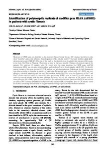
Fig. 1. Schematic illustration of the BaMV genome, the deletion mutants constructed within TGBp1 and their ability to bind ssRNA at various pH values. (a) The relative positions and the molecular masses of proteins encoded by the five open reading frames are depicted along the BaMV genome. (b) The deleted amino acids of TGBp1 are given above the dashed lines. The remaining portions are represented by rectangles, with the calculated molecular masses (in kDa) shown on the right. The ‘ j ’ sign indicates positive RNA-binding activity, whereas the ‘ k ’ sign indicates absence of RNA-binding activity.
deleted in the mutant proteins (M1–M7) are shown in Fig. 1. The substitution mutants including R11A, R16A, R21A and R16,21A were created by the same PCR method. Oligonucleotides used are also shown in Table 1. Each of the mutant derivatives of both types was shown to have the predicted structure by DNA sequencing and restriction enzyme digestion.
Overexpression and purification of the wild-type and mutant TGBp1. E. coli BL21(DE3) harbouring a plasmid containing a mutated ORF2 was grown at 37 mC in 2iYT medium (16 g tryptone, 10 g yeast extract, 5 g NaCl per litre) and induced with 0n4 mM IPTG when the culture had reached an optical density at 550 nm of 0n6–0n8 ; 50 min after IPTG induction, rifampicin was added to the culture at a final concentration of 100 µg\ml. The cells were harvested 4 h later by centrifugation at 5000 r.p.m. for 15 min and washed twice with buffer L (10 mM Tris–HCl, pH 8n0, 200 mM NaCl, 1 mM EDTA, 10 % glycerol, 1 mM PMSF, 1 mM DTT). The washed cells were then homogenized with a French press at 18 000 p.s.i. (ca. 124 MPa) followed by centrifugation at 12 000 r.p.m. for 10 min. The cell debris, which contained inclusions of the target protein, was washed several times with buffer L and suspended in the same buffer. The target protein was present to about 70 % homogeneity at this step. To further purify the protein, an equal volume of sample buffer (0n125 M Tris–HCl, pH 6n8, 4 % SDS, 20 % glycerol, 0n002 % bromophenol blue) was added to the protein suspension. The protein sample was then run in SDS–polyacrylamide gels and stained with 0n25 M KCl (4 mC). Gel fragments containing the target protein were excised and chopped into small pieces. The target protein was electroeluted with a Blue tank (Isco) and precipitated with 80 % acetone by centrifugation at 10 000 r.p.m., 4 mC for 15 min. The recovered protein was washed twice with a solution containing 20 % (v\v) buffer A (50 mM Tris–HCl, pH 8n0, 0n1 mM EDTA, 150 mM NaCl) and 80 % (v\v) acetone followed by denaturation with buffer L supplemented with 6 M urea. Refolding of target protein was performed by dialysis against crosslinking buffer (10 mM Tris–HCl, 50 mM NaCl, 5 mM MgCl , # 1 mM EDTA, 1 mM DTT, 0n1 mM PMSF, 10 % glycerol) at appropriate pH using a microdialysis apparatus (BRL).
Photochemical crosslinking of protein and RNA. Transcripts of BaMV, which were synthesized and labelled with [α-$#P]CTP (3000 Ci\mmol ; Amersham) in vitro, were used as templates for RNA binding
by the UV crosslinking method. Basically, 10 ng of $#P-labelled RNA transcript (1i10( c.p.m.\µg) and an appropriate amount of renatured TGBp1 were mixed in buffer containing 10 mM Tris–HCl, 50 mM NaCl, 5 mM MgCl , 1 mM EDTA, 1 mM DTT, 0n1 mM PMSF, 10 % glycerol. # The final volume of binding mixture was 12 µl. The mixture was incubated on ice for 15 min before irradiation for about 8 min in a Stratalinker (Stratagene) at 8 cm from the light source (0n78 J\cm#). After UV crosslinking, 10 µg of RNase A was added to the mixture and the sample was further incubated at 37 mC for 30 min to digest uncrosslinked RNA. The resulting sample was then boiled for 5 min with an equal volume of sample buffer (0n125 M Tris–HCl, pH 6n8, 4 % SDS, 20 % glycerol, 0n002 % bromophenol blue) and electrophoresed through a 12n5 % SDS–polyacrylamide gel. The gel was stained with Coomassie blue, dried, and autoradiographed after electrophoresis.
Results RNA-binding properties of TGBp1
Unlike the TGBp1 homologue of FMV (Rouleau et al., 1994), the TGBp1 encoded by ORF2 of BaMV (Fig. 1 a) was fairly low in solubility when overexpressed in E. coli (Chang et al., 1997). The solubility of the refolded TGBp1 was also low, about 0n06–0n1 µg\µl depending on buffer pH and salt concentration. RNA-binding activity has been observed for the insoluble aggregates of CaMV P1 protein (Thomas & Maule, 1995) and the 66 kDa cytoplasmic inclusion of tamarillo mosaic potyvirus (TamMV) (Eagles et al., 1994). In order to determine the RNA-binding activity of TGBp1 in both the pelleted and soluble states, the method of photochemical crosslinking between protein and RNA was adopted. As shown in Fig. 2 (a), both monomeric (M) and dimeric (D) forms of the TGBp1 crosslinked to the $#P-labelled RNA transcript of the 3h noncoding region of BaMV were observed for the suspension of refolded protein (Sus). Moreover, the soluble fraction (Sup1) possessed most of the RNA-binding activity of BBCB
C.-H. Wung and others
Fig. 2. RNA-binding properties of the BaMV TGBp1. (a)RNA-binding activity of the soluble and pellet fractions of TGBp1. Each fraction was incubated with the 32P-labelled transcript (220 bases) of the 3h noncoding region of BaMV. The RNA-binding activity of protein was analysed by the photochemical crosslinking method as described in Methods. Sus, 4 µg of the refolded TGBp1 suspended in crosslinking buffer containing 50 mM NaCl ; P1, the precipitate of the refolded protein suspension (Sus) after centrifugation at 100 g for 1 min ; Sup1, the soluble fraction of the refolded protein ; Sup2 and Sup3, the soluble fractions of the resuspended P1 and P2, respectively ; P3, the pellet fraction of the resuspended P2. C, RNA only. M and D indicate the monomeric and dimeric forms of TGBp1. (b) Effect of NaCl concentration on the stability of RNA–protein complexes. One µg of the refolded TGBp1 suspended in crosslinking buffer was used for the assay. The RNA-binding activity was analysed as in (a) except that experiments were carried out at different NaCl concentrations (mM). (c) Interaction of the refolded TGBp1 with different RNA transcripts. Formation of protein and RNA complexes was analysed as in (a). The RNA transcripts of ORF2 (759 nucleotides), ORF3 (357 nucleotides), ORF4 (156 nucleotides) and the 3h noncoding sequence (220 nucleotides) of BaMV were synthesized using T7 RNA polymerase and BamHI-linearized pJP1, pCT14, pWC1 and pBaHB plasmids, respectively. The complementary sequence of the 3h noncoding region was synthesized using SP6 RNA polymerase and HindIII-linearized pBaHB plasmid. The numbers in the right margin of each panel indicate the molecular masses (kDa) of protein markers.
the refolded protein (Sus) compared with the pellet (P1). Resuspension of P1 did not further solubilize the refolded protein (data not shown) ; only trace RNA-binding activity was detected in both supernatant (Sup2 and Sup3) and pellet (P3) fractions of the resuspended sample, indicating that only a certain portion of the refolded TGBp1 was soluble and active. The stability of TGBp1 and RNA complexes was analysed at different salt concentrations (Fig. 2 b). The TGBp1 bound RNA maximally at 0 mM NaCl ; the binding stability decreased as salt concentration was increased. At 200 mM NaCl, the protein–RNA complexes were almost abolished. We also investigated whether TGBp1 bound preferentially to particular nucleic acid sequences. To accomplish this, four more RNA probes including ORF2, ORF3, ORF4 and the complementary sequence of the 3h noncoding region were prepared. The similarity in binding strength among different RNA transcripts (Fig. 2 c) clearly indicated that TGBp1 had no sequence specificity in interaction with BaMV RNA. Mapping of the RNA-binding domain(s) of TGBp1
To map the RNA-binding domain(s) of TGBp1, we expressed and purified a series of mutant proteins with deletions spanning different regions from the N- to the Cterminal end of the protein (Fig. 1 b). All of the mutant proteins overexpressed in E. coli BL21(DE3) had observed molecular masses ranging from 22 to 25 kDa, comparable to those calculated from their amino acid sequences. Moreover, all of them were able to react with antiserum raised against the wildBBCC
type TGBp1 (data not shown), suggesting that they were all in the correct reading frame. Since the wild-type TGBp1 overexpressed in E. coli in the form of inclusion bodies was capable of binding to RNA after urea denaturation and subsequent refolding processes (see Methods), the same strategy was adopted to prepare mutant proteins for RNA-binding assays. Furthermore, the decrease in the RNA-binding activity of TGBp1 at a relatively high salt condition (Fig. 2 b) suggested that ionic interaction was one of the major force involved in TGBp1 and RNA binding. Since the surface charge of a protein might be influenced by buffer pH (the lower the buffer pH, the higher the net surface positive charge), we located the RNA-binding site by analysing the effects of buffer pH and deletion sites on binding of the mutant proteins to RNA (Figs 1 and 3). Clearly, all the mutant proteins were able to bind RNA molecules when the buffer pH was as low as 6n0 or 6n6 (Fig. 3 a, b). However, M1 failed to bind RNA as the pH was raised to 7n4 (Fig. 3 c) ; M2, M3 and M7 also lost their RNA-binding activities as the pH was further increased to 8n0. Only M5 and M6, which had relatively higher pI values (9n14 and 9n11, respectively), and M4, with a pI of 8n01, retained their RNA-binding activity at the highest pH (Fig. 3 d ). The loss of RNA-binding activity of M1, M2, M3 and M7 at pH 8n0 suggested that multiple positively charged regions were responsible for the RNA binding of TGBp1 at elevated pH. However, the exclusive loss of RNA-binding activity of M1 at pH 7n4 indicated that amino acids 3–24 in the Nterminal region of TGBp1 were more critical than those in other regions for RNA binding at physiological pH.
RNA-binding sites of BaMV movement protein
Fig. 3. Mapping of the RNA-binding domain of the BaMV TGBp1 at different pH values. (a) pH 6n0 ; (b) pH 6n6 ; (c) pH 7n4 ; (d) pH 8n0. About 2n5 µg of the wild-type or deletion mutant proteins (M1 to M7) dialysed against the crosslinking buffer containing 50 mM NaCl at the indicated pH was incubated with the 32P-labelled transcript of the 3h noncoding region of BaMV followed by UV radiation and RNase A digestion. The RNA-binding activity of each deletion mutant was analysed as described in Methods. Lane WT, RNA binding with the wild-type TGBp1 ; lane C, RNA only. M and D indicate the monomeric and dimeric forms of TGBp1. The numbers in the right margin of each panel indicate the molecular masses (kDa) of protein markers.
Fig. 4. Alignment of amino acid sequences at the N-terminal regions of the TGBp1 homologues of potexviruses and the four Arg-to-Ala substitutions for identification of RNA-binding sites. (a) The amino acid residues which are identical at the N-terminal regions of the TGBp1 homologues of BaMV, FMV and PVX are indicated by stars ; those which are homologous are indicated by dots. BaMV, bamboo mosaic virus ; FMV, foxtail mosaic virus ; PVX, potato virus ; PMA, papaya mosaic virus ; WClMV, white clove mosaic virus ; NMV, narcissus mosaic virus. (b)The Arg-to-Ala substitutions in TGBp1. The positions of amino acid substitutions are underlined and indicated in the left margin of the panel.
Involvement of three arginine residues in RNA binding
To identify amino acid residues which were responsible for binding of TGBp1 to RNA at physiological pH, the N-terminal 25 amino acids of six TGBp1 homologues of potexviruses were aligned. Besides certain conserved hydrophobic and uncharged amino acids (such as isoleucine, leucine, threonine
and glycine), the basic amino acid at position 16 was also conserved among the homologues ; it was either arginine or lysine (Fig. 4). We suspected that Arg-16 and possibly also Arg-11 and Arg-21 in the neighbourhood were involved in RNA binding. To test this idea, four mutant proteins with either single (residues 11, 16 or 21) or double (both residues 16 and 21) Arg-to-Ala substitutions were constructed. These BBCD
C.-H. Wung and others
Fig. 5. Identification of amino acid residues involved in RNA binding of TGBp1. About 0n2 µg of either wild-type or substitution mutant protein, solubilized in crosslinking buffer containing 50 mM NaCl at pH 7n4, was assayed for RNA-binding activities by the photochemical crosslinking method described in Methods. Lane C, RNA only ; lane WT, RNA binding with wild-type TGBp1 ; lane M1, RNA binding with M1 protein. R11A, R16A, R21A and R16, 21A are the four mutant proteins with Arg-to-Ala substitutions shown in Fig. 4 (b). M indicates the monomeric form of TGBp1. The numbers in the right margin indicate the molecular masses (kDa) of protein markers.
proteins were overexpressed and purified (data not shown). Effects of Arg-to-Ala substitutions on RNA-binding activity of the mutant proteins were analysed. Similar to the loss of RNAbinding activity of the M1 protein at pH 7n4, the RNA-binding activities of R16A and R21A were almost abolished (Fig. 5), and only trace activity was retained by R11A and R16,21A. These results indicate that all three arginine residues are essential for RNA-binding activity of the TGBp1 of BaMV.
Discussion We have been able to test the RNA-binding activity of the BaMV TGBp1 overexpressed in E. coli in the form of inclusion bodies. UV crosslinking assays show that most of the RNAbinding activity resides in the soluble fraction of the refolded TGBp1. Studies on the RNA-binding activities of the deletion and substitution mutant proteins have enabled us to locate the RNA-binding domain to the N-terminal region between amino acids 3–24 of TGBp1, within which three arginine residues were shown to be important to RNA binding. Several approaches such as photochemical crosslinking, gel mobility shifting, filter binding and Northwestern blotting have been adopted to detect the interaction between protein and RNA, but preliminary experiments revealed that not all of them were suitable for our purpose. However, we found that the photochemical crosslinking method, which trapped the binding complexes, worked well in our case. The stability of protein–RNA complexes with respect to salt concentration is often a criterion to evaluate the binding strength of the complexes (Li & Palukaitis, 1996). The stability of the RNA–BaMV TGBp1 complex is lower than that formed with BBCE
the more water-soluble TGBp1 homologue of FMV, which remains strongly bound to RNA at 0n2 M NaCl (Rouleau et al., 1994). It is also not as stable as complexes formed with the movement proteins of RCNMV (Osman et al., 1992) and TMV (Citovsky et al., 1990), which are stable even at 0n4 and 0n6 M NaCl, respectively. However, it is similar to those reported for the TGBp1 homologue of PVX (Kalinina et al., 1996 ; Karpova et al., 1997). The physiological significance of the solubility of movement proteins or the stability of RNA–movement protein complexes remains unknown. The proximity of virus aggregates to the cytoplasmic inclusions formed by the TGBp1 homologues in PVX-, FMVand BaMV-infected tissues (Davies et al., 1993 ; Rouleau et al., 1994 ; Chang et al., 1997) raises the possibility that these inclusions are the source of active TGBp1. Possibly, active TGBp1 might be continuously released from protein inclusions in soluble form due to chemical equilibration under certain circumstances. Our finding that most of the RNA-binding activity resides in the soluble fraction of the refolded TGBp1 is consistent with this idea (Fig. 2 a). However, the detection of only a very low amount of soluble protein and a very low level of RNA-binding activity after resuspension of the pelleted TGBp1 is not consistent with this hypothesis (Fig. 2 a). Possibly the protein inclusions in BaMV-infected tissues do not function in the same manner as reported for the insoluble P1 protein of CaMV (Thomas & Maule, 1995) or the 66 kDa cytoplasmic inclusions of TamMV (Eagles et al., 1994). Alternatively, the pelleted TGBp1 may not possess the same structural properties as the protein inclusions in infected tissues due to the improper folding during renaturation. This is the first report on mapping the RNA-binding site in the TGBp1 homologues of potexviruses. Many different structure motifs responsible for the interaction between protein and RNA have been reported (Burd & Dreyfuss, 1994). They include the RNP motif, the arginine-rich motif (ARM), the K homology (KH) motif, the RGG box and the double-stranded RNA-binding motif (DSRM). The RNA-binding domain of TGBp1 was mapped to the N-terminal 24 amino acids, a region within which no such typical structure motif was observed. However, the involvement of three neighbouring arginine residues in RNA binding (Fig. 5) suggests that the TGBp1 of BaMV possesses an ARM-like structure. There are probably nonspecific ionic interactions between the positively charged arginines of TGBp1 and the negatively charged phosphate groups on RNA, as well as specific hydrogen bonding networks between arginines and the RNA sugar-phosphate backbone and bases (Burd & Dreyfuss, 1994). These interactions appear to require a certain structure(s), rather than just particular amino acids, since each of the four Arg-to-Ala substitutions tested destroys most of the RNA-binding activity of TGBp1 (Fig. 5). The predicted absence of α-helix or βstructure in the N-terminal 24 amino acids (Chou & Fasman, 1974) suggests that a loop or bulge structure(s) is present in this region and is important to RNA binding.
RNA-binding sites of BaMV movement protein This research was supported by National Science Council of the Republic of China grant NSC 86-2311-B-005-025-B11.
References Bancroft, J. B., Rouleau, M., Johnston, R., Prins, L. & Mackie, G. A. (1991). The entire nucleotide sequence of foxtail mosaic virus RNA.
Journal of General Virology 72, 2173–2181. Beck, D. L., Guilford, P. J., Voot, D. M., Andersen, M. T. & Forster, R. L. S. (1991). Triple gene block proteins of white clover mosaic
potexvirus are required for transport. Virology 183, 695–702. Bleykasten, C., Gilmer, D., Guilley, H., Richards, K. E. & Jonard, C. (1996). Beet necrotic yellow vein virus 42 kDa triple gene block protein
binds nucleic acid in vitro. Journal of General virology 77, 889–897. Burd, C. G. & Dreyfuss, G. (1994). Conserved structures and diversity of functions of RNA-binding proteins. Science 265, 615–621. Chang, B. Y., Lin, N. S., Liou, D. Y., Chen, J. P., Liou, G. G. & Hsu, Y. H. (1997). Subcellular localization of the 28 kDa protein of the triple-
gene-block of bamboo mosaic potexvirus. Journal of General Virology 78, 1175–1179. Chou, P. Y. & Fasman, G. D. (1974). Empirical predictions of protein conformation. Biochemistry 13, 222–245. Citovsky, V., Knorr, D., Schuster, G. & Zambrisky, P. (1990). The P30 movement protein of tobacco mosaic virus is a single-stranded nucleic acid binding protein. Cell 60, 637–647. Citovsky, V., Wong, M. L., Shaw, A. L., Prasad, B. V. V. & Zambrisky, P. (1992). Visualization and characterization of tobacco mosaic virus
movement protein binding to single-stranded nucleic acid. Plant Cell 4, 397–411. Davies, C., Hills, G. & Baulcombe, D. C. (1993). Sub-cellular localization of the 25 kDa protein encoded in the triple gene block of potato virus X. Virology 197, 166–175. Deom, C. M., Lapidot, M. & Beachy, R. N. (1992). Plant virus movement proteins. Cell 69, 221–224. Eagles, R. M., Balmori-Melian, E., Beck, D. L., Gardner, R. C. & Forster, R. L. S. (1994). Characterization of NTPase, RNA-binding and RNA-
helicase activities of the cytoplasmic inclusion protein of tamarillo mosaic potyvirus. European Journal of Biochemistry 224, 677–684. Fujita, M., Mise, K., Kajiura, Y., Dohi, K. & Furusawa, I. (1998). Nucleic acid-binding properties and subcellular localization of the 3a protein for brome mosaic bromovirus. Journal of General Virology 79, 1273–1280. Giesman-Cookmayer, D. & Lommel, S. A. (1993). Alanine scanning mutagenesis of a plant virus movement protein identifies three functional domains. Plant Cell 5, 973–982. Gilmer, D., Bouzouba, S., Hehn, A., Guilley, H., Richards, K. & Jonard, G. (1992). Efficient cell-to-cell movement of beet necrotic yellow vein
virus requires 3h proximal gene located on RNA2. Virology 189, 40–47. Ho, S. N., Hunt, H. D., Horton, R. M., Pullen, J. K. & Pease, L. R. (1989). Site-directed mutagenesis by overlapping extension using the
polymerase chain reaction. Gene 77, 51–79. Horton, R. M., Cai, Z., Ho, S. N. & Pease, L. R. (1990). Gene splicing by
overlap extension : tailor-made genes using the polymerase chain reaction. Biotechniques 8, 528–535. Ivanov, K. I., Ivanov, P. A., Timofeeva, E. K., Dorokhov, Y. L. & Atabekov, J. G. (1994). The immobilized movement proteins of two
tobamoviruses form stable ribonucleoprotein complexes with full length viral genome RNA. FEBS Letters 346, 217–220. Kalinina, N. O., Fedokin, O. N., Samuilova, O. V., Maiss, E., Korpela, T.,
Morozov, S. Yu. & Atabekov, J. G. (1996). Expression and biochemical analyses of the recombinant potato virus X 25K movement protein. FEBS Letters 397, 75–78. Karpova, O. V., Ivanov, K. I., Rodionova, P. P., Dorokhov, Yu. L. & Atabekov, J. G. (1997). Nontranslatability and dissimilar behavior in
plants and protoplasts of viral RNA and movement protein complexes formed in vitro. Virology 230, 11–21. Li, Q. & Palukaitis, P. (1996). Comparison of the nucleic acid- and NTPbinding properties of the movement protein of cucumber mosaic cucumovirus and tobacco mosaic tobamovirus. Virology 216, 71–79. Lin, N. S., Chen, C. C. & Hsu, Y. H. (1993). Post-embedding in situ hybridization for localization of viral nucleic acid in ultra-thin sections. Journal of Histochemistry and Cytochemistry 41, 1513–1519. Lin, N. S., Lin, B. Y., Lo, N. W., Hu, C. C., Chow, T. Y. & Hsu, Y. H. (1994). Nucleotide sequence of the genomic RNA of bamboo mosaic
potexvirus. Journal of General Virology 75, 2513–2518. Linstead, P. J., Hills, G. J., Plaskitt, K. A., Wilson, I. G., Harker, C. L. & Maule, A. J. (1988). The subcellular location of the gene 1 product of
cauliflower mosaic virus is consistent with a function associated with virus spread. Journal of General Virology 69, 1809–1818. Lough, T. J., Shash, K., Xoconostle-Cazares, B., Hofstra, K. R., Beck, D. L., Balmori, E., Forster, R. L. S. & Lucas, W. J. (1998). Molecular
dissection of the mechanism by which potexvirus triple gene block proteins mediate cell-to-cell transport of infectious RNA. Molecular Plant–Microbe Interactions 11, 801–814. Maule, A. J. (1991). Virus movement in infected plants. Critical Reviews in Plant Sciences 9, 457–473. Osman, T. A. M., Hayes, R. J. & Buck, K. W. (1992). Cooperative binding of the red clover necrotic mosaic virus movement protein to single-stranded nucleic acids. Journal of General Virology 73, 223–227. Petty, I. T. D. & Jackson, A. O. (1990). Mutational analysis of barley stripe mosaic virus RNAβ. Virology 179, 712–718. Rouleau, M., Smith, R. J., Bancroft, J. B. & Mackie, G. A. (1994).
Purification, properties, and subcellular localization of foxtail mosaic potexvirus 26 kDa protein. Virology 204, 254–265. Schoumacher, F., Enry, C., Berna, A., Godefroy-Colburn, T. & StussiGaraud, C. (1992). Nucleic acid-binding properties of the alfalfa mosaic
virus movement protein produced in yeast. Virology 188, 654–667. Schoumacher, F., Giovane, C., Maira, M., Poirson, A., GodefroyColburn, T. & Berna, A. (1994). Mapping of the RNA-binding domain
of the alfalfa mosaic virus movement protein. Journal of General Virology 75, 3199–3202. Shanks, M., Tomenius, K., Clapham, D., Huskisson, N., Barker, P. J., Wilson, I. G., Maule, A. J. & Lomonossoff, G. P. (1989). Identification
and subcellular localization of a putative cell-to-cell transport protein from red clover mottle virus. Virology 173, 400–407. Skryabin, K. G., Morozov, S. Yu., Kraev, A. S., Rozanov, M. N., Chernov, B. K., Lukasheva, L. I. & Atabekov, J. G. (1988). Conserved
and variable elements in RNA genomes of potexviruses. FEBS Letters 240, 33–40. Tacke, E., Prufer, D., Schmitz, J. & Rohde, W. (1991). The potato leafroll luteovirus 17K protein is a single-stranded nucleic acid-binding protein. Journal of General Virology 72, 2035–2038. Thomas, C. L. & Maule, A. J. (1995). Identification of the cauliflower mosaic virus movement protein RNA-binding domain. Virology 206, 1145–1149. van Lent, J., Wellink, J. & Goldbach, R. (1990). Evidence for the involvement of the 58K and 48K proteins in the intercellular movement of cowpea mosaic virus. Journal of General Virology 71, 219–223. BBCF
C.-H. Wung and others Vaquero, C., Liao, Y.-C., Nahring, J. & Fischer, R. (1997). Mapping of the RNA-binding domain of the cucumber mosaic virus movement protein. Journal of General Virology 78, 2095–2099. Wieczorek, A. & Sanfaçon, H. (1993). Characterization and subcellular localization of tomato ringspot nepovirus putative movement protein. Virology 194, 734–742. Wolf, S., Deom, C. M., Beachy, R. N. & Lucas, W. J. (1989). Movement
BBCG
protein of tobacco mosaic virus modifies plasmodesmatal size exclusion limit. Science 246, 377–379. Zuidema, D., Linthorst, H. J. M., Huisman, M. J., Asie, C. J. & Bol, J. F. (1989). Nucleotide sequence of narcissus mosaic virus RNA. Journal of
General Virology 70, 267–276. Received 19 November 1998 ; Accepted 26 January 1999