This is an enhanced PDF from The Journal of Bone and Joint Surgery The PDF of the article you requested follows this cover page.
Evidence for a humoral mechanism for enhanced osteogenesis after head injury SM Bidner, IM Rubins, JV Desjardins, DJ Zukor and D Goltzman J. Bone Joint Surg. Am. 72:1144-1149, 1990.
This information is current as of November 21, 2006 Reprints and Permissions
Click here to order reprints or request permission to use material from this article, or locate the article citation on jbjs.org and click on the [Reprints and Permissions] link.
Publisher Information
The Journal of Bone and Joint Surgery 20 Pickering Street, Needham, MA 02492-3157 www.jbjs.org
Downloaded from www.ejbjs.org on November 21, 2006
Copyright
1990
Evidence
by The Journal
of Bone
after BY SANDY
M. BIDNER,
J. ZUKOR,
M.D4,
M.D.,
From
the
AND
Calcium
Head GOLTZMAN,
Research
Laboratory,
The rate of fracture-healing is acceland abundant callus develops in patients who have a head injury and fractures. The mechanism undenying this is unclear. We studied the possibility that increased circulating growth factors or circulating factors that stimulate local release ofgrowth factors mediate the increased osteogenesis. Samples of serum were obtamed from thirty-two subjects: patients who had a head injury alone, those who had a head injury and fractures of the lower extremities, those who had only fractures, and control subjects who had neither a head injury nor a fracture. Severe head injury was defined as that producing coma of at least three days’ duration. Growth-factor activity was determined by assessing the effect of serum on the incorporation of [3H]thymidine and on cell counts of osteoblastic
cells
from
the calvaria
of fetal patients
rats. Samples of serum from the two groups of who had a head injury had higher mitogenic
activity
and
produced
a greater
increase
in the
number
of cells than did the samples from the other two groups. The mean levels of activity were not statistically different between the first two groups or between the patients who had fractures only and the control subjects. Dilution studies
showed
that
increased
mitogenic
activity
in the
serum from the patients who had a head injury was dosedependent. In three patients in whom it was studied, the mitogenic activity peaked approximately thirty-seven days after the head injury was sustained. CLINICAL RELEVANCE: This study demonstrated that, in the serum from patients who have a head injury, growth-factor activity increased for cells ofthe osteoblast phenotype. There may be a humoral mechanism for the enhanced osteogenesis that accompanies head injury. 4 No benefits in any form have been received or will be received from a commercial party related directly or indirectly to the subject ofthis article. Funds were received in total or partial support of the research or clinical study presented in this article. The funding sources were Grant MT-5775 from the Medical Research Council of Canada and Public Health Service Grant CA37126 from the National Cancer Institute. t Read in part at the Annual Meeting of the Canadian Orthopaedic Association, Toronto, Ontario, Canada, May 29, 1989.
Department
of
K1Y 4E9, Canada. § Calcium Research Avenue West, Montreal reprints to Dr. Goltzman,
Orthopaedics,
Ottawa
Civic
Hospital,
Ottawa,
On-
1144
Mechanism
M.D.,
JOHANNE
M.D.,
MONTREAL,
Royal
Victoria
Victoria Please
Hospital, 687 address requests
Pine for
V. DESJARDINS,
Hospital,
QUEBEC,
CANADA
Montreal
It has been reported that fractures heal with excessive formation of callus and faster than normal in patients who have a head injury4’4’6”7. However, the mechanism that stimulates osteogenesis remains obscure. ports of small, uncontrolled series were
demonstrated
no evidence
formation
of callus4.
problem.
of the femoral
reor
union
or increased
recent
have
clarified
studies
of forty-four
shaft,
Early clinical inconclusiv&
of accelerated
Two
In a study
patients
Perkins
who
and Skirving
this
had a fracture
found
that
the
calculated volume of callus was significantly greater and the average time to union was almost four weeks shorter (sixteen compared with twelve weeks) in the patients who had a head injury than in patients who did not. In both
groups, the fracture of the femoral shaft had been fixed with intramedullary nailing, which suggests that mobility was not a factor Similar
in the excess formation findings were reported
of callus. by Spencer,
in a study
of fifty-three adults who had a severe injury of the head and thirty matched control subjects. He noted a more abundant healing response (as demonstrated by the size of the fracture callus) severe.
in the patients The healing
in whom response
the head and the
injury was accelerated
the most time to
union of the fractures were directly correlated in the patients who had a head injury. Histological analysis of the callus revealed findings characteristic of mature woven bone at the periphery. Thus, this study, as well as that by Perkins and Skirving, demonstrated that a fracture of a long bone heals more quickly and with more callus in patients who have a head injury than in those who do not. Despite the increasing support for this clinical observation, no study has addressed the underlying mechanisms, to our knowledge. We assessed the possibility that the increased osseous formation is mediated by increased circulating growth-factor activity for osseous cells. Materials Samples
of blood
and
were
pitalized patients. The mean seven women was forty-two enty-seven
years).
those
had
who
extremities;
tario
Laboratory, Royal H3A lAl , Canada. Room H4.67.
Incorporated
Injury*t
IAN M. RUBINS, DAVID
ABSTRACT:
cultures
Surgery,
Osteogenesis
erated
in primary
Join:
for a Humoral
for Enhanced
DAVID
and
those
who
head injury;
those had
The
obtained
injury
who
of the
JOURNAL
four
fractures
injury
lower
who had no head
Downloaded from www.ejbjs.org on November 21, 2006 ThE
thirty-two
comprised and
had a head
fractures
from
hos-
age of the twenty-five men and years (range, nineteen to sev-
patients
a head
and those
Methods
OF BONE
of the
groups: lower
and no fracture;
extremities
injury,
and
no
fracture,
AND JOINT
SURGERY
or
EVIDENCE
FOR A HUMORAL
MECHANISM
FOR ENHANCED
neural or osseous disorder (control subjects). The mean ages of the patients in the four groups were forty-four, thirtyfive, forty-six, and forty-three years. A head injury was defined as trauma to the head that resulted in a coma of at least three days’ duration. Samples
of blood
utes
after
was
immediately
were
withdrawal
and frozen
centrifuged
were and
within
separated,
was
thirty
and
stored
at
mm-
the serum
-
II; Worthington Biochemical, Freehold, New per milliliter in RPMI 1640 medium
two milligrams Grand
Island,
New
York)9”3”9’21.
Cells
ter Electronics,
with
0.05
EDTA
Dickinson
Labware,
Lincoln
that were
Park,
New
in calcium Statistical of variance,
ysis t
for forty-eight
to seventy-two
hours
Jersey), (Gibco,
filter
(Millipore,
Bedford,
sets of cells were
then exposed
hours
in a final
concentration
cent.
[3H]thymidine
per millimole; Canada)
(specific
New
was
(two
Jersey)
(1.5
Massachusetts).
Nuclear,
of trichloroacetic incorporated
To correlate DNA,
midine, also
as
the effect
determined.
with
the effect
demonstrated
by
Markham,
Ontario,
per well)
of the serum After
five
days
of the serum
and,
after
on synthesis of
[3H]thy-
on proliferation
of cells was
of incubation
of the cells
with the serum from the patients, the number of cells was estimated with an automated cell-counter (Model ZM; CoulVOL. 72-A, NO. 8, SEPTEMBER
1990
Hanks
balanced
salt
with one-way analtest, and the Student
I OF THE
SUBJECTS
Mitogenic Activity in Serum4 (Per cent)
Age (Yrs.)
Sex
1 2 3
70 77 27
M F M
1050 1089 837
4
29
M
849
5 6 7 8
19 34 33 66
M M M M
813 722 985 1120
Head
injury
only
Head injury and fractures
9
38
M
1052
10 11 12 13 14 15
20 30 42 34 33 56
M F M M M
16
24
M M
782 985 588 1120 1075 907
52 38
M F
19
62
M
202
20 21 22 23 24
36 42 29 68 38
F M M M M
442 334 450 467 252
66 42 46 26 34 59 28 45
F M M F F M M M
202 350 246 374 225 410 425 582
Fractures 17 18
acid. The precipitated [3H]thymidine was dis-
incorporation
dispersion
of one millimolar
test.
Case
Both
solved in 0.6N sodium hydroxide, and a 400-microliter aliquot was used to determine 3H radioactivity by liquid scintillation spectrometry. The intra-assay and interassay variability of incorporation of [3Hlthymidine is 7 per cent and 14 per cent, respectively. of
analysis was performed the least-significance
TABLE
two hours of incubation, all wells were washed twice with cold Hanks balanced salt solution and once with an ice-cold 10 per cent solution DNA that contained
and magnesium-free
CHARACTERISTICS
in a 10 per cent
for twenty-two of 10, 1 , 0. 1 , or 0.01 per activity, eighty to ninety curies
microcuries
enzymatic
The age, sex, and clinical characteristics of all of the subjects and the activity of the sera on the osteoblastic cells in the mitogenic assay are shown in Table I. Mitogenic activity was increased in the serum from each patient who had only a head injury compared with the serum from any patient who did not have a head injury. The serum from only one patient who had a head injury and several fractures (Case 12) displayed mitogenic activity that overlapped with
to the serum
England
added
after
in a solution
released
solution of fetal bovine serum and then for twenty-four hours in serum-free medium. To determine the mitogenic activity of the serum from the patients, at the time of assay, samples of serum were snap-thawed and were filtered with a 0.22micrometer
Florida)
trypsin
1145
INJURY
Results
x 10 cells per well) in RPMI medium containing 10 per cent fetal bovine serum (Gibco) at 37 degrees Celsius in an atmosphere of 5 per cent carbon dioxide and 95 per cent air. The osteoblast-like cells and skin fibroblasts were grown
Hialeah,
per cent
HEAD
solution.
between thirty and 120 minutes of incubation were collected and were passed through a thirty-five-micrometer bolting mesh to remove larger particles and clumps of cells. The filtrate was centrifuged at 500 times gravity to pellet the cells. Fibroblasts from the skin of fetal rats were obtained simultaneously, as previously described9. The cells were grown in twenty-four-well tissue-culture plates (Falcon; Becton
AFTER
70 degrees
-
Celsius. After determination of individual time and dosedependent bioactivity, the samples from the control subjects were pooled, divided, and frozen at 70 degrees Celsius. Aliquots of this pool were used in subsequent bioassays. Cells of the osteoblast phenotype were isolated from nineteen-day-old Sprague-Dawley rat fetuses by digestion of the calvaria with collagenase ( 168 units per milligram) (CLS
OSTEOGENESIS
Control 25 26 27 28 29 30 31 32
Mitogenic
osteoblastic
ration serum
595 282
subjects
4
of
1555
only
activity cells,
of serum
expressed
of [‘H]thymidine (the basal value).
compared
Downloaded from www.ejbjs.org on November 21, 2006
(10
as the
with
per cent concentration) in cultures percentage of increase of incorpothe incorporation in the absence of
1146
S.
M.
BIDNER
ET
AL.
C
0
0
CO 2c >,,..-
II Ce,
HEAD
CONTROLS
HEAD.#
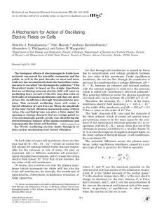
Serum FIG.
Groups I
Mitogenic
activity on osteoblastic cells of serum from patients who had only a head injury (HEAD), those who had a head injury and fractures those who had fractures only (##), and control subjects. Mitogenic activity is expressed as the percentage of increase of incorporation of [‘Hithymidine compared with the incorporation in the absence of serum (the basal value) and is shown as the mean and the standard error of the mean of triplicate determinations. The final concentration of serum was 10 per cent. The basal incorporation of [3H]thymidine was 5419 ± 561 counts per minute per 150,000 cells. The asterisks represent significant differences from the control value at p < 0.001 , as determined with analysis of variance and the least-significance test. No significant differences were noted between the values for the patients who had fractures only and the control subjects or between the values for the patients who had a head injury and fractures and the patients who had only a head injury.
(HEAD
+
#),
the levels
of activity
in the sera
from
the patients
who
did
not have a head injury. The mean levels of serum-induced
incorporation
[3H]thymidine
in the two groups
that had a head
were
significantly
injury
did not (Fig. 1). The significantly between
compared
increased
with
the two groups
activity in the serum the patients who had
of that
did not differ a head injury
only and the patients who had a head injury and fractures or between the control subjects and the patients who had fractures only. The number of osteoblastic cells increased more than 100 per cent after exposure to serum from patients who had a head injury compared with the number after exposure to serum
from
patients
who
did
not
have
a head
injury
(Fig.
E#{176} z =.O
Serum FIG.
Groups
2
Influence on the number of osteoblast-like cells of serum from patients who had only a head injury (HEAD) and from the control subjects. The number of cells is depicted as the percentage of increase compared with the number of cells in the absence of serum (the basal value) and is expressed as the mean and the standard error of the mean of triplicate determinations. The basal number of cells was 296,890 ± 14,890 per well. The asterisk represents a significant difference between the two groups at p < 0.03, as determined by the Student t test. The final concentration of serum was 10
per cent.
Downloaded from www.ejbjs.org on November 21, 2006 ThE
JOURNAL
OF BONE
AND
JOINT
SURGERY
EVIDENCE
FOR
A HUMORAL
MECHANISM
FOR
ENHANCED
OSTEOGENESIS
AFTER
HEAD
INJURY
I 147
1800 1600
C
0
1400 0
1200
a) a)>
-
1000
CO
800
E ..,.-, I-.
600
I
400 200
0
0.01%
1%
0.1%
Serum
Concentration
10%
(%)
FIG. 3 Relationship between the concentrations of serum that were added and the mitogenic response on osteoblastic cells. Serum from three patients who had a head injury (diamond, circle, and triangle) and from a control subject (square) was added separately (in a final incubation volume of one milliliter) at the final concentrations that are shown. Mitogenic activity is expressed as the percentage of increase of incorporation of [‘Hjthymidine compared with the incorporation observed in the absence of serum (the basal value) and is shown as the mean and the standard error of the mean of triplicate detenninations. The basal incorporation of [3H]thymidine was 3778 ± 502 counts per minute per 150,000 cells.
injury only and in the group that had such an injury and fractures. The mitogenesis of osteoblast-like cells was dose-
[3Hjthymidine increased (Fig. 3). The sera from all patients who had a head injury promoted greater growth of osteoblastic cells than of skin fi-
dependent. increased
broblasts. patients
2).
Similar
values
As from
were
seen
in the
the concentration 0 to 10 per
group
of the
cent,
that
had
added
the
a head
serum
was
incorporation
of
The sera from who had fractures
the control subjects and from the only stimulated statistically sim-
1200
C
1000
0 5-
0
800 00
0 C
;
600’
CO .-.O 0
.
>;
400
.C I-
I
Ce)
200
0
HEAD
CONTROLS
HEAD.#
Serum
Groups
FIG. 4 Comparative mitogenic effects those who had fractures only Mitogenic activity is expressed as (the basal Value) and is shown as per cent. The basal incorporation minute per 150,000 skin fibroblasts. p < 0.001.
#),
VOL.
72-A,
NO. 8, SEPTEMBER
of serum
who had a (black bars) the percentage of increase of incorporation of [3H]thymidine compared with the the mean and the standard error of the mean of triplicate determinations. The of [3H]thymidine was 6995 ± 959 counts per minute per 150,000 osteoblastic The asterisks represent significant differences between mitogenic activity in
1990
(##),
from
and
patients
control
who
subjects
had
only
a head
injury
on
cells
of
osteoblast
the
(HEAD),
those
phenotype
Downloaded from www.ejbjs.org on November 21, 2006
head injury and fractures (HEAD + and on skin fibroblasts (gray bars). incorporation in the absence of serum final concentration of serum was 10 cells and 4700 ± 332 counts per osteoblastic cells and in fibroblasts at
1 148
S.
M.
BIDNER
ET
AL.
1400#{149}
1200
C 0 (8
s’-..iooo 00
800
#{163} 0> CO
i-
H-H----
4
600 >,-
I-
400
200#{149}
10
20
30 Time FIG.
Relationship between the time after Specimens of serum from three patients
injury were
percentage
of
of
increase
of
incorporation
(day 0) and mitogenic activity assayed and all determinations
[3H]thymidine
compared
with
the
40
60
70
(days)
5 for were
osteoblastic performed
incorporation
cells in the in the
in the serum of patients who same assay. Mitogenic activity absence
of
serum
(the
basal
the mean and the standard error of the mean of triplicate determinations. The upper limit of normal values for the control subjects The basal incorporation of [‘H]thymidine in the assays for each of the three patients was 10,987 ± 1354 counts per minute per 6995 ± 959 counts per minute per 150,000 cells (triangle), and 6995 ± 959 counts per minute per 150,000 cells (square).
ilar growth of osseous and non-osseous (Fig. 4). The stimulation ofosteoblastic was related to the length of time after
patients,
all of whom
admission
to the
remained
hospital
to the
time
from
that
as responsiveness
the time of
the
study
was
completed, were studied for as long as sixty-three days. In two independent assays, the activity of the sera from each patient was maximum at approximately thirty-five to thirtyseven days after injury (Fig. 5). The osteoblast-stimulating activity control
then decreased subjects.
and
approached
the
levels
in the
Discussion
a humoral
associated
with
mechanism head
injury.
in the The
influence
on osseous growth has not, in general, studied, and our work does not exclude an additional
neural
mechanism
osteogenesis
that
of neural
is
activity
been thoroughly the possibility of
in the genesis
of this
phe-
nomenon. Both accelerated fracture-healing and excess formation of callus are associated with increased osteoblastic activity and, almost certainly, with proliferation of increased numbers of osteoblasts, whether by stimulation of so-called determined osteoprogenitor cells or by induction of noncommitted mesenchymal cells12. We used mitogenic assays in cells possible
who
have
of the osteoblast phenotype humoral mechanism for
a head
injury.
in order osteogenesis
The population
to explore a in patients
of calvarial
injury. as the
is shown
as
(diamond) is shown. 150,000 cells (circle),
cells
to parathyroid
hormone,
alkaline
phos-
phatase activity, synthesis of type-I collagen, and synthesis of bone-matrix proteins (notably osteocalcin). This population of cells is known to respond to mitogens either as a result
of the presence
of isolated these
cells
of precursor
or because
differentiated
cells,
cells
within
the mixture
of the replicative or both.
Interestingly,
capacity
of
in our
pa-
tients who had a head injury, the mitogenic activity of the serum was preferential for these osseous cells compared with skin fibroblasts. The mitogenic activity in prostatic tissue has been
Accelerated fracture-healing and increased formation of callus after a coma-producing head injury may involve either humoral or neural mechanisms, or both. The results in this study demonstrated increased circulating growthfactor activity for cells of the osteoblast phenotype in the serum of patients who had a head injury. The results implicate
and
that was used has been clearly demonstrated to display phenotypic characteristics of differentiated osteoblasts9”#{176}, such
(skin fibroblast) cells growth by the serum the head injury. Three
in a coma
had a head is expressed
value)
reported
to be similarly
preferential
for osteoblastic
cells9”0. However, no such activity in the brain has yet been reported. This characteristic may distinguish this growthpromoting activity from a variety of previously defined growth
factors.
The interaction of systemic and local humoral growth factors with skeletal tissue has been the subject of considerable interest in the past few years2’15. Circulating peptides, such
as epidermal
factor, fibroblast and transforming stimulate
growth
factor,
platelet-derived
growth factors, insulin-like growth factor beta have
synthesis
of DNA
and
replication
growth
growth factors, been shown to of cells
and,
in
some cases, to stimulate synthesis of collagen in osseous cell and organ cultures in vitro. Other humoral growth factors are believed to be elaborated, released, and active locally
in bone.
This
category
includes
cytokines
such
as
interleukin I, prostaglandins of the E series, and bone morphogenetic protein&8. Indeed, the isolation (from the skeleton), cloning, and sequencing of bone morphogenetic proteins has recently been reported, and these peptides have been demonstrated to be members of the super family of transforming growth factor beta-like molecules#{176}. Although
Downloaded from www.ejbjs.org on November 21, 2006 ThE
JOURNAL
OF BONE
AND
JOINT
SURGERY
EVIDENCE
the
presence
strated bound bring
FOR
of systemic
growth
factors
and
MECHANISM
factors
has
FOR
been
ENHANCED
several
biological
have
led
efficacy
that
local
was
concentrations. In our patients
who
brain barrier. ulated from
teoblast-stimulating
activity
than baseline related
levels
with
dynamic
the duration
increase
impact
over
a head
were
injury,
clearly
a defined
on bone-stimulating
activity activity
to more
of time that corinsult.
Such
could
have
a major
might
enhance
and
a any
existing activity that had been basally active in the serum. The reduction in circulating activity after approximately one month might clearance of whether
the
of patients circulating a serum cells,
be due activity, actual
who
to a fall in production or both. It remains growth-promoting
have
a head
injury
mitogen or are indirect factor of growth factors
which
act locally
in an autocrine
effects
or to increased to be resolved of the
serum
are direct effects of a effects due to release by from the osseous target or paracrine
manner.
described.
brain
of Os-
elevated
period
of the neurological
in circulating
levels
exclude
HEAD
1149
INJURY
the possibility
that
a circulating
activity is reduced after to the increased mitogenic
of the serum that was observed in our study. know the source of the osteoblast-stimulating
pro-
duction of even these agents may have more of an impact on modulation of cellular growth than do their circulating had
AFTER
inhibitor of growth-factor jury, and that this leads
of circulating
to speculation
OSTEOGENESIS
We cannot
demon-
are believed to circulate ‘ . These and other considerations
in the circulation, to plasma into question the
growth
A HUMORAL
The
tissue,
activity
might
in association
with
We do not activity that
be stimulated
disruption
Alternatively, release might peripheral sites or it might
directly
be neurally be stimulated
organ pituitary
to be rich
of growth
factors
inflammatory
cells
fibroblast
growth
of production factors,
and
recruited to damaged sites in the brain of growth-promoting cytokines3. Irrespective of the precise nature and growth-promoting activity that we observed,
injury. the
that
such that
could
as are
be the
origin of the results
the of
increased circulating growth-factor cells in patients who had a head
This is an important
mechanisms
stimby
such as the pituitary gland6 are known
being source
our study demonstrated activity for osteoblastic
by
of the blood-
damaged tissue from an adjacent gland. Both the brain7 and the
sites
head inactivity
underlie
initial the
step in the elucidation enhanced
after head injury. Future studies will further characterization of this activity.
of
osteogenesis
be
directed
toward
References 1. 2. 3. 4. 5. 6. 7. 8. 9. 10. 11. 12. 13. 14. 15. 16. 17. 18. 19. 20. 21.
VOL.
CALANDRIELLO, BRUNO: Callus Formation in Severe Brain Injuries. Bull. Hosp. Joint Dis., 25: 170-175, 1964. CANALIS, ERNESTO: Effect of Growth Factors on Bone Cell Replication and Differentiation. Clin. Orthop. , 193: 246-263, DURUM, S. K.; SCHMIDT, J. A.; and OPPENHEIM, J. J.: An Immunological Perspective. Ann. Rev. Immunol. , 3: 263-287, GARLAND, D. E. , and TODER, LAWRENCE: Fractures of the Tibial Diaphysis in Adults with Head Injuries. Clin. Orthop.,
1985.
1985.
198-202, 1980. BRIAN; and WATERS, R. L.: Femoral Fractures in Head-Injured Adults. Clin. Orthop., 166: 219-225, 1982. DENI5: Purification of a Fibroblast Growth Factor from Bovine Pituitary. J. Biol. Chem. , 250: 2515-2520, 1975. DENIS; BIALECKI, HUGH; and GREENBURG, GARY: Purification of the Fibroblast Growth Factor Activity from Bovine Brain. J. Biol. Chem. , 253: 3736-3743, 1978. KNAUER, D. J. , and SruTH, G. L.: Inhibition of Biological Activity of Multiplication-Stimulating Activity by Binding to Its Carrier Protein. Proc. Nat. Acad. Sci. , 77: 7252-7256, 1980. K0UT5ILIEiu5, M. ; RABBANI, S. A. ; and GOLTZMAN, D. : Effects of Human Prostatic Mitogens on Rat Bone Cells and Fibroblasts. J. Endocrinol., 115: 447-454, 1987. K0UT5ILIEiu5, MICHAEL; RABBANI, S. A. ; BENNET, H. P. J. ; and GOLTZMAN, D. : Characteristics of Prostate-Derived Growth Factors for Cells of the Osteoblast Phenotype. J. Clin. Invest. , 80: 941-946, 1987. MIYAz0N0, KOHEI; HELLMAN, ULF; WERNSTEDT, CHRISTER; and HELDIN, C. H.: Latent High Molecular Weight Complex ofTransforming Growth Factor 3l. Purification from Human Platelets and Structural Characterization. J. Biol. Chem. , 263: 6407-6415, 1988. OWEN, M. : Lineage of Osteogenic Cells and Their Relationship to the Stromal System. In Bone and Mineral Research Annual 3: A Yearly Survey of Development in the Field of Bone and Mineral Metabolism, pp. 1-25. Edited by W. A. Peck. New York, Elsevier, 1985. PECK, W. A.; BIRGE, S. J., JR.; and FEDAK, S. A.: Bone Cells: Biochemical and Biological Studies after Enzymatic Isolation. Science, 146: 1476-1477, 1964. PERKINS, R. , and SIURVING, A. P. : Callus Formation and the Rate of Healing of Femoral Fractures in Patients with Head Injuries. J. Bone and Joint Surg. , 69-B(4): 521-524, 1987. RAI5z, L. G.; CANALIS, E. M. ; DIETRICH, J. W. ; KREAM, B. E. ; and GWOREK, S. C. : Hormonal Regulation of Bone Formation. Rec. Prog. Horm. Res. , 34: 335-356, 1978. SMITH, ROGER: Head Injury, Fracture Healing and Callus. J. Bone and Joint Surg., 69-B(4): 518-520, 1987. SPENCER, R. F.: The Effect of Head Injury on Fracture Healing. A Quantitative Assessment. J. Bone and Joint Surg. , 69-B(4): 525-528, 1987. URIST, M. R.; DELANGE, R. J.; and FINERMAN, G. A. M.: Bone Cell Differentiation and Growth Factors. Science, 220: 680-686, 1982. WONG, G. L. , and COHN, D. V.: Target Cells in Bone for Parathormone and Calcitonin Are Different. Enrichment for Each Cell Type by Sequential Digestion of Mouse Calvaria and Selective Adhesion to Polymeric Surfaces. Proc. Nat. Acad. Sci., 72: 3167-3171, 1975. WOZNEY, J. M. ; ROSEN, VICiu; CELESTE, A. J. ; MITSOCK, L. M. ; WHITTERS, M. J. ; Kmz, R. W. ; HEWICK, R. M. ; and WANG, E. A. : Novel Regulators of Bone Formation: Molecular Clones and Activities. Science, 242: 1528-1534, 1988. YAGIELA, J. A. , and WOODBURY, D. M.: Enzymatic Isolation of Osteoblasts from Fetal Rat Calvaria. Anat. Rec. , 188: 287-305, 1977.
GARLAND, D. GOSPODAROWICZ, GOSPODAROWICZ,
72-A,
E.;
ROTHI,
NO. 8, SEPTEMBER
1990
Downloaded from www.ejbjs.org on November 21, 2006
150: