Arthropod Systematics & Phylogeny 133–148 64 (2)
133 © Museum für Tierkunde Dresden, ISSN 1863-7221, 01.12.2006
Insect Interordinal Relationships: Evidence from the Visual System MARKUS FRIEDRICH 1, 2 *, YING DONG 3 & MAGDALENA JACKOWSKA 1 1
2
3
*
Department of Biological Sciences, Wayne State University, 5047 Gullen Mall, Detroit, MI 48202, USA [
[email protected]] Department of Anatomy and Cell Biology, Wayne State University, School of Medicine, 540 East Canfield Avenue, Detroit MI 48201, USA [
[email protected]] Simmons Comprehensive Cancer Center, University of Texas Southwestern Medical Center at Dallas, 5323 Harry Hines Blvd, Dallas, TX 75390, USA Corresponding author
Received 1.iii.2006, accepted 2.ix.2006. Available online at www.arthropod-systematics.de
>
Abstract
Insects are by far the most speciose and also one of the most intensively studied animal groups on earth. To contribute to a recent effort in reviewing and revalidating morphological and molecular data sets for the reconstruction of insect interordinal phylogeny, we turned our attention to structural and ontogenetic traits of the visual system. Discussed is a suite of characters, nine of which are proposed to show phylogenetically informative differences between insect orders. Of these, three (second mitotic wave, retina blood border, indirect ocellus innervation) relate to basal diversification events in the Pterygota. Four character states represent autapomorphies of the Endopterygota (optic lobe invagination, possession of stemma, stemmata derived adult brain photoreceptors, and postembryonic progressive eye development). Lastly, the spatially undissociated lobula plate in hymenopteran representatives like honey bee, which contrasts with the well separated lobula plate in other endopterygotan orders, is discussed as possibly indicating a basal position of the Hymenoptera in the Endopterygota.
>
Key words
Insect visual system, Strepsiptera, evolution of development, eye development, ocellus, stemma, Bolwig organ.
1.
Introduction
1.1.
Insect phylogeny
Insects stand out as the largest animal group, and exhibit a vast diversity in morphological, physiological, and ecological traits. These very features cause major challenges to attempts towards resolving genealogical relationships among the currently 30 recognized orders. High rates of evolutionary change have diluted and blurred phylogenetically informative traits. Decades of phylogenetic research produced a widely used consensus framework (for recent reviews and discussion see GRIMALDI & ENGEL 2005; KRISTENSEN 1999, 1995; WHITING 2004; WILLMANN 2004), which however is still peppered with weakly supported nodes and unresolved areas (Fig. 1). The relationships between the most primitive insect orders are relatively stable. As taxonomic entity, insects comprise the whole of ectognathous hexapods. Within these, Archaeognatha are widely considered
the most basal lineage, rendering the other primitively wingless insect order, the Zygentoma, the second oldest insect lineage. The traditional Zygentoma may be paraphyletic, as both some morphological and some molecular data support Tricholepidon gertschi (Lepidotrichidae) to be located outside the Zygentoma, either at the base of Dicondylia (KRISTENSEN 1991) or at the base of the Pterygota (KJER 2004). The next higher up nodes in the tree initiate the realm of the Pterygota. A large number of winged insects has been structured into four superclades. The most basal of these is the most likely paraphyletic Palaeoptera, which include Ephemeroptera (mayflies) and Odonata (dragon- and damselflies). Several lines of morphological and molecular evidence speak for Ephemeroptera as the more basal branch of the Palaeoptera (BEUTEL & GORB 2006; TERRY & WHITING 2005). Alternative configurations such as a monophyletic Palaeoptera have been considered as well (HENNIG 1981; KJER et al. 2006). The most problematic area of the insect tree concerns the relationships among the remaining 26 orders of winged insects which together form the Neoptera.
134
FRIEDRICH et al.: Insect visual system
Fig. 1. Current status of insect phylogeny. Topology adapted from KRISTENSEN (1991) and WILLMANN (2004).
While some affinities are relatively well understood, such as those among Isoptera, Mantodea and the paraphyletic Blattodea (Fig. 1), relationships of most orders are still unresolved. The scope of the problem has recently been widened by the discovery of the Mantophasmatodea, a new neopteran order (KLASS et al. 2002). Within the Neoptera, the Paraneoptera, which are also known as Acercaria, represent a tentatively supported subclade uniting Psocodea (bark lice), Hemiptera (true bugs), and Thysanoptera (thrips) (KRISTENSEN 1991; WILLMANN 2004). The Paraneoptera may constitute the sister clade of the Endopterygota. Putative ancestors of a clade uniting Paraneoptera and Endopterygota may have originated from within a specific but yet unidentified neopteran lineage. Alternatively, the remaining neopteran lineages may constitute a sister monophylum of the Paraneoptera +
Endopterygota, which is referred to by the name Polyneoptera (WILLMANN 2004; GRIMALDI & ENGEL 2005). Relationships among Endopterygota, i.e. Holometabola, are better resolved. The current view holds that Endopterygota split into a superclade Neuropterida (Neuroptera [lacewings], Raphidioptera [snakeflies], Megaloptera [alderflies] and Coleoptera [beetles]), and a superclade Hymenoptera + Mecopterida (Mecoptera [scorpion flies], Siphonaptera [fleas], Diptera [true flies], Lepidoptera [butterflies and moths] and Trichoptera [caddiesflies]) (WHITING 2004). The most weakly supported node is that associating the Hymenoptera (wasp like insects) with the Mecopterida (KRISTENSEN 1991). The “Hymenoptera problem” however is topped by the provocative Strepsiptera (twisted-wing insects) problem. This uniquely derived group of obligatorily parasitic insects has been offered various branches
Arthropod Systematics & Phylogeny 64 (2)
135
Fig. 2. Schematic of components of the insect visual system. Left hemisphere represents species with spatially distinct lobula plate with direct axonal connections to medulla. Right hemisphere represents species with lobula plate closely connected to lobula s.str. (after SINAKEVITCH et al. 2003). Light blue: compound eye retina. Dark blue: median (unpaired) and lateral (paired) ocelli. Red: optic lobe anlagen components. Green: stemmata modified into extraretinal photoreceptors of the adult. lam = lamina, med = medulla, lob = lobula s.str., lbp = lobula plate.
within the Holometabola, although some authors have voiced concern that the Strepsiptera may actually not belong into the Holometabola (KRISTENSEN 1999). In summary, a tentative consensus framework of insect phylogeny exists, but with question marks. The challenge is highlighted by the fact that the past 10 years of insect phylogenetics has not seen a substantial increase in better resolved nodes, despite a large body of systematic work (KRISTENSEN 1991; WILLMANN 2004). Moreover, even generally accepted nodes such as a monophyletic Holometabola are based on surprisingly little character support (KRISTENSEN 1999). The quest to extract more phylogenetic information from morphology and molecules is thus still standing.
1.2.
The insect visual system
The visual system is one of the most long-term and hence, best studied organ complexes in insects. Several authoritative reviews have become classic references (BATE 1978; GOODMAN 1981; MEINERTZHAGEN 1991; PAULUS 1979, 2000). More recently, a new surge of papers appeared which investigate the origin of vi-
sual system structures in arthropods (BITSCH & BITSCH 2005; OAKLEY 2003). This renaissance was in part triggered by the advancement of molecular genetic investigations in Drosophila, which deepened the understanding of the development and, consequently, structural organization of the visual system (MOSES 2002). Five major external visual system components can be identified in most insects (Fig. 2): the prominent pair of lateral compound eyes and a trio of simpler lens eyes, the ocelli. The compound eyes are serially connected to three optic neuropils: the lamina, medulla and lobula; the latter consisting of lobula s.str. and lobula plate. The ocelli do not project into dedicated processing neuropils, but instead connect to different regions in the brain (GOODMAN 1981). One ocellus, the median ocellus, is unpaired and located on the midline of the frons. The pair of lateral ocelli is situated usually more dorsally in the region of the vertex, but can take many different positions in relation to the compound eyes. The ancestral set of five external eyes increased by two during the evolution of the Holometabola, which introduced specialized larval eyes or stemmata to the world of modern insects (GILBERT 1994). A fascinating
136
FRIEDRICH et al.: Insect visual system
discovery of the past years was that these larval stemmata survive into the adult serving as remodeled extraretinal photoreceptors (HELFRICH-FORSTER et al. 2002; YASUYAMA & MEINERTZHAGEN 1999). Thus, in a very literal sense, holometabolous species like Drosophila utilize seven eyes (HOFBAUER & BUCHNER 1989). This paper investigates the phylogenetic information content of major components of the visual system. Rather than attempting exhaustive analysis of character state systems, potentially informative aspects of the visual system are highlighted. In most cases, the preliminary quality of this information is due to the lack of data from a sufficiently wide sample of species. Where possible, character state evolution is discussed considering the mechanics of development as they have been elucidated in Drosophila. Not only will the phylogenetic value of final structures be evaluated but also that of the developmental processes leading to their differentiation and realization.
2.
Ommatidial morphology and development: conservation and convergence
2.1.
Conservation
Ommatidia are the ground unit of the arthropod compound eye (for reviews see BITSCH & BITSCH 2005; MEINERTZHAGEN 1991; OAKLEY 2003). There is a great deal of variation in the cellular architecture of the ommatidia in the Arthropoda. A major divide exists between the types of ommatidia found in Hexapoda/ Crustacea, which have been united as Pancrustacea or Tetraconata (DOHLE 1997), versus those of Myriapoda/ Chelicerata, which have been dubbed Paradoxopoda (MALLATT et al. 2004). The hexapod/crustacean type ommatidium exhibits an extraordinary degree in conservation of cellular organization. This is consistent with the strong evidence that crustaceans represent the most closely related clade to hexapods (BOORE et al. 1998; GIRIBET et al. 2001; HWANG et al. 2001; REGIER et al. 2005). The core of the hexapod/crustacean type ommatidium consists of eight photoreceptor cells. These are topped by four lens cells and two primary cone [Hexapoda] or corneagenous [Crustacea] cells (PAULUS 2000). This cell arrangement is embedded in a phylogenetically more variable number of secondary and tertiary pigment cells, as well as bristle cells. In addition to the conservation of cell type numbers, various examples of notable conservation of structural aspects have been discovered. The most recent finding concerns the spatial distribution of cone cell processes, which are sent as fine projections from the distally located main body of each cone cell towards
the floor of the ommatidium (WOLFF & READY 1993). Every cone cell process extends between the borders of two specific photoreceptor cells. A survey in a sample of distantly related hexapod and crustacean species revealed identical relative positions of cone cell projections between the photoreceptor cells (MELZER et al. 1997). This subtle feature has thus been conserved for more than 500 million years. Interestingly, the recent re-examination of the ommatidial structure in the common house centipede Scutigera coleoptrata revealed comparable cone cell processes (MÜLLER et al. 2003). Their positional homology in relation to that in hexapod/crustacean type ommatidium is less straightforward to establish due to the major differences in the number and distribution of photoreceptor cells in this species. Variations in ommatidial structure have been utilized in malacostracan systematics (RICHTER 1999). Among hexapods, the variation in ommatidial architecture has rarely been investigated for phylogenetic purposes, which is not very surprising, considering the high degree of conservation. Extensive cladistic analyses have been carried out on the visual system within Hemiptera and polyphagan coleopterans (CAVENEY 1986; FISCHER et al. 2000). Most of the major differences in ommatidial structure, such as lens construction or rhabdome organization, are too variable for phylogenetic inference at the interordinal level. Open rhabdomes, for instance, evolved independently in distantly related groups including Diptera, Heteroptera, Dermaptera and Coleoptera-Cucujiformia (FISCHER et al. 2000).
2.2.
Convergence
A case of remarkable convergence at the interordinal level concerns similarity of unusual photoreceptor cell numbers and projection patterns in Hymenoptera and Lepidoptera. Departures from the eight photoreceptor cells comprising ommatidial ground plan have been described in the Crustacea as well as Hexapoda (OAKLEY 2003; TRUJILLO-CENOZ 1985). Photoreceptor cells can be reduced down to five or considerably increased exceeding 10 (OAKLEY 2003). There are only a few species, however, which feature nine photoreceptor cells. Such examples are found in Hymenoptera and Lepidoptera (OAKLEY 2003). The ommatidia of these Hymenoptera and Lepidoptera match also in terms of axonal projection patterns. In the hexapod groundplan visual system, six photoreceptor cells project into the first optic neuropil, the lamina. These photoreceptors are called peripheral short fiber receptors. The two remaining photoreceptors project through the lamina into the second optic neuropil, the medulla. These long fiber photoreceptors are also unique in contributing rhabdomeres to the center of the ommatidium. In
Arthropod Systematics & Phylogeny 64 (2)
both the hymenopteran and lepidopteran species, the extra photoreceptor cell number nine is a long fiber photoreceptor cell with a centrally located rhabdomere (MEINECKE 1981; WELSCH 1977). Assuming a monophyletic Mecopterida (Fig. 1) and considering the presence of eight photoreceptor cells in the ommatidia of Diptera and Mecoptera, the extra photoreceptor cell may have evolved once in an ancestral linage preceding Diptera and Hymenoptera followed by secondary reduction in at least the last common ancestral lineage of the Diptera and Mecoptera. Alternatively, the possession of three long fiber photoreceptors in the ommatidia of hymenopteran and lepidopteran species resulted from convergent evolution. This can be further tested by investigating if the ommatidia of basal species in these orders are equipped with the ancestral number of eight photoreceptors. Investigations on ommatidial structure of basal hymenopteran species seem to be missing. However, the larval eyes of representatives of one of the most basal hymenopteran families, the Tenthredinidae, have been described as ommatidia-like cell arrangements with eight photoreceptor cells per unit (PAULUS 1979). Assuming perfect correspondence of larval and adult eye cell architecture in this species, one may tentatively conclude that eight photoreceptors per adult eye ommatidium is the ancestral condition in the Hymenoptera. A caveat to keep in mind, however, is that larval and adult eye morphology possibly diverged which is commonly observed (GILBERT 1994). One such example is the reduction of photoreceptor and cone cells in the stemmata of Trichoptera and Lepidoptera discussed below. Unfortunately, information on ommatidial structure is also scarce in lepidopteran lineages basal to Macrolepidoptera. It is possible that nine photoreceptor cells is the ancestral state in the Macrolepidoptera. Ninephotoreceptor cell ommatidia have been reported from various butterfly species (Papilionoidea) as well as from moth families like the Sphingidae (KELBER et al. 2003; KITAMOTO et al. 2000; WELSCH 1977). Eight photoreceptors however have been described for the Noctuoidea (MEINECKE 1981). Further down the lepidopteran tree, nine and more photoreceptor cells have been found in the wax moth Ephestia kuehniella (FISCHER & HORSTMANN 1971). Ephestia gives a hint at the developmental plasticity in photoreceptor number differentiation. The minimal number of nine photoreceptors in this species could be interpreted as evidence that this number is ancestral at least for the Ditrysia. Obviously, a more comprehensive taxon sampling of ommatidial structure in basal Hymenoptera or Trichoptera/Lepidoptera is required to clarify the evolutionary time point of the emergence of ommatidia with one additional long fiber retina cell in the Hymenoptera and Lepidoptera.
137
From the perspective of what we know about the regulation of photoreceptor cell specification in Drosophila, convergent evolution of surplus long fiber retina cells is highly plausible (READY 1989; WOLFF & READY 1993). The ommatidial cell fates in the developing Drosophila retina are determined along a tightly controlled time line. The first cell to differentiate is the central photoreceptor cell R8, which is one of the two long fiber photoreceptors. Its specification is followed by induction of three pairs of outer photoreceptor cells (R2/R5, R3/R4 and R1/R6). The last photoreceptor cell to join is R7, which, like R8, is a long fiber photoreceptor. Both R8 and R7 contribute rhabdomeres into the center of the ommatidium. An important difference between the two cells is that the R8 rhabdomere is restricted to the bottom of the ommatidium, while that of R7 lies on top of it in the distal half. As has been noted by READY (1989) in the honeybee eye, the long fiber extra photoreceptor cell in the Hymenoptera has characteristics of an R7 cell based on its centrally positioned, apically restricted rhabdomere. Remarkably, position and morphology of the lepidopteran extra cells satisfy the same R7 cell type criteria. In sphingid moths for instance, two cells share the unique R7 morphology, i.e. a distal rhabdomere (SCHLECHT 1979). The occurrence of extra R7 cells is not surprising. The genetic dissection of ommatidial patterning in Drosophila revealed that photoreceptors R1 and R6 as well as all four cone cells have the potential to differentiate into extra R7 type cells. These cells have therefore been referred to as the R7 equivalence group (DICKSON & HAFEN 1993) (Fig. 3). Receptor Tyrosine Kinase (RTK) signaling is a key regulator of cell fate induction in this process. The R7 cell expresses two receptors, which when ligand bound, mediate activation of RTK signaling: the Epidermal Growth Factor Receptor (EGFR) RTK and the Sevenless (Sev) RTK. Importantly, manipulating the timing and strength of RTK signaling in the R7 equivalence group can produce extra R7 cells (FREEMAN 1996). At the so-called “seven cell stage” of normal ommatidial development, all photoreceptor cell fates except for that of R7 have been induced (WOLFF & READY 1993). The R7 equivalence group cells R1 and R6 have already adopted their fates, making them non-responsive to R7 induction at later stages of patterning. The cells, however, immediately adjacent to the developing ommatium, i.e. the cone precursor cells, do have the potential to differentiate into R7. This step requires contact formation with the R8 cell, the only cell expressing Bride of Sevenless (Boss) protein, the activating ligand of the Sev RTK (DICKSON & HAFEN 1993). Contact of one of the remaining R7 equivalence group cells with R8 is thus the critical spatial cue for correct induction of R7 fate. In Drosophila, the spatial properties of photoreceptor cells in the developing ommatidium guarantee that only one cell contacts R8.
138
FRIEDRICH et al.: Insect visual system
Fig. 3. Ommatidial patterning and spatial regulation of cell proliferation in the differentiating insect compound eye retina. In all panels anterior to the left. Scale bar in A corresponds to 10 µm. Scale bars in C and D correspond to 1 μm. A: Schematic representation of evolutionarily conserved ommatidial assembly stages. Specified photoreceptor cell fates indicated by numbers. Blue: founder photoreceptor cell R8. Green: Peripheral photoreceptor cells 1–6. Strong pink: R7 cell fate. Light pink: R7 equivalence group. Light blue: cone cells. Grey: mitotic cells. 1ʼ: domain of first mitotic wave. 2ʼ: domain of second mitotic wave. mf: morphogenetic furrow. B: Confocal image of anti-armadillo antibody (RUEL et al. 1999) labeled embryonic grasshopper retina. Mitotic cells anterior and posterior to the morphogenetic furrow indicated with arrows. C: Five cell cluster ommatidial assembly stage. Specified photoreceptor cell fates indicated by numbers. R7 equivalence group cells labeled with asterisk. D: Five cell cluster ommatidial assembly stage. R7 equivalence group cells from panel B have now adopted cells fates R1, R6 and R7.
The sequence of ommatidial cell recruitment is highly conserved in other insects ranging from Lepidoptera to beetles and grasshopper (CHAMPLIN & TRUMAN 1998; EGELHAAF 1988; FRIEDRICH et al. 1996) (Fig. 3). It is thus likely that the EGFR and Sevenless mediated induction of R7 cells applies to insects in general. Lepidopteran and hymenopteran extra R7 cells may thus have evolved by independent changes in RTK signaling intensity. It is tempting to speculate that changes in the expression of the surface ligand Boss or in the regulation of intercellular contacts facilitated the evolution of additional R7 cells. Both Manduca and Apis are experimentally well accessible systems inviting to test some of these ideas in the lab.
3.
Spatial control of cell proliferation
While the cellular and molecular dynamics of retinal differentiation help to understand character state
transition in the insect ommatidium, the high degree of conservation renders the recovery of phylogenetically informative aspects at this level unlikely. Indeed, histological analysis of cellular patterning in the retina of the tadpole shrimp Triops suggest that the sequence of cell determination and intercellular contacts during ommatidial development has been conserved for a remarkable 500 million years (MELZER et al. 2000). Similar cellular cluster arrangements have been found in crayfish (HAFNER & TOKARSKI 1998). There is, however, a second aspect of retinal patterning which may be phylogenetically informative at a deep level of insect or hexapod evolution. The development of the retina is lead by a progressive front of differentiation, the morphogenetic furrow (READY et al. 1976). The furrow like epithelial surface structure is produced by the transient shortening of the cells at the onset of differentiation all along the polar axis of the Drosophila eye disc (Fig. 3). In the furrow, the retinal precursor cells undergo concerted changes in cytoskeletal organization and cell cycle regulation.
139
Arthropod Systematics & Phylogeny 64 (2)
Mitotic activity is suppressed in the furrow but concentrated in two narrow domains immediately anterior and posterior to the furrow, which are referred to as primary and secondary mitotic waves respectively. The same pattern of spatially regulated cell proliferation is documented for many insect species including holometabolous representatives in the Coleoptera and Lepidoptera and the more ancestrally organized hemimetabolous grasshopper (CHAMPLIN & TRUMAN 1998; EGELHAAF 1988; FRIEDRICH & BENZER 2000; FRIEDRICH et al. 1996). Interestingly, studies of retinal development in a diverse range of Crustacea including Branchiopoda and Malacostraca reported only a single proliferation zone in front of the differentiating retina (HAFNER & TOKARSKI 1998; HARZSCH et al. 1999; HARZSCH & WALOSSEK 2001; WILDT & HARZSCH 2002). Its position in front of the differentiating retina suggests that it most likely corresponds to the first mitotic wave in insects. As no data on retinal patterning and proliferation control are available for basal Pterygota besides Orthoptera, it is open whether this character state evolved within the insects or dates back to an earlier point in arthropod evolution. It would therefore be interesting to investigate the control of cell proliferation in insect taxa outside the Neoptera, importantly Archaeognatha, and potentially in a wider range of the Pterygota (Tab. 1). Of course, not too much expectation can be tied to this character. Apterygote species with elaborate compound eyes are only found in the Archaeognatha. The eyes of entognathan orders and of Zygentoma are highly reduced, making it likely that cell proliferation mechanisms have been secondarily reduced as well.
4.
Progressive versus expansive retinal differentiation
The morphogenetic furrow is the hallmark of progressive retinal differentiation in the Drosophila eye-antennal imaginal disc (Fig. 3). Similar morphogenetic furrow driven progressive differentiation has been demonstrated in the postembryonic retina of diverse holometabolous orders such as Coleoptera and Lepidoptera (CHAMPLIN & TRUMAN 1998; EGELHAAF 1988; FRIEDRICH & BENZER 2000). Strikingly, a progressing, morphogenetic furrow like dynamic cell constriction front is present in the developing embryonic retina of orthopteran, hemimetabolous insects such as grasshopper and cricket (FRIEDRICH & BENZER 2000; INOUE et al. 2004). In the latter case, the progressive front of differentiation transforms into a standing zone of differentiation during the transition from embryogenesis to postembryogenesis. In grasshopper nymphs, the eye continues to develop, but by differentiation of newly
born cells from a stem cell niche in front of the juvenile eye (BODENSTEIN 1953). This may be considered an expansive form of differentiation in the eye of the nymphs (ANDERSON 1978; FRIEDRICH 2006). A similar mode of postembryonic retinal development has been reported for other hemimetabolous species such as blattodeans (NOWEL & SHELTON 1980). Moreover, the description of retinal differentiation in representatives of the Crustacea, Myriapoda and Chelicerata correspond to an expansive type of retinal differentiation further suggesting that this organization of retinal development is ancestral in the Insecta (ENGHOFF et al. 1993; HARZSCH et al. 2006; WILDT & HARZSCH 2002). In regards to insect phylogeny, the difference in the timing of furrow driven progressive retinal differentiation between the holometabolous and hemimetabolous species is clear and striking. It therefore seems justified to consider the postembryonic onset of progressive retinal differentiation an autapomorphy of the Holometabola. To confirm this idea, it will be necessary to study the cellular dynamics of larval and adult eye development in the less derived visual systems of holometabolan species such as scorpion flies (Mecoptera).
5.
Organization of the retina-hemolymph border
The cells in the insect retina require connection with the circulatory system for supply with energy, removal of metabolized materials, and interaction with the hormonal control system. Neuronal cell function, on the other hand, also requires a constant electrolyte environment. The interface between retina and hemolymph must therefore be a regulated border. Two different types of retina-hemolymph borders appear to exist in hexapods. In primitively wingless hexapods such as Collembola or Archaeognatha, the border between retina and hemolymph is furnished with a basal membrane and a loose mesh of glia cells. These structures provide free passage for ions or small molecules, as measured by uptake of tracer fluids (SHAW & VARNEY 1999). In most winged insects, the basal membrane on the floor of the retina is followed by a contiguous glia derived cell sheet that provides a barrier between retina and hemolymph. This type of organization is referred to as blood-retina barrier, and is analogous to the likewise glia derived blood-brain barrier (SHAW 1977, 1978). A functional blood-retina barrier can be detected by delayed or blocked uptake of tracer dye fluids injected into the hemocoel. SHAW & VARNEY (1999) investigated the presence of a blood-retina barrier for representatives of 31 hexapod species representing 16 orders. Lack of tracer dye uptake was found in all Neoptera tested while apterygote representatives, in-
140
FRIEDRICH et al.: Insect visual system
cluding the archaeognathan Petrobius brevistylus, exhibited features compatible with an open blood-retina border. The latter type of organization is also found in crustaceans, suggesting that it is ancestral in hexapods (SHAW & VARNEY 1999). This survey therefore indicates that the blood-retina border is an autapomorphy for the Neoptera or a clade including Neoptera and Odonata. In the odonatans, only small traces of tracer dye were observed in the retina (SHAW & VARNEY 1999). In ephemeropteran species examined, presence of dye in the optic lobes and in the retina suggested the ancestral state of free passage between hemocoel and retina (SHAW & VARNEY 1999). Future studies on the blood-retina organization in Odonata and basal Neoptera may be valuable to corroborate if this character state has a bearing on the relationships between the palaeopteran orders and Neoptera.
6.
Optic neuropil organization
As recently reviewed by HARZSCH (2006), optic lobe neuropil organization has served early as character complex in the discussion of arthropod relationships. This is due to the striking similiarity in the structure of the optic neuropils in insects and crustaceans, particularly malacostracan crustaceans. In both taxa, the optic neuropil system of most species is tripartite consisting of lamina, medulla and lobula (Fig. 2). Axon chiasmata connect the lamina with the medulla neuropil, and the medulla with the lobula. Even the organization of the neuropils exhibits similarities of extreme subtleness such as identical photoreceptor projection patterns. Optic lobe structure has thus been used as strong evidence for a close phylogenetic relationship between insects and malacostracans. Particularly pointed out was the neuroanatomical similarity in the cellular architecture of lamina and medulla (OSORIO & BACON 1994). More recently, similarities in the third neuropil of malacostracan crustaceans and insects have added a homologous lobula compartment to the discussion (HARZSCH 2002). Considering the deep conservation of optic lobe morphology, it does not come as a surprise that their structures are strongly conserved among insects. However, some aspects of lobula neuropil variation may deserve attention with regards to potential phylogenetic implications. In some endopterygotan orders, such as Diptera, Coleoptera and Lepidoptera, the lobula is composed of two distinct elements: the lobula s.str. and the lobula plate (SINAKEVITCH et al. 2003). The lobula s.str. receives retinotopic input from the medulla via a chiasm. The lobula plate receives non-chiasmatic connections being the center of higher order neurons integrating motion detection information. The structure
of the lobula complex is less distinctly developed in the Hymenoptera (SINAKEVITCH et al. 2003). The twolayered but undivided lobula of Apis is more similar to that found in more basal Pterygota such as cockroaches and grasshoppers (SINAKEVITCH et al. 2003; STRAUSFELD 1998). Current data raise the possibility that an undivided lobula complex is ancestral in the Pterygota. It may be derived from the compact lobula emersed in the protocerebrum of primitive wingless insects such as Archaeognatha and Zygentoma (SINAKEVITCH et al. 2003). In discussing the lobula plate as potential synapomorphy which separates the main part of Holometabola from Hymenoptera, it must be cautioned that there is evidence for convergent evolution of lobula neuroanatomy. Lobula plate like secondary lobula neuropils are also found in isopods (Crustacea-Isopoda). Yet another possibility has to be considered in the light of a recent suggestion to revise homology assignments of arthropod optic neuropils (STRAUSFELD 2005). Accordingly, the presence of the lobula plate is considered plesiomorphic for Tetraconata. It would follow that the compact lobula complex seen in many contemporary insects evolved later. Also this scenario, which deserves to be investigated in a wider range of species, could explain the striking similarity of a lobula plate in some insects and isopods (SINAKEVITCH et al. 2003). If the lobula plate neuropil as such is not a derived character in insects, one may have to consider the potential significance of the spatial arrangement of the two lobula compartments to each other. These seem to be contiguously adjacent in primitive insects, but dissociated in some Endopterygota. Considering the uncertainty with phylogenetic positioning of the Hymenoptera, more comprehensive taxon sampling of this character complex may turn out worthwhile.
7.
Optic neuropil development
Besides the potential significance of lobula plate morphology variation in endopterygotan insect orders, the structurally rich optic neuropil system offers little information regarding the interordinal level of insect phylogeny. There is, however, a dramatic difference in early optic lobe morphogenesis, which is likely to differentiate the Holometabola from the rest of insects. In Drosophila, the precursor cells of the outer optic lobe anlagen segregate from the embryonic head neuroectoderm as an invaginating contiguous cell sheet (GREEN et al. 1993) (Fig. 4B). An equivalent mode of optic lobe morphogenesis is also seen in less derived Holometabola. In the Coleoptera, the outer optic lobe anlage invaginates from neuroectoderm in the embryonic head lobes (HEMING 1982; ULLMANN 1966). Optic
141
Arthropod Systematics & Phylogeny 64 (2)
lobe invagination is substantially different from the way the outer optic lobe anlagen develop in representatives of non-endopterygotan orders. In the grasshopper Schistocerca americana, retinal and outer optic lobe precursor cells originate within the lateral head lobes of the embryonic head. The optic lobe precursor cells have been described to separate individually from the neuroectoderm by delamination (ROONWAL 1936) (Fig. 4A). Re-examination of embryonic eye lobe development in the American desert locust also revealed no evidence for the involvement of invaginating cell sheets, consistent with the delamination model of optic lobe anlagen development (FRIEDRICH & BENZER 2000). Delamination is the general mechanism with which central nervous system precursor neuroblasts segregate from neuroectoderm in insects (HEMING 2003). It is therefore reasonable to assume that the development of the outer optic lobe anlagen by delamination represents the ancestral condition. This renders outer optic lobe anlagen invagination a putative autapomorphy of the Endopterygota. It will be necessary to test this idea in a wider number of endopterygotan orders. Most important will be to examine ancestrally organized orders such as the Mecoptera, which retained a very primitive status of visual system structure and development. Similarly important will be a more detailed investigation of outer optic lobe development in non-endopterygotan species to confirm the delamination of optic lobe anlagen precursors with modern tools.
8.
Ocelli
Insects from many orders are equipped with three ocelli as accessory visual organs (for review see GOODMAN 1981). There is no difference between the cellular architecture of median and lateral ocelli. Each type contains a large lawn of several hundreds of photoreceptor cells, which form an irregular array of partially fused rhabdomes. The photoreceptor cells are covered by a single cuticular lens. A pigment cell sheath layer provides optical isolation to the entire ocellus. While ocellus structure is fairly conserved, there is considerable variation regarding presence or absence of ocelli. This character, however, exhibits no clear phylogenetic patterns. It has been suggested to be correlated with flight activity (KALMUS 1945). Furthermore, the absence of externally visible ocelli has in some cases been shown to be caused by reduction of accessory cells and retraction of the photoreceptor component from the outer ectoderm in direction of the central nervous system (DICKENS & EATON 1973). External ocellus reduction thus does not appear to be a valuable character state system, at least at the interordinal level.
The most primitive and at the same time elaborate system of ocelli is known from the Collembola. According to the seminal study by PAULUS (1972), the ancestral number of hexapod ocelli, still conserved in some Collembola, is six. Two of these, the distal and proximal median ocelli, are resting on the midline of the frontal head cuticle. They are followed in the more dorsal head by the bilateral pairs of vertex- and frontal ocelli. Following the terminology of PAULUS (1972), the vertex ocelli and median ocelli are conserved as the lateral and median ocelli in the Ectognatha. The ancestrally separate distal and proximal median ocelli of the Collembola are fused to the single median ocellus in ectognathan species. Homologs of the collembolan dorsal ocelli may exist as derived neurohemal organs or internal neurons in Insecta (PAULUS 1972). A search for such homologous organs might yield phylogenetically valuable information. One potentially significant interordinal difference concerns the ocellar innervation. In the collembolan case, the ocellar photoreceptor cells project directly into their target regions in the brain (PAULUS 1972). In the few examined Pterygota, the ocellar photoreceptors are contacted by intermediate neurons, which then fasciculate into the ocellar nerve that establishes connections to downstream targets in the brain (GOODMAN 1981). In order to interpret this character, it will be necessary to establish if the collembolan situation is ancestral. According to PAULUS (1979) the neuronal connectivity of the Nauplius eyes, which he considers homologous to the median and lateral ocelli of Insecta also represents a case of direct targeting of the protocerebral neuropils by ocellar axons. Assuming the collembolan situation is ancestral, it might be worthwhile to investigate neuronal connectivity in representatives of apterygote insects (Archaeognatha, the zygentoman Tricholepidion) and the ephemeropterans to determine which major hexapod assemblage is characterized by indirect ocellar innervation.
9.
Insect larval eyes
A major difference in visual system organization between Endopterygota and less derived insects is the utilization of juvenile specific eyes and optic neuropils in the former. The larval eyes of endopterygotan species look in most cases dramatically different from the final compound eye, by which they are replaced as major peripheral visual organ during metamorphosis. Their morphological diversity ranges from compound eye-like organs in the Mecoptera to the internalized 12 photoreceptor cell strong Bolwig organ of Drosophila (BOLWIG 1946; MELZER et al. 1994). The most frequent morphology observed is the concentration
142
FRIEDRICH et al.: Insect visual system
A
B
Fig. 4. Comparison of outer optic lobe anlagen segregation mechanisms. A: Delamination of single outer optic lobe neuroblast cells (red) from the embryonic visual primordium neuroectoderm (green). B: Invagination of a continuous sheet of outer optic lobe neuroblast precursor cells from the visual primordium neuroectoderm.
of small groups of photoreceptor cells into stemmata. Reduction of the larval eyes is a pervasive trend in the Holometabola (for excellent review see GILBERT 1994). However, stemmata can also acquire larger field size and more sophisticated optics, such as those of the predatory cicindelid beetles (FRIEDRICHS 1931) or of sawflies (MEYER-ROCHOW 1974). Some species possess only a single stemma. A familiar example is the Bolwig organ of Drosophila. In most more basal species, however, five or six discrete stemmata are present in the lateral head capsule (GILBERT 1994). Interestingly, some of the variation of stemmata cell architecture has been found to be phylogenetically informative at the interordinal level. As pointed out by PAULUS & SCHMIDT (1978), shared derived features of stemmatal anatomy in Trichoptera and Lepidoptera include the unusual assortment of seven retinal cells, three cone cells and three pigment cells. This type of stemma was therefore suggested as a further synapomorphy of Trichoptera and Lepidoptera (PAULUS & SCHMIDT 1978). Setting the vast morphological diversity of stemmata aside, the possession of juvenile stage specific lateral eyes can be considered a homologous trait of different holometabolous insect orders. The evolutionary origin of the stemmata dates back to the partition of the compound eye in non-holometabolous insects that is formed during embryogenesis and functional in both the juvenile and adult form. This conclusion is based on similarities of position and morphogenesis (MELZER & PAULUS 1989; PAULUS 2000). HEMING (1982) noted the similarity between larval eye development in the embryos of the blister beetle Lytta viridana and embryonic compound eye development in stick insects and other hemimetabolous insects. These conclusions were confirmed by comparative analysis of embryonic
visual system development in the grasshopper Schistocerca americana and the red flour beetle Tribolium castaneum using molecular markers (LIU & FRIEDRICH 2004). In a list of tentative synapomorphies of the Holometabola, KRISTENSEN (1999) correctly included the possession of larva specific eyes consistent with earlier such notions (NUESCH 1987). This definition embraces both the more derived stemmata type eyes in higher species as well as the small compound eyes of mecopteran larvae, both of which are specifically used as peripheral photoreceptor in the larval stages (PAULUS 1989). It is thus the shared life history aspect of the larval eyes which constitutes their autapomorphic status for the Endoperygota. The possession of larva specific visual organs is not the only synapomorpy of endopterygotan insects that can be extracted form the visual system. In many cases, including Drosophila, it has been assumed that the stemmata simply degenerate during metamorphosis (TIX et al. 1989). However, investigations into the developmental origin of a unique extraretinal photoreceptor bundle located at the posterior margin of the Drosophila imago lamina resulted in a revision of this assumption (HOFBAUER & BUCHNER 1989). Its ontogenetic origin could be traced back to the Bolwig organs, the larval eyes of Drosophila (HELFRICH-FORSTER et al. 2002). The transformation of stemmata to adult extraretinal photoreceptors during Drosophila metamorphosis involves complex cell relocation, reduction of photoreceptor cell number from 12 to eight and changes in photoreceptor cell morphology (HELFRICH-FORSTER et al. 2002). Significant for the phylogenetic context is that similar redeployment of stemmata into brain photoreceptors has been observed in e.g. lepidopterans and trichopterans (HAGBERG 1986; ICHIKAWA 1991). Stemmata re-localization has
143
Arthropod Systematics & Phylogeny 64 (2)
ders and the Holometabola, other scenarios shifting Thysanoptera closer to the base of Holometabola may deserve discussion once a broader base of comparative data has become available.
10.
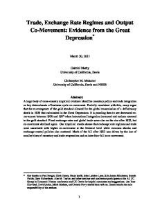
Fig. 5. Larval stemmata in the pupal brain of the red flour beetle Tribolium castaneum. Darkfield stereomicroscope image of frontal view of brain dissected from 48 hours old Tribolium pupa. Dorsal is up. The stemmata (white arrowheads) have organized into clusters of screening pigment expressing photoreceptors, which relocated into the optic neuropils in the lateral pupal brain. Oesophageal passage opening indicated by black arrowhead. lam = lamina, lob = lobula, med = mdeulla, one = optic neuropil, spg = supraesophageal ganglion, sbg = subesophageal ganglion, vng = ventral nervous system ganglion. Scale bar corresponds to 100 μm.
also been found in beetles such as Tribolium (FELISBERTI et al. 1997; SCHULZ et al. 1984; SOKOLOFF 1972). Preliminary investigation of the pupal brain in Tribolium confirms that the stemmata indeed retract into the optic neuropils (Fig. 5). In sum, these data suggest that the reutilization of juvenile instar eyes as extraretinal photoreceptors in the adult is yet another derived ground plan feature of the Holometabola. This trait is of course linked to the evolution of larval stage specific eyes in the ancestor of this clade. Nonetheless, it seems justified to consider juvenile eye derived extraretinal photoreceptors a likely autapomorphy of the Holometabola. Final confirmation awaits an investigation of the adult fate of the larval compound eyes in endopterygotan species with a primitive state of visual system morphology and development such as panorpid and nannochoristid Mecoptera. In addition, it will be extremely interesting to investigate how the larva specific eyes of thrips (Thysanoptera) relate to the stemmata of classically considered holometabolous insect orders. The few data published on visual system development in the Thysanoptera raise the possibility that the larval eyes relocate into the brain as has been described for the Holometabola (KUMM & MORITZ 1999). In the current view of insect phylogeny, the larval eyes of thrips and Holometabola represent convergent juvenile stage specific organs (Fig. 1). However, considering the debated position of Thysanoptera combined with the tentative evidence for a closer relationship between paraneopteran or-
The visual system and the Strepsiptera problem
The examination of phylogenetically informative aspects in the insect visual system yields several hitherto unrecognized shared derived character states of the Holometabola. This is useful, considering the surprisingly short list of autapomorphies supporting this otherwise uncontested major group (KRISTENSEN 1991, 1999). One obvious aspect to explore further is if these character states can also contribute to determining the phylogenetic position of the enigmatic Strepsiptera. A plethora of hypotheses have been voiced regarding the phylogenetic position of this group of extremely specialized parasitic insects. The major challenge to establishing strepsipteran relationships is the rampant frequency of extreme modifications of ancestral character states such as the reduction of the mesothoracic wings to haltere-like structures or obligatory neotenism of the females in most families. Although Strepsiptera are often viewed as an order most closely related to Coleoptera or Diptera, even their association with Holometabola as such has been questioned (KINZELBACH 1990; KRISTENSEN 1991; WHITING et al. 1997). Four new synapomorphies of the Holometabola can now be offered for discussion: (1) postembryonic onset of progressive retina differentiation, (2) possession of larval eyes, (3) larval eye derived extraretinal photoreceptors in the adult, and (4) outer optic lobe anlagen invagination. Can these character states be used to assess the phylogenetic position of the Strepsiptera? Strepsipteran males are equipped with very capable eyes necessary for the challenging task of locating a partner within only a few hours of nuptial pursuit. As might be expected, the strepsipteran visual system is also one of the most unique and unusual among insects. The elaborate lateral eyes have been referred to as compound lens eyes (BUSCHBECK et al. 1999). A single strepsipteran eye is composed of up to 150 large optical subunits. The cellular architecture of these subunits resembles that of the ocelli rather than the ommatidia (BUSCHBECK et al. 1999; KINZELBACH 1971). However, unlike ocelli the optical subunits of the strepsipteran eye form axonal projections into a clearly identifiable lamina neuropil from where visual information is relayed further to the medulla and lobula neuropils. Unfortunately, not enough information is available on strepsipteran embryology. For this reason, the development of the outer optic lobe anlagen
144
FRIEDRICH et al.: Insect visual system
Tab. 1. List of visual system characters with interordinal differences of potential phylogenetic significance for insects. Crustacea and Collembola are included for outgroup comparison. The eyeless Protura and Diplura are omitted. Grey background shading indicates hypothesized group that is supported by respective character state (partly very tentative given the sparse available data). Presence (+) or absence (–) of character state is indicated when published data exist for an insect order; –/+ = intermediate state; +,– = both states occur in taxon.
cannot be evaluated his point. Valuable information on other characters has been provided by a recent study of visual development in the strepsipteran species Xenos peckii (BUSCHBECK 2005). According to this analysis, the onset of adult eye differentiation is postembryonic and progressive (BUSCHBECK 2005). A region equivalent to the morphogenetic furrow could not be described
based on light microscopic analysis. Also, the assembly of strepsipteran eye subunits appears very different from the highly conserved ommatidial assembly program. Yet the array of lens clusters develops in an anterioposterior gradient, which is compatible with a progressive mode of adult eye development.
145
Arthropod Systematics & Phylogeny 64 (2)
Secondly, Xenos males develop larval eyes, stemmata, which retract during postembryogenesis to make room to the developing adult eye, like in other Holometabola (BUSCHBECK 2005). Stemmata have also been described in Mengenilla chobauti (BEUTEL et al. 2005), but their final fate was not established. However, there is data in the literature which suggests that the larval eyes are maintained (KINZELBACH 1971). Moreover, the available description of stemma migration during larval development is highly reminiscent to that described in Coleoptera and Lepidoptera. It thus seems very likely that the larval eyes differentiate further into brain photoreceptors in male Strepsiptera. These data are significant, considering that the evolutionary origin of the adult strepsipteran eye has been controversial. Some authors favored an origin of the strepsipteran lens eyes from larval stemmata (KINZELBACH 1971; PAULUS 1979). This possibility is ruled out be the new data from Xenos. In summary, two out of four character states are clearly consistent with a position of the Strepsiptera within Endopterygota: progressive differentiation of the adult retina during postembryonesis and possession of stemmata. The adult fate of the latter has the potential to further substantiate this issue.
11.
Summary and perspectives
While the visual system enables insects to view their environment at high resolution, it appears of limited value for improving our view of phylogenetic relationships between insect orders. This review identifies nine aspects of the visual system which show structural or developmental differences between orders, and may thus be phylogenetically informative (Tab. 1). Many of these require further, more comprehensive analysis in a wider sample of insect species. Unfortunately, all of these characters exhibit differences that concern relatively well established parts of the insect phylogenetic tree (compare Fig. 1 and Tab. 1). Ocellus innervation separates Collembola from Insecta or a subgroup thereof. The elaboration of a retina-blood border seems to be a synapmorphy of Odonata + Neoptera, a constellation which is widely favored (Fig. 1). While that remains to be determined in more primitive Neoptera and apterygotes, it seems likely that the occurrence of two mitotic waves in the developing retina is a derived character of insects in general, if not an autapomorphy of the Hexapoda. These three character states that relate to the deepest branches in the insect tree are complemented with four characters that very clearly speak for the monophyly of the Holometabola. While this superclade is probably one of the most widely accepted and known, the above character
states are valuable as the number of characters which do support endopterygotan monophyly is relatively small (KRISTENSEN 1999). The most significant shared derived character state is the possession of larval eye derived extraretinal photoreceptors in the adult optic neuropils, which represents the first autapomorphy in the adult body plan of the Holometabola. These characters prove also valuable for consolidating the endopterygotan position of the Strepsiptera. The phylogenetically most interesting trait is also the least substantiated in terms of character state polarity and consistency. It concerns the absence of a spatial separation of the lobula plate optic neuropil in the Hymenoptera in contrast to most other holometabolous insect orders for which optic neuropil anatomy has been studied. While it remains a plausible possibility that Hymenoptera represent the most basal branch of the Endopterygota, the elaboration of the lobula plate needs to be investigated in a wider sample of species. What are the future prospects of looking for phylogenetic information in the visual system? While it is impossible to predict the number of yet to be discovered informative structural or developmental differences between orders, the generally high degree of conservation of basic components does make it seem unlikely that a large number of useful character state differences will surface. It seems more realistic to hope for subtle differences that can be used in the analysis at the intraordinal level as previous studies in the Hemiptera, Diptera and Coleoptera have demonstrated (FISCHER et al. 2000).
12.
Acknowledgements
I am grateful to Dr. Klaus-Dieter Klass, Dr. Niels Peder Kristensen and Dr. Matthias Nuss for the opportunity to participate at the 2nd Dresden Meeting on Insect Phylogeny. Kudos are extended to Dr. James Woodgett for generously providing anti-Armadillo antiserum, and to Ivanna Yavorenko for diligent proofreading. The detailed comments and corrections by Dr. Klaus-Dieter Klass and an anonymous reviewer on the manuscript were highly appreciated. A Career Development Chair Award from Wayne State University provided the time resources necessary for completing this review.
13.
References
ANDERSON, H. 1978. Postembryonic development of the visual system of the locust, Schistocerca gregaria. I. Pattern of growth and developmental interactions in the retina and optic lobe. – Journal of Embryology and Experimental Morphology 45: 55–83.
146
BATE, C.M. 1978. Development of sensory systems in arthropods. Pp. 2–53 in: M. JACOBSON (ed.), Development of Sensory Systems. – Springer, Heidelberg. BEUTEL, R.G. & S.N. GORB 2006. A revised interpretation of the evolution of attachment structures in Hexapoda (Arthropoda), with special emphasis on Mantophasmatodea. – Arthropod Systematics & Phylogeny 64(1): 3–25. BEUTEL, R.G., H. POHL & F. HUENEFELD 2005. Strepsipteran brains and effects of miniaturization (Insecta). – Arthropod Structure & Development 34: 301–313. BITSCH, C. & J. BITSCH 2005. Evolution of eye structure and arthropod phylogeny. – Crustacean Issues 16: 185–214. BODENSTEIN, D. 1953. Postembryonic development. Pp. 275– 367 in: K.D. ROEDER (ed.), Insect Physiology. – Wiley, New York. BOLWIG, N. 1946. Senses and sense organs of the anterior end of the house fly larvae. – Videnskabelige Meddelelser fra Dansk Naturhistorik Forening 109: 81–217. BOORE, J.L., D.V. LAVROV & W.M. BROWN 1998. Gene translocation links insects and crustaceans. – Nature 392: 667–668. BUSCHBECK, E., B. EHMER & R. HOY 1999. Chunk versus point sampling: visual imaging in a small insect. – Science 286: 1178–1180. BUSCHBECK, E.K. 2005. The compound lens eye of Strepsiptera: morphological development of larvae and pupae. – Arthropod Structure & Development 34: 315–326. CAVENEY, S. 1986. The phylogenetic significance of ommatidium structure in the compound eyes of polyphagan beetles. – Canadian Journal of Zoology 64: 1787–1819. CHAMPLIN, D.T. & J.W. TRUMAN 1998. Ecdysteroids govern two phases of eye development during metamorphosis of the moth Manduca sexta. – Development 125: 2009– 2018. DICKENS, J.C. & J.L. EATON 1973. External ocelli in Lepidoptera previously considered to be anocellate. – Nature 242: 205–206. DICKSON, B. & E. HAFEN 1993. Genetic dissection of eye development in Drosophila. Pp. 1327–1362 in: P. LAWRENCE & A.M. MARTINEZ (eds.), The Development of Drosophila melanogaster. – Cold Spring Harbour Laboratory Press, New York. DOHLE, W. 1997. Myriapod-insect relationships as opposed to an insect-crustacean sister group relationship. Pp. 305–315 in: R.A. FORTEY & R.H. THOMAS (eds.), Arthropod Relationships. – Chapman & Hall, London. EGELHAAF, A. 1988. Evidence for the priming role of the central retinula cell in ommatidium differentiation of Ephestia kuehniella. – Rouxʼs Archive of Developmental Biology 197: 184–189. ENGHOFF, H., W. DOHLE & G.J. BLOWER 1993. Anamorphosis in millipedes (Diplopoda) – the present state of knowledge with some developmental and phylogenetic considerations. – Zoological Journal of the Linnean Society 109: 103–234. FELISBERTI, F., D.F. VENTRURA & H. HERTEL 1997. Cerebral extraocular photoreceptors in beetles. – Comparative Biochemistry and Physiology 118A: 1353–1357. FISCHER, A. & G. HORSTMANN 1971. Der Feinbau des Auges der Mehlmotte, Ephestia kuehniella (Lepidoptera, Pyralidae). – Zeitschrift für Zellforschung 116: 275–304. FISCHER, C., M. MAHNER & E. WACHMANN 2000. The rhabdom structure in the ommatidia of the Heteroptera (Insecta),
FRIEDRICH et al.: Insect visual system
and its phylogenetic significance. – Zoomorphology 120: 1–13. FREEMAN, M. 1996. Reiterative use of the EGF receptor triggers differentiation of all cell types in the Drosophila eye. – Cell 87: 651–660. FRIEDRICH, M. 2006. Continuity versus split and reconstitution: exploring the molecular developmental corollaries of insect eye primordium evolution. – Developmental Biology. doi: 10.1016/j.ydbio.2006.08.027. FRIEDRICH, M. & S. BENZER 2000. Divergent decapentaplegic expression patterns in compound eye development and the evolution of insect metamorphosis. – Journal of Experimental Zoology (Molecular and Developmental Evolution) 288: 39–55. FRIEDRICH, M., I. RAMBOLD & R.R. MELZER 1996. The early stages of ommatidial development in the flour beetle Tribolium castaneum (Coleoptera, Tenebrionidae). – Development Genes and Evolution 206: 136–146. FRIEDRICHS, H.F. 1931. Beiträge zur Morphologie und Physiologie der Sehorgane der Cicindeliden (Col.). – Zeitschrift für Morphologie und Ökologie der Tiere 21: 1–172. GILBERT, C. 1994. Form and function of stemmata in larvae of holometabolous insects. – Annual Review of Entomology 39: 323–349. GIRIBET, G., G.D. EDGECOMBE & W.C. WHEELER 2001. Arthropod phylogeny based on eight molecular loci and morphology. – Nature 413: 157–161. GOODMAN, L.J. 1981. Organisation and physiology of the insect dorsal ocellar system. Pp. 201–286 in: H. AUTRUM (ed.), Comparative Physiology and Evolution of Vision in Invertebrates. – Springer, Heidelberg. GREEN, P., A.Y. HARTENSTEIN & V. HARTENSTEIN 1993. The embryonic development of the Drosophila visual system. – Cell and Tissue Research 273: 583–598. GRIMALDI, D. & M.S. ENGEL 2005. Evolution of the Insects. – Cambridge University Press, New York. HAFNER, G.S. & T.R. TOKARSKI 1998. Morphogenesis and pattern formation in the retina of the crayfish Procambarus clarkii. – Cell and Tissue Research 293: 535–550. HAGBERG, M. 1986. Ultrastructure and central projections of extraocular photoreceptors in caddiesflies (Insecta, Trichoptera). – Cell and Tissue Research 245: 643–648. HARZSCH, S. 2002. The phylogenetic significance of crustacean optic neuropils and chiasmata: a re-examination. – Journal of Comparative Neurology 453: 10–21. HARZSCH, S. 2006. Neurophylogeny. Architecture of the nervous system and a fresh view on arthropod phylogeny. – Integrative and Comparative Biology 46: 162–194. HARZSCH, S., J. BENTON, R.R. DAWIRS & B. BELTZ 1999. A new look at embryonic development of the visual system in decapod crustaceans: Neuropil formation, neurogenesis, and apoptotic cell death. – Journal of Neurobiology 39: 294–306. HARZSCH, S., K. VILPOUX, D.C. BLACKBURN, D. PLATCHETZKI, N.L. BROWN, R. MELZER, K.E. KEMPLER & B.A. BATTELLE 2006. Evolution of arthropod visual systems development of the eyes and central visual pathways in the horseshoe crab Limulus polyphemus Linnaeus, 1758 (Chelicerata, Xiphosura). – Developmental Dynamics 235: 2641–2655. HARZSCH, S. & D. WALOSSEK 2001. Neurogenesis in the developing visual system of the branchiopod crustacean Triops longicaudatus (LeConte, 1846): corresponding
Arthropod Systematics & Phylogeny 64 (2)
patterns of compound-eye formation in Crustacea and Insecta? – Development Genes and Evolution 211: 37–43. HELFRICH-FORSTER, C., T. EDWARDS, K. YASUYAMA, B. WISOTZKI, S. SCHNEUWLY, R. STANEWSKY, I.A. MEINERTZHAGEN & A. HOFBAUER 2002. The extraretinal eyelet of Drosophila: development, ultrastructure, and putative circadian function. – Journal of Neuroscience 22: 9255–9266. HEMING, B.S. 1982. Structure and development of the larval visual system in embryos of Lytta viridana Leconte (Coleoptera, Meloidae). – Journal of Morphology 172: 23–43. HEMING, B.S. 2003. Insect Development and Evolution. – Cornell University Press, Ithaca, New York. HENNIG, W. 1981. Insect Phylogeny. – Wiley & Sons, New York. HOFBAUER, A. & E. BUCHNER 1989. Does Drosophila have seven eyes? – Naturwissenschaften 76: 335–336. HWANG, U.W., M. FRIEDRICH, D. TAUTZ, C.J. PARK & W. KIM 2001. Mitochondrial protein phylogeny joins myriapods with chelicerates. – Nature 413: 154–157. ICHIKAWA, T. 1991. Brain photoreceptors in the pupal and adult butterfly: fate of the larval ocelli. – Zoological Sciences 8: 471–476. INOUE, Y., K. MIYAWAKI, T. TERASAWA, K. MATSUSHIMA, Y. SHINMYO, N. NIWA, T. MITO, H. OHUCHI & S. NOJI 2004. Expression patterns of dachshund during head development of Gryllus bimaculatus (cricket). – Gene Expression Patterns 4: 725–731. KALMUS, H. 1945. Correlations between flight and vision, and particularly between wings and ocelli in insects. – Proceedings of the Royal Entomological Society London A 20: 84–96. KELBER, A., A. BALKENIUS & E.J. WARRANT 2003. Colour vision in diurnal and nocturnal hawkmoths. – Integrative and Comparative Biology 43: 571–579. KINZELBACH, R.K. 1971. Morphologische Befunde an Fächerflüglern und ihre phylogenetische Bedeutung. – Zoologica 41: 1–256. KINZELBACH, R. 1990. The systematic position of Strepsiptera (Insecta). – American Entomologist 36: 292–303. KITAMOTO, J., K. OZAKI & K. ARIKAWA 2000. Ultraviolet and violet receptors express identical mRNA encoding an ultraviolet-absorbing opsin: identification and histological localization of two mRNAs encoding short-wavelengthabsorbing opsins in the retina of the butterfly Papilio xuthus. – Journal of Experimental Biology 203 Pt 19: 2887–2894. KJER, K.M. 2004. Aligned 18S and insect phylogeny. – Systematic Biology 53: 506–514. KJER, K.M., F.L. CARLE, J. LITMAN & J. WARE 2006. A molecular phylogeny of Insecta. – Arthropod Systematics & Phylogeny 64(1): 35–44. KLASS, K.-D., O. ZOMPRO, N.P. KRISTENSEN & J. ADIS 2002. Mantophasmatodea: a new insect order with extant members in the Afrotropics. – Science 296: 1456–1459. KRISTENSEN, N.P. 1991. Phylogeny of extant hexapods. Pp. 125–140 in: I.D. NAUMANN, P.B. CARNE, J.F. LAWRENCE, E.S. NIELSEN, J.P. SPRADBERRY, R.W. TAYLOR, M.J. WHITTEN & M.J. LITTLEJOHN (eds.), The Insects of Australia: A textbook for students and research workers. – CSIRO, Melbourne University Press, Melbourne. KRISTENSEN, N.P. 1995. Forty yearsʼ insect phylogenetics. – Zoologische Beiträge N.F. 36: 83–124.
147
KRISTENSEN, N.P. 1999. Phylogeny of endopterygote insects, the most successful lineage of living organisms. – European Journal of Entomology 96: 237–253. KUMM, S. & G. MORITZ 1999. Development of the visual system in important thrips species. In: 6. Symposium of Thysanoptera. Antalya, 1999. LIU, Z. & M. FRIEDRICH 2004. The Tribolium homologue of glass and the evolution of insect larval eyes. – Developmental Biology 269: 36–54. MALLATT, J.M., J.R. GAREY & J.W. SHULTZ 2004. Ecdysozoan phylogeny and Bayesian inference: first use of nearly complete 28S and 18S rRNA gene sequences to classify the arthropods and their kin. – Molecular Phylogenetics and Evolution 31: 178–191. MEINECKE, C.C. 1981. The fine structure of the compound eye of the African armyworm moth, Spodoptera exempta Walk. (Lepidoptera, Noctuidae). – Cell And Tissue Research 216: 333–347. MEINERTZHAGEN, I.A. 1991. Evolution of the cellular organization of the arthropod compound eye and optic lobe. Pp. 341–362 in: J.R. CRONLY-DILLON & R.L. GREGORY (eds.), Vision and Visual Dysfunction. – Macmillan Press, Boston. MELZER, R.R., R. DIERSCH, D. NICASTRO & U. SMOLA 1997. Compound eye evolution: Highly conserved retinula and cone cell patterns indicate a common origin of the insect and crustacean ommatidium. – Naturwissenschaften 84: 542–544. MELZER, R.R., C. MICHALKE & U. SMOLA 2000. Walking on insect paths? Early ommatidial development in the compound eye of the ancestral crustacean, Triops cancriformis. – Naturwissenschaften 87: 308–311. MELZER, R.R. & H.F. PAULUS 1989. Evolutionswege zum Larvalauge der Insekten – Die Stemmata der höheren Dipteren und ihre Abwandlung zum Bolwig-Organ. – Zeitschrift für Zoologische Systematik und Evolutionsforschung 27: 200–245. MELZER, R.R., H.F. PAULUS & N.P. KRISTENSEN 1994. The larval eye of nannochoristid scorpionflies (Insecta, Mecoptera). – Acta Zoologica 75: 201–208. MEYER-ROCHOW, V.B. 1974. The structure and function of the larval eye of the sawfly Perga. – Journal of Insect Physiology 20: 1565–1591. MOSES, K. 2002. Drosophila Visual System Development. – Springer Verlag, Berlin, Heidelberg. MÜLLER, C.H.G., J. ROSENBERG, S. RICHTER & V.B. MEYERROCHOW 2003. The compound eye of Scutigera coleoptrata (Linnaeus, 1758) (Chilopoda: Notostigmophora): an ultrastructural reinvestigation that adds support to the Mandibulata concept. – Zoomorphology 122: 191–209. NOWEL, M.S. & P.M. SHELTON 1980. The eye margin and compound-eye development in the cockroach: evidence against recuitment. – Journal of Embryology and Experimental Morphology 60: 329–343. NUESCH, H. 1987. Metamorphose bei Insekten. Direkte und indirekte Entwicklung bei Apterygoten und Exopterygoten. – Zoologische Jahrbücher Anatomie 115: 453–487. OAKLEY, T.H. 2003. On homology of arthropod compound eyes. – Integrative and Comparative Biology 43: 522–530. OSORIO, D. & J.P. BACON 1994. A good eye for arthropod evolution. – BioEssays 16: 419–424.
148
PAULUS, H.F. 1972. Die Feinstruktur der Stirnaugen einiger Collembolen (Insecta, Entognatha) und ihre Bedeutung für die Stammesgeschichte der Insekten. – Zeitschrift für Zoologische Systematik und Evolutionsforschung 10: 81–122. PAULUS, H.F. 1979. Eye structure and the monophyly of the Arthropoda. Pp. 299–371 in: A.P. GUPTA (ed.), Arthropod Phylogeny. – Van Nostrand Reinhold Company, New York. PAULUS, H.F. 1989. Das Homologisieren in der Feinstrukturforschung: Das Bolwig-Organ der höheren Dipteren und seine Homologisierung mit Stemmata und Ommatidien eines ursprünglichen Facettenauges der Mandibulata. – Zoologische Beiträge N.F. 32: 437–478. PAULUS, H.F. 2000. Phylogeny of the Myriapoda-CrustaceaInsecta: a new attempt using photoreceptor structure. – Journal of Zoological Systematics and Evolutionary Research 38: 189–208. PAULUS, H.F. & M. SCHMIDT 1978. Evolutionswege zum Larvalauge der Insekten: Die Stemmata der Trichoptera und Lepidoptera. – Zeitschrift für Zoologische Systematik und Evolutionsforschung 16: 188–216. READY, D.F. 1989. A multifaceted approach to neural development. – Trends in Neurosciences 12: 102–110. READY, D.F., T.E. HANSON & S. BENZER 1976. Development of the Drosophila retina, a neurocrystalline lattice. – Developmental Biology 53: 217–240. REGIER, J.C., J.W. SHULTZ & R.E. KAMBIC 2005. Pancrustacean phylogeny: hexapods are terrestrial crustaceans and maxillopods are not monophyletic. – Proceedings of Biological Sciences 272: 395–401. RICHTER, S. 1999. The structure of the ommatidia of the Malacostraca (Crustacea) – a phylogenetic approach. – Verhandlungen des Naturwissenschaftlichen Vereins Hamburg (NF) 38: 161–204. ROONWAL, M.L. 1936. Studies on the embryology of the African migratory locust, Locusta migratoria migratorioides Reiche and Frm. (Orthoptera, Acrididae) II-Organogeny. – Philosophical Transactions of the Royal Society London B 227: 175–244. RUEL, L., V. STAMBOLIC, A. ALI, A.S. MANOUKIAN & J.R. WOODGETT 1999. Regulation of the protein kinase activity of Shaggy (Zeste-white3) by components of the wingless pathway in Drosophila cells and embryos. – Journal of Biological Chemistry 274: 21790–21796. SCHLECHT, P. 1979. Colour discrimination in dim light. An analysis of the photoreceptor arrangement in the moth Deilephila. – Journal of Comparative Physiology 129: 257–267. SCHULZ, W.-D., U. SCHLUETER & G. SEIFERT 1984. Extraocular photoreceptors in the brain of Epilachna varivestis (Coleoptera, Coccinellidae). – Cell and Tissue Research 129: 257–267. SHAW, S.R. 1977. Restricted diffusion and extracellular space in the insect retina. – Journal of Comparative Physiology 113: 257–282. SHAW, S.R. 1978. The extracellular space and blood-eye barrier in an insect retina: an ultrastructural study. – Cell and Tissue Research 188: 35–61. SHAW, S.R. & L.P. VARNEY 1999. Primitive, crustacean-like state of blood-brain barrier in the eye of the apterygote insect Petrobius (Archaeognatha) determined from uptake of fluorescent tracers. – Journal of Neurobiology 41: 452–470.
FRIEDRICH et al.: Insect visual system
SINAKEVITCH, I., J.K. DOUGLAS, G. SCHOLTZ, R. LOESEL & N.J. STRAUSFELD 2003. Conserved and convergent organization in the optic lobes of insects and isopods, with reference to the other crustacean taxa. – Journal of Comparative Neurology 467: 150–172. SOKOLOFF, A. 1972. The Biology of Tribolium. – Clarendon Press, Oxford. STRAUSFELD, N.J. 1998. Crustacean-insect relationships: The use of brain characters to derive phylogeny amongst segmented invertebrates. – Brain Behavior and Evolution 52: 186–206. STRAUSFELD, N.J. 2005. The evolution of crustacean and insect optic lobes and the origins of chiasmata. – Arthropod Structure & Development 34: 235–256. TERRY, M.D. & M.F. WHITING 2005. Mantophasmatodea and phylogeny of the lower neopterous insects. – Cladistics 21: 240–257. TIX, S., J.S. MINDEN & G.M. TECHNAU 1989. Pre-existing neuronal pathways in the developing optic lobes of Drosophila. – Development 105: 739–746. TRUJILLO-CENOZ, O. 1985. The eye: development, structure and neural connections. Pp. 171–223 in: G.A. KERKUT & L.I. GILBERT (eds.), Comprehensive Insect Physiology, Biochemistry and Pharmacology. – Pergamon Press, Oxford. ULLMANN, S.L. 1966. The development of the nervous system and other ectodermal derivatives in Tenebrio molitor L. (Insecta, Coleoptera). – Philosophical Transactions of the Royal Society London B 248: 245–277. WELSCH, B. 1977. Ultrastruktur und funktionelle Morphologie der Augen des Nachtfalters Deilephila elpenor (Lepidoptera, Sphingidae). – Cytobiologie 14: 376–400. WHITING, M.F. 2004. Phylogeny of holometabolous insects. Pp. 345–361 in: J. CRACRAFT & M.J. DONOGHUE (eds.), Assembling the Tree of Life. – Oxford University Press, New York. WHITING, M.F., J.C. CARPENTER, Q.D. WHEELER & W.C. WHEELER 1997. The Strepsiptera problem: Phylogeny of the holometabolous insect orders inferred from 18S and 28S ribosomal DNA sequences and morphology. – Systematic Biology 46: 1–68. WILDT, M. & S. HARZSCH 2002. A new look at an old visual system: structure and development of the compound eyes and optic ganglia of the brine shrimp Artemia salina Linnaeus, 1758 (Branchiopoda, Anostraca). – Journal of Neurobiology 52: 117–132. WILLMANN, R. 2004. Phylogenetic relationships and evolution of insects. Pp. 330–344 in: J. CRACRAFT & M.J. DONOGHUE (eds.), Assembling the Tree of Life. – Oxford University Press, New York. WOLFF, T. & D. READY 1993. Pattern formation in the Drosophila retina. Pp. 1277–1326 in: P. LAWRENCE & A.M. MARTINEZ (eds.), The development of Drosophila melanogaster. – Cold Spring Harbour Laboratory Press, New York. YASUYAMA, K. & I.A. MEINERTZHAGEN 1999. Extraretinal photoreceptors at the compound eyeʼs posterior margin in Drosophila melanogaster. – The Journal of Comparative Neurology 412: 193–202.