TECHNICAL NOTES J Oral Maxillofac Surg 65:1840-1842, 2007
Modified Osteotome for Inferior Border Sagittal Split Osteotomy José Nazareno Gil, DDS, MS, PhD,* Charles Marin, DDS,† Jonathas Daniel Paggi Claus, DDS,‡ and Sergio Monteiro Lima Júnior, DDS§ Bilateral sagittal split osteotomy of the ramus (BSSO) is a surgical procedure used widely for the correction of mandibular deformities because of its versatility for both advancement and setback of the mandible. Despite its frequent use and numerous modifications, the BSSO demands technical precision and is not without associated intraoperative complications.1 To decrease the frequency of intraoperative and postoperative complications, many modifications in the BSSO technique were reported with an improvement in stability and predictability of the postsurgical results.2-5 The buccal cortex of the mandible normally is thin posterior to the second molar, weakening this region, and this can contribute to fracture of the proximal segment.6,7 Fractures of either proximal or distal segments range from 2% to 18% of the cases.1,8-10 According to Merha et al,11 the occurrence of unfavorable splits is infrequent, irrespective of the presence or absence of third molars if an inferior basilar bone osteotomy is made during the BSSO. The difficulty in separating the inferior basilar bone led to the development of a modification in the BSSO and an osteotome to divide precisely this basilar bone into 2 similar segments. The objective of this study was to show the efficacy of a basilar osteotome to properly separate the mandible and
Received from the Department of Oral and Maxillofacial Surgery, University Hospital, College of Dentistry, Santa Catarina Federal University, Florianopolis, Brazil. *Professor. †Resident. ‡Resident. §Resident. Address correspondence and reprint requests to Dr Gil: Rua Tenente Silveira 293, sala 1001, edifício Reflex. Florianópolis, SC, Brasil, CEP: 88010-310; e-mail:
[email protected]
prevent the bad split of the segments during the BSSO.
Technique Description The authors carried out the modified sagittal osteotomy described by Epker2 using a modified osteotome that was designed specifically for inferior border osteotomy of the mandible (Fig 1). A 45° bevel was included anterior to the vertical cut at molar region through the cortical bone (Fig 2A). This maneuver will allow the introduction of the instrument properly (Fig 2B; Fig 3). This osteotome is used to initiate the split in the inferior border (Fig 4), followed by a series of small to large osteotomes and a Smith spreader to complete the BSSO. The surgical assistants provide extra oral forward manual support at the posterior border of the mandible to facilitate the split and avoid injury to the temporomandibular joint (TMJ). Two parallel cortical surfaces at the inferior border should result. In addition, separation of the medial and lateral segments in similar surfaces would help in moving the proximal and distal segments and applying less force
0278-2391/07/6509-0029$32.00/0
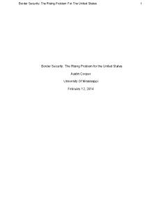
FIGURE 1. Design of the modified osteotome used to make the osteotomy of the inferior border. The tip of the osteotome is supported below the inferior border of the mandible.
doi:10.1016/j.joms.2005.12.045
Gil et al. Modified BSSO. J Oral Maxillofac Surg 2007.
© 2007 American Association of Oral and Maxillofacial Surgeons
1840
1841
GIL ET AL
FIGURE 2. A, Bevel included anterior to the vertical cut at molar region in 45° through cortical bone. B, Correct position of the osteotome for inferior border osteotomy.
FIGURE 4. Basilar bone divided into 2 similar segments. Gil et al. Modified BSSO. J Oral Maxillofac Surg 2007.
Gil et al. Modified BSSO. J Oral Maxillofac Surg 2007.
to the condyles, which should decrease the risk of relapse.
Discussion Fracture of the buccal plate most often occurs when the inferior border of the mandible is incompletely transected. In this situation, the force used to split the mandible fractures the thin buccal plate. The task of completing the split is made technically difficult because the fracture leaves the buccal plate shortened and with less bone to pry against.12 Precious et al1 recommend the completion of the split with spreaders instead of mallet and chisel. However, the inferior border osteotomy in the BSSO technique minimizes unfavorable splits and decreases trauma to the TMJ by using an osteotome to partially section the inferior border of the mandible.13 TMJ edema and hemarthrosis could displace the condyle in the glenoid fossa contributing to mandibular relapse. Posterior manual support of the
mandible is carried out to prevent TMJ injuries caused by the osteotome in posterior direction. Using an osteotome to split the inferior border will prevent fractures that may occur on the lingual aspect of the mandible, between the inferior border and the inferior aspect of the inferior alveolar canal.14 The proposed modification attempts to obtain 2 parallel cortical surfaces at the inferior border, and allows an easy mobilization of the fragments and a better fixation. Twenty-five patients were evaluated, with no bad splits of the proximal segment in the 50 osteotomies made. This split produces 2 similar parallel surfaces at the inferior border that will result in less force applied to the condyles during fixation, with a better area at the inferior border for screw introduction. In this study, the osteotome efficiently separates the basilar bone in 2 similar segments. The BSSO is more predictable, making the repositioning of the segments easier due to the parallelism that results from the split and applying the internal fixation is facilitated, because the basilar bone is kept intact and creates a bigger area of contact between segments.
References
FIGURE 3. Intraoperative view. Note the bevel at the anterior border of the cut to allow osteotome introduction. Gil et al. Modified BSSO. J Oral Maxillofac Surg 2007.
1. Precious DS, Lung KE, Pynn BR, Goodday RH: Presence of impacted teeth as a determining factor of unfavorable splits in 1256 sagittal-split osteotomies. Oral Surg Oral Med Oral Pathol Oral Radiol Endod 85:362, 1998 2. Epker BN: Modification in the sagittal osteotomy of the mandible. J Oral Surg 35:157, 1977 3. Marquez IM, Stella JP: Modification of sagittal split ramus osteotomy to avoid unfavorable fracture around impacted third molars. Int J Adult Orthod Orthognath Surg 13:183, 1998 4. Wolford LM, Davis WM: The mandibular inferior border split: A modification in the sagittal split osteotomy. J Oral Maxillofac Surg 48:92, 1990 5. Max D, Rotskoff K: A modified technique for the sagittal split osteotomy. J Oral Maxillofac Surg 51:1050, 1993 6. Smith BR, Rajchel JL, Waite DE, et al: Mandibular ramus anatomy as it relates to the medial osteotomy of the sagittal split ramus osteotomy. J Oral Maxillofac Surg 49:12, 1991 7. Schubert W, Kobienia BJ, Pollock RA: Cross-sectional area of the mandible. J Oral Maxillofac Surg 55:689, 1997
1842 8. Guernsey LH, DeChamplain RW: Sequelae and complications of the intraoral sagittal osteotomy of the mandibular rami. Oral Surg Oral Med Oral Pathol 32:176, 1971 9. Martis CS: Complications after mandibular sagittal split osteotomy. J Oral Maxillofac Surg 42:101, 1984 10. Turvey TA: Intraoperative complications of sagittal osteotomy of the mandibular ramus: Incidence and management. J Oral Maxillofac Surg 43:504, 1985 11. Mehra P, Castro V, Freitas R, et al: Complications of the mandibular sagittal split ramus osteotomy associated with the pres-
MODIFIED BSSO ence or absence of third molars. J Oral Maxillofac Surg 59:854, 2001 12. O’Ryan F, Poor DB: Completing sagittal split osteotomy of the mandible after fracture of the buccal plate. J Oral Maxillofac Surg 62:1175, 2004. 13. Bell WH, Proffit WR, White RP: Surgical Correction of Dentofacial Deformities. Philadelphia, PA, Saunders, 1980, p 715 14. Cillo JE, Stella JP: Selection of sagittal split ramus osteotomy technique based on skeletal anatomy and planned distal segment movement: current therapy. J Oral Maxillofac Surg 63:109, 2005