505
NFAT signaling in vertebrate development Isabella A Graef, Feng Chen and Gerald R Crabtree* NFATc proteins transduce Ca2+ signals to the nucleus and then pair with other proteins on DNA to generate NFAT complexes that activate transcription in response to both electrical and tyrosine kinase signaling. The four NFATc genes arose at the origin of vertebrates, implying that they have evolved for the development of vertebrate-specific functions, such as a complex nervous system, a recombinational immune system, and a vascular system with a complex heart. These speculations are borne out by studies of mice with null mutations in the different family members. Addresses Departments of Developmental Biology and Pathology, Stanford University Medical School, Stanford, California 94305, USA *e-mail:
[email protected] Current Opinion in Genetics & Development 2001, 11:505–512 0959-437X/01/$ — see front matter © 2001 Elsevier Science Ltd. All rights reserved. Abbreviations BNP b-type natriuretic peptide CHP calcineurin homologous protein CnA calcineurin A CnB calcineurin B CsA Cyclosporin A DSCR Down syndrome critical region GSK-3 glycogen-synthase kinase-3 IGF insulin-like growth factor IL2 interleukin 2 inositol 1,4,5-trisphosphate IP3 IP3R1 IP3 receptor, type-1 NFATc nuclear-factor-of-activated-T-cells cytoplasmic NMDA N-methyl-D-aspartate VSCC voltage-sensitive calcium channel
Introduction The genes that encode the cytoplasmic, calcium-sensitive subunits of NFAT transcription complexes arose at the time of the origin of vertebrates and are not found in the genomes of invertebrates [1]. It was probably for this reason that NFAT signaling was not initially recognized as an important developmental pathway. Recent genetic evidence in mice, however, has indicated that these proteins play critical roles in the development of the immune system, the heart and blood vessels as well as the muscular and nervous systems in vertebrates. The latter observations are of particular interest because control of the NFAT pathway is exerted at the subtle interface of electrical and receptorcontrolled signaling resulting in greatly expanded regulatory capacity. We will briefly review the complex levels of control of this signaling pathway and then discuss recent data implicating NFAT signaling in development.
Multiple levels of positive and negative controls over NFAT signaling Before reviewing the role of NFAT signaling in development, it is worth pointing out the complex positive and negative
feedback controls that modulate this pathway and hence make simple genetic analysis difficult. The NFAT signaling pathway was first described in lymphocytes as a pathway carrying signals from the polymorphic T-cell receptor to genes that coordinate an immune response. The pathway was defined by a strategy of working backward from the regulatory regions of an early activation gene (IL-2) to the cell membrane [2,3]. This approach resulted in the delineation of the pathway shown in Figure 1. Sustained, low-amplitude Ca2+ signals lead to the activation of the heterodimeric phosphatase calcineurin [4]. Calcineurin binds directly to NFATc proteins (NFATc1–c4, Hugo Nomenclature Committee http://www.gene.ucl.ac.uk/ nomenclature/) through a conserved motif and dephosphorylates serines within the SP repeats and serine rich motifs (SRR and SP-repeats) in the amino-terminus of NFATc family members. This dephosphorylation unmasks nuclear localization sequences in NFATc proteins and triggers their cytoplasmic→nuclear translocation [5]. The immunosupressive drugs CsA and FK506 block the nuclear localization of NFAT transcription complexes by inhibiting calcineurin phosphatase activity. These chemically distinct natural products bind at subnanomolar affinity to intracellular receptors thus forming inhibitory complexes. The resultant drug/protein composite surface binds tightly to calcineurin and prevents substrate access [6,7]. One very intriguing and surprising result published this year was the finding that most Ca2+ regulated gene expression in lymphocytes is dependent on calcineurin activity [8••]. NFAT signaling is opposed by at least three negative regulatory mechanisms. The first is exerted by a group of proteins that inhibit calcineurin phosphatase activity. These endogenous inhibitors directly bind to calcineurin and thereby block dephosphorylation of substrates such as NFATc proteins. One class of these inhibitory proteins includes DSCR1 (MCIP1) [9,10], DSCR1L2 (MCIP2) and DSCR1L1 (ZAK-4) [11] which act as competitive inhibitors with nanomolar binding affinity. Cabin (Cain) is a noncompetitive inhibitor of calcineurin activity [12,13]. CHP proteins appear to compete with binding of the regulatory subunit CnB to the catalytic subunit, CnA and thereby inhibit the Ca2+-dependent activation of the phosphatase [14]. The scaffolding protein AKAP is also a calcineurin inhibitor which binds both calcineurin and PKA and may anchor calcineurin at specific sites that allow the protein to engage the proper substrates when activated [15]. The second negative regulatory mechanism is the rapid regulated export of the NFATc family members from the nucleus by kinases such as GSK-3 [16–18]. Rephosphorylation of NFATc proteins leads to rapid export of NFATc family members from the nucleus and
506
Differentiation and gene regulation
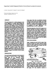
Figure 1 Ca2+
Receptor
CRAC
ra mb me l l Ce
ne
Ca2+ Ca2+
IP3R
Ras, Rac, PKC
CsA/CpH FK506/FKBP
PO3 AKAP-79 DSCR1 Cain CHP
NFATc1–c4
urin
e lcin
Ca
ses
ina
k AT
NF
a cle Nu
em rm
e bran NFATn
NFATc1–c4
Cytokines, calcium channels cell–cell interaction molecules
Current Opinion in Genetics & Development
Signal transduction by Ca2+, calcineurin, and NFAT. A rise in intracellular Ca2+ following receptor stimulation leads to activation of the Ca2+/calmodulin-regulated serine/threonine phosphatase calcineurin. Calcineurin directly dephosphorylates NFATc proteins, which rapidly translocate to the nucleus upon dephosphorylation. Calcineurin activity is controlled by endogenous inhibitory proteins such as DSCR1, AKAP-79, Cain or CHP and can be inhibited by the
immunosupressive drugs CsA and FK506. The term NFAT-kinases is used for a group of kinases such as GSK-3 that oppose calcineurin by rephosphorylation of NFATc proteins. Ras, Rac or PKC signaling must be coincident with the Ca2+/calcineurin signaling to assemble the NFAT transcriptional complex. NFATn is used to indicate tissue-specific transcription factors that act together with NFATc to activate transcription.
termination of transcription. Preliminary data indicate that GSK-3 in the nucleus might be controlled independently from GSK-3 in the cytoplasm (K Stankunas, GR Crabtree, unpublished data).
addition, Ca2+ release from IP3-sensitive stores can cooperate with Ca2+ influx via voltage-gated and receptor-operated calcium channels [20]. In neurons, activation of NFAT signaling leads to induction of the IP3R1 gene and thus provides a positive feedback mechanism that could alter the amplitude or spatial organization of Ca2+ signals. These levels of regulatory control are summarized in Figure 1 and provide an explanation for some of the unanticipated results of disruption of genes within the pathway. They also point to the need for new genetic approaches if we are to understand the mechanistic aspects of this pathway in vivo.
The third mechanism of negative inhibition involves the transcriptional activation of the DSCR1 (MCIP) gene by NFAT signaling and the modulation of forward signaling by the accumulation of a calcineurin inhibitor [19•]. In addition, NFAT signaling is probably also subject to at least one level of positive feedback control. Many receptors respond to stimulation by generating the second messenger IP3. IP3 triggers the release of Ca2+ from the endoplasmic reticulum by binding to the IP3 receptor, thereby causing the rapid influx of extracellular Ca2+ via calcium release-activated calcium (CRAC) channels. In
DNA binding by NFATc proteins is quite weak and therefore NFATc family members probably never act alone but rather need a partner protein, NFATn, to bind to DNA at physiologic concentrations [21–23]. Thus, cooperative
NFAT signaling in vertebrate development Graef et al.
507
Figure 2 Morphogenesis of the heart valves appears to involve an NFATc-dependent cross talk between endocardium and local mycocardium. The blue cells in the upper left hand corner are the NFATc1-expressing endocardium that will be transformed into components of a heart valve. The signal appears to come from the myocardium (pink cells) and is likely to be transmitted through the gray cardiac jelly to interact with receptors that are on the endocardial cells leading to calcineurin activation and the nuclear import of NFATc1. A few days later, valves (yellow) appear as a result of this developmental inductive event.
Valves E9–12
Lumen Lumen
?Receptor
Cn
Ca2+
?Signal
NFATc1
Valve morphogens
Endocardial cells Myocardial Cells Current Opinion in Genetics & Development
binding of NFATc proteins with diverse NFATn proteins, such as AP-1, GATA, cMAF and MEF2 family members makes Ca2+ signaling dependent on coincident activation of other signaling pathways and as such contributes to the activation of the NFAT pathway by a variety of upstream signals in many cell types and the control of transcription of very diverse target genes [3,24]. NFAT signaling thus acts as a signal integrator and coincidence detector rather than as a master-control pathway.
NFAT signaling in lymphoid development Although the NFAT signaling pathway was first defined in lymphocytes, ironically it has been most difficult to study the role of individual components in these cells because of the high degree of genetic redundancy at each level of the pathway. Lymphoid cells express all four family members. Although all family members are able to interact with the same core DNA-binding element, it now appears that they have at least some degree of specificity in DNA binding, which may be a product of their associated nuclear partners. Deletion of NFATc2 results in minor defects in lymphoid development and a mild paradoxical hyperproliferation of lymphocytes, which is
likely the result of the complex positive and negative feedback mechanisms mentioned above [25,26]. Insight into the lymphoid roles of NFATc1 has been difficult because of its early essential role in heart valve development (see below) but, using RAG-deficient blastocyst complementation, NFATc1 was found to play a role in the proliferative expansion of immature thymocytes as well as of peripheral, mature T- and B-lymphocytes [27,28]. Recently, NFATc1/c2 double mutant T-lymphocytes were found to pass through early thymic development and have profound defects in cytokine production and differentiation, whereas double mutant B-lymphocytes were hyperactive [29•]. These cells were derived from RAGdeficient mice in which the immune system was reconstituted with fetal liver cells from NFATc1/c2 doublemutant embryos. In contrast, NFATc2/c3 mice are viable but develop a severe lymphoproliferative disorder with dramatic increase of cytokines and IgE levels [30]; this results in the development of inflammatory symptoms resembling a severe allergic response. At this point, it is not clear whether these hyperallergic responses reflect early developmental roles for NFATc2 and NFATc3 or a role in mature T-cell responses.
508
Differentiation and gene regulation
Figure 3
Somite
Somite
Neural tube
Somite
Somite
Somite
Somite
Current Opinion in Genetics & Development
NFAT signaling at E8.5 patterns the mammalian vascular system (red). Local calcineurin/NFAT signaling in the somites and neural tubes leads to the production of an inhibitory signal (blocking symbol) that prevents aberrant branching of vessels into the areas where NFATc4 is highly expressed.
NFAT signaling is essential for the morphogenesis of vertebrate heart valves NFATc1-deficient mice have defects in the formationof heart valves and the interventricular septum [31,32] that resemble congenital heart defects seen in humans. Congenital heart valve and septal defects occur in ~1% of the human population, making it one of the most common serious congenital defects. Despite their vital importance, very little is known about the molecular mechanisms governing valve development. The heart valve forms by a unique developmental process in which endothelial cells delaminate from the endocardial layer and are transformed into mesenchymal cells that accumulate in the endocardial cushion area. The cushion then undergoes a complex series of morphogenic steps that lead to the formation of the valve. The mitral and tricuspid valves also have to connect with papillary muscles that provide precise control of apposition of the valve leaflets to prevent regurgitation. NFATc1-expressing cells are initially distributed over the entire endocardium but by E11.5 NFATc1 expression becomes restricted to regions of the future heart valves (Figure 2). It is unknown at present whether NFATc1-expressing endocardial cells migrate to the valve primordia or whether specific signals regulate the restricted expression of NFATc1. The nuclear localization of NFATc1 is reversed rapidly by CsA treatment, indicating that calcineurin is essential for NFATc1 function in the
nucleus. Mice with mutations in calcineurin B also show a failure of nuclear localization of NFATc1, providing genetic evidence that Ca2+/calcineurin signaling is indeed essential for NFAT localization [33•], as first discovered in lymphocytes by biochemical reconstitution. But what regulates Ca2+ and calcineurin at the sites where cardiac valves will eventually form? The observation that NFATc1 is directed to the nucleus of heart valve precursor cells by a signaling process makes it possible to look for upstream regulators of NFATc1 localization in this essential process. One of the most exciting developments in signaling in recent years has been the discovery of the unusual role of the connexins [34] — transmembrane proteins that form gap junctions between cells, allowing the passage of ions and other second messengers between cells. These gap junctions synchronize connected cells both electrically and biochemically, thereby coordinating intercellular communication. Mutation of connexin-45 results in defects in endocardial cushion formation that resemble those of NFATc1 null mice [35••]. In connexin-45-deficient mice, NFATc1 is cytoplasmic, indicating that a potential role of connexin-45 is to allow Ca2+ signals to be transmitted within a sheet of synchronized valvular precursor cells. This is also underscored by the earlier finding that NFATc1 is preferentially nuclear in endocardial cells that are adjacent to each other. The developmental implications of this are profound, in that it allows a morphogenic Ca2+ signal to spread very rapidly through a population of cells. However, these exciting findings raise as many questions as they answer. For example, which receptor initiates the Ca2+ signal? Previous work has shown that Ca2+ signals are constrained to ~1/10th a cell diameter by Ca2+ buffering molecules. How is it that the Ca2+ signal is transmitted between cells? What is the time course of the movement of the hypothetical intercellular Ca2+ wave and how does this timing regulate valvular morphogenesis?
The role of NFATc3/c4 signaling in the organization of the vascular system Early in embryonic life, NFATc3 and c4 function to organize the peripheral vascular system. During the first stage of vascular development, VEGF signaling directs the differentiation of endothelial cells from mesodermally derived precursors and the assembly of a relatively uniform network of endothelial cells. This is followed by remodeling of these primary vessels into the anatomically highly stereotyped hierarchical network of mature vessels composed of endothelial cells and perivascular supporting cells — i.e. angiogenesis. Mice with null mutations in both NFATc3 and c4, but neither alone, show both disorganization of growing vessels and a failure to fully assemble mature vessels resulting in death at ~E11.5 [33•]. A similar defect is seen in mice bearing a mutation in the regulatory subunit of calcineurin, CnB [33•]. This mutation prevents the interaction of CnB with CnA and hence specifically interferes with the Ca2+-dependent activation
NFAT signaling in vertebrate development Graef et al.
509
Figure 4 Interplay of dephosphorylation and phosphorylation in the regulation of NF-ATc4 in hippocampal neurons. Synaptic transmission driven by NMDA-receptors leads to Ca2+ influx via L-type voltage-gated Ca2+ channels and activation of calcineurin. Calcineurin dephosphorylates NFATc4, thereby triggering its cytoplasmic→nuclear translocation. GSK-3β acts in the nucleus of hippocampal neurons to re-phosphorylate NF-ATc4, leading to nuclear export of NFATc4 and termination of NFAT-dependent transcription. NFATn denotes an unidentified nuclear partner protein (distinct from AP-1) that co-operates with NFATc4 in the activation of IP3R1 transcription.
L type channels
NMDA Receptor Ca2+
Ca2+
CsA, FK506
Calcineurin NF-ATc4 P
K-3
NF-ATn
NF-ATc4
β
GS
IP3R1
Current Opinion in Genetics & Development
of CnA catalytic activity, resulting in hyperphosphorylation and cytoplasmic localization of NFATc proteins. Calcineurin’s activity towards all of its substrates is expected to be inhibited by the introduced mutation but the phenotype of the NFATc3/c4 null mice and the CnB mice is so similar that it appears that the major function of calcineurin in early development is to dephosphorylate NFATc3 and c4. Remarkably, administration of the calcineurin inhibitors, cyclosporin A or FK506 to pregnant mice phenocopies the developmental defect of the NFATc3/c4 and CnB mutants. This observation permitted an experiment that underlines the power of small molecules in deciphering complex development phenotypes. CsA administration to pregnant females was used to produce brief pulses of CnA inhibition during development and it was demonstrated that calcineurin/NFAT signaling was only required between E7.5 and E8.5. At E 8.5, NFATc4 is expressed most prominently in the tissues surrounding vessels rather than the newly differentiated endothelial cells. When signals transmitted by calcineurin/NFATc3/c4 are missing, the newly formed vessels lose guidance cues and begin to invade the somites and the neural tube where NFATc3 and c4 are expressed at the highest levels (Figure 3). A vascular patterning defect reminiscent of the one seen in the NFATc3/c4 null and CnB mutant mice has been described in ephrinB2 as well as EphB2/EphB3 mutant embryos [36,37], but not in other knockout animals with angiogenic defects. Interestingly, VEGF is overproduced in NFATc3/c4 double mutants, CnB mutants and
CsA-treated embryos. The most parsimonious explanation of these observations is that NFAT signaling in the somites and neural tube either represses the expression of molecules that promote vascular growth, such as VEGF, or that NFAT signaling induces anti-angiogenic molecules that prevent the formation of aberrant sprouts into to the neural tube and somites. Additional experiments will be necessary to distinguish these possible mechanisms. NFAT signaling may function concurrently in the endothelium as well as surrounding tissues. Recent work has shown that NFATc1 is expressed in endothelial cells and is regulated by VEGF [38]. The same group also demonstrated that VEGF induces vascular growth in adult mice is blocked by systemic administration of CsA [39•]. Although VEGF-dependent endothelial differentiation is normal in the NFATc3/c4 and CnB mutant mice, it is possible that NFAT mediates only a subset of specific outcomes of VEGF signaling. Such observations raise the possibility that NFAT signaling is critical, in both the cells producing the signal and the cells responding to it. If this is correct, a number of critical questions arise. What are the receptors that regulate NFAT in cooperating cell types? Are the same NFATc family members used in both cell types? Does the use of the same pathway to carry out responses in both tissues have some particular advantage in coordinating the responses of the two communicating tissues? Do the NFAT-dependent genes focus on regulating the cooperating cell types or do they also control other as
510
Differentiation and gene regulation
yet unknown processes that must be brought into play to form and guide a vessel along stereotypic paths? The discovery that NFAT signaling is critical for angiogenesis will provide a starting point that will be useful in defining the molecules critical for understanding the interplay of signals leading to the development of an organized vascular system.
The role of NFAT in the development of cardiac and skeletal muscle hypertrophy Hypertrophic growth of cardiac and skeletal muscle, in response to mechanical overload and a variety of other stimuli, has long been known to be dependent on Ca2+. It has been discovered only recently, however, that some of these responses are mediated by calcineurin/NFAT signaling. As the role of this pathway in muscle development and growth has been the topic of several excellent recent reviews [40,41•,42•], we will only highlight a few major points. Transgenic overexpression of constitutively active calcineurin or truncated, nuclear NFATc4 leads to severe cardiac hypertrophy. Nuclear NFATc4 associates with the zinc-finger protein GATA-4 and they synergistically activate fetal cardiac genes such as BNP [43]. Furthermore, in several models of cardiac hypertrophy, the hypertrophic response is blocked by treatment with either FK506 or CsA [40,42•]. Similarly, it has been shown that calcineurin/ NFATc1 mediate hypertrophic growth of skeletal muscle in response to IGF-1 [44,45]. Moreover, IGF-1 stimulation of skeletal myocytes induces the association of NFATc1 with GATA-2, indicating that cardiac and skeletal myocytes utilize similar effectors to regulate hypertrophic growth.
A possible role for NFAT signaling in brain development The topographic pattern of some developing neural circuits is fine-tuned by use-dependent modifications in connectivity, and therefore requires neural activity. This process is thought to occur during the first few weeks of life and is best studied in the development of the visual system [46,47]. Needless to say, the underlying molecular events during this period are of utmost importance and the subject of intense investigation. One of these molecular events is the induction of the IP3R1 gene. Expression of IP3R1 increases during the time of synaptogenesis in the central nervous system and axonogenesis in the peripheral nervous system [48,49]. The regulation of IP3R1 expression may therefore have interesting implications for developmental plasticity. IP3R1-induced Ca2+ signals have been shown to cooperate with Ca2+ influx via voltagegated and receptor-operated calcium channels to amplify and shape the intracellular Ca2+ signal [20]. In addition, the induction of long-term depression in the cerebellum and hippocampus has been shown to dependent on Ca2+ release from IP3-sensitive stores [50,51]. This raises the possibility that this channel could be involved in a regulatory feedback loop that regulates synaptic strength and thereby influences the refinement of functional connections during development. The expression of IP3R1 in
cultured neonatal neurons is regulated by Ca2+ influx through L-type voltage-sensitive calcium channels (VSCCs) and NMDA (N-methyl-D-aspartate) receptors at the transcriptional level and this induction requires calcineurin activity [52•]. Calcineurin has been shown to have important roles in neuronal function [53] and has been implicated in plasticity in the mature nervous system [54,55]. In general, these functions of calcineurin have been thought to be independent of transcription but recent evidence indicates that NFATc family members may have critical roles in neuronal gene transcription in response to electrical activity [56]. The activation of NFAT-dependent transcription in neurons requires Ca2+ influx via L-type VSCCs and NMDA receptors. NFAT transcriptional complexes bind to the IP3R1 promoter at several sites and hence is likely to be one of the transcription factors that regulate activity-dependent expression of this receptor during development. In addition, mice with mutations in NFATc4 or double mutant for the NFATc2 and c4 genes have reduced levels of IP3R1 expression in the brain (IA Graef, unpublished observation). These observations raise a number of questions. Does the Ca2+/calcineurin/NFATc4 pathway, in response to neural activity, indeed initiate a positive feedback loop by inducing the IP3R1 gene? Does the newly formed IP3R1, in turn, enhance Ca2+ signaling initiated by L-type VSCCs and NMDA receptors in a synapse-specific manner? Although local regulation of Ca2+ beneath a synapse would be an attractive way to regulate its efficacy, such a mechanism would require that the newly induced IP3R1 be targeted to the active synapse. Finally, to what degree do developmental plasticity and plasticity in the mature nervous system share molecular mechanisms and is the Ca2+/ calcineurin/NFATc4 pathway involved in both?
Conclusions and perspectives NFATc proteins are major regulators of Ca2+-dependent gene transcription in many cells types and progress in understanding their role in development has been greatly accelerated in recent years. Although much has been added to our knowledge about the role of NFAT-dependent gene transcription, many questions remain unanswered. It will be important to identify additional cis- and trans- regulatory elements that dictate transcriptional specificity and cross-regulation of this gene family. The identification of nuclear partner proteins involved in NFAT-dependent transcription will be critical to testing the hypothesis that that combinatorial association of the different NFAT subunits underlies the biologic specificity of this pathway. We also anticipate exciting progress as more upstream receptors that regulate gene transcription via NFAT proteins and NFAT-dependent target genes are identified. A major challenge will be to understand how different signals are integrated at the level of NFAT-dependent gene regulation and how they contribute to developmental processes in vivo. Analysis of the full spectrum of NFAT function, both developmental and adult, will require conditional inactivation of the components of this pathway.
NFAT signaling in vertebrate development Graef et al.
References and recommended reading Papers of particular interest, published within the annual period of review, have been highlighted as:
• of special interest •• of outstanding interest
511
This report shows that NFAT-dependent transcription controls the expression of a calcineurin inhibitory protein, thus demonstrating the first negative autoregulatory loop of this signaling pathway. 20. Berridge MJ: Neuronal calcium signaling. Neuron 1998, 21:13-26. 21. Wolfe SA, Zhou P, Dotsch V, Chen L, You A, Ho SN, Crabtree GR, Wagner G, Verdine GL: Unusual Rel-like architecture in the DNA-binding domain of the transcription factor NFATc. Nature 1997, 385:172-176.
1.
Graef IA, Gastier JM, Francke U, Crabtree GR: Evolutionary relationships among Rel domains indicate functional diversification by recombination. Proc Natl Acad Sci USA 2001, 98:5740-5745.
2.
Shaw JP, Utz PJ, Durand DB, Toole JJ, Emmel EA, Crabtree GR: Identification of a putative regulator of early T-cell activation genes. Science 1988, 241:202-205.
3.
Crabtree GR: Generic signals and specific outcomes: signaling through Ca2+, calcineurin, and NF-AT. Cell 1999, 96:611-614.
23. Chen L, Glover JN, Hogan PG, Rao A, Harrison SC: Structure of the DNA-binding domains from NFAT, Fos and Jun bound specifically to DNA. Nature 1998, 392:42-48.
4.
Dolmetsch RE, Lewis RS, Goodnow CC, Healy JI: Differential activation of transcription factors induced by Ca2+ response amplitude and duration. Nature 1997, 386:855-858.
24. Rao A, Luo C, Hogan PG: Transcription factors of the NFAT family: regulation and function. Annu Rev Immunol 1997, 15:707-747.
5.
Beals CR, Clipstone NA, Ho SN, Crabtree GR: Nuclear localization of NF-ATc by a calcineurin-dependent, cyclosporin-sensitive Intramolecular interaction. Genes Dev 1997, 11:824-834.
25. Xanthoudakis S, Viola JPB, Shaw KTY, Luo C, Wallace JD, Bozza PT, Curran T, Rao A: An enhanced immune response in mice lacking the transcription factor NFAT1. Science 1996, 272:892-895.
6.
Clipstone NA, Crabtree GR: Identification of calcineurin as a key signalling enzyme in T-cell activation. Nature 1992, 357:695-697.
7.
Liu J, Farmer JD Jr, Lane WS, Friedman J, Weissman I, Schreiber SL: Calcineurin is a common target of cyclophilin–cyclosporin A and FKBP–FK506 complexes. Cell 1991, 66:807-815.
8. ••
Feske S, Giltnane J, Dolmetsch R, Staudt LM, Rao A: Gene regulation mediated by calcium signals in T-lymphocytes. Nat Immunol 2001, 2:316-324. In this paper, transcript arrays from CsA-treated lymphocytes and calcium entry-deficient lymphocytes were compared to control lymphocytes. The authors present the suprising finding that most Ca2+-regulated genes in lymphocytes are calcineurin-dependent. 9.
Fuentes JJ, Genesca L, Kingsbury TJ, Cunningham KW, Perez-Riba M, Estivill X, Luna S: DSCR1, overexpressed in down syndrome, is an inhibitor of calcineurin-mediated signaling pathways. Hum Mol Genet 2000, 9:1681-1690.
10. Rothermel B, Vega RB, Yang J, Wu H, Bassel-Duby R, Williams RS: A protein encoded within the Down syndrome critical region is enriched in striated muscles and inhibits calcineurin signaling. J Biol Chem 2000, 275:8719-8725. 11. Miyazaki T, Kanou Y, Murata Y, Ohmori S, Niwa T, Maeda K, Yamamura H, Seo H: Molecular cloning of a novel thyroid hormone-responsive gene, ZAKI-4, in human skin fibroblasts. J Biol Chem 1996, 271:14567-14571. 12. Sun L, Youn HD, Loh C, Stolow M, He W, Liu JO: Cabin 1, a negative regulator for calcineurin signaling in T-lymphocytes. Immunity 1998, 8:703-711. 13. Lai MM, Burnett PE, Wolosker H, Blackshaw S, Snyder SH: Cain, a novel physiologic protein inhibitor of calcineurin. J Biol Chem 1998, 273:18325-18331. 14. Lin X, Sikkink RA, Rusnak F, Barber DL: Inhibition of calcineurin phosphatase activity by a calcineurin B homologous protein. J Biol Chem 1999, 274:36125-36131. 15. Coghlan VM, Perrino BA, Howard M, Langeberg LK, Hicks JB, Gallatin WM, Scott JD: Association of protein kinase A and protein phosphatase 2B with a common anchoring protein. Science 1995, 267:108-111. 16. Beals CR, Sheridan CM, Turck CW, Gardner P, Crabtree GR: Nuclear export of NF-ATc enhanced by glycogen synthase kinase-3. Science 1997, 275:1930-1934. 17.
Zhu J, Shibasaki F, Price R, Guillemot JC, Yano T, Dotsch V, Wagner G, Ferrara P, McKeon F: Intramolecular masking of nuclear import signal on NF-AT4 by casein kinase I and MEKK1. Cell 1998, 93:851-861.
18. Chow CW, Rincon M, Cavanagh J, Dickens M, Davis RJ: Nuclear accumulation of NFAT4 opposed by the JNK signal transduction pathway. Science 1997, 278:1638-1641. 19. Yang J, Rothermel B, Vega RB, Frey N, McKinsey TA, Olson EN, • Bassel-Duby R, Williams RS: Independent signals control expression of the calcineurin inhibitory proteins MCIP1 and MCIP2 in striated muscles. Circ Res 2000, 87:E61-E68.
22. Zhou P, Sun LJ, Dotsch V, Wagner G, Verdine GL: Solution structure of the core NFATC1/DNA complex. Cell 1998, 92:687-696.
26. Hodge MR, Ranger AM, Charles de la Brousse F, Hoey T, Grusby MJ, Glimcher LH: Hyperproliferation and dysregulation of IL-4 expression in NF-ATp-deficient mice. Immunity 1996, 4:397-405. 27.
Yoshida H, Nishina H, Takimoto H, Marengere LE, Wakeham AC, Bouchard D, Kong YY, Ohteki T, Shahinian A, Bachmann M et al.: The transcription factor NF-ATc1 regulates lymphocyte proliferation and Th2 cytokine production. Immunity 1998, 8:115-124.
28. Ranger AM, Hodge MR, Gravallese EM, Oukka M, Davidson L, Alt FW, de la Brousse FC, Hoey T, Grusby M, Glimcher LH: Delayed lymphoid repopulation with defects in IL-4-driven responses produced by inactivation of NF-ATc. Immunity 1998, 8:125-134. 29. Peng SL, Gerth AJ, Ranger AM, Glimcher LH: NFATc1 and NFATc2 • together control both T and B-cell activation and differentiation. Immunity 2001, 14:13-20. Here the authors have averted the embryonic lethality of the NFATc1 knockout mice by reconstituting the immune system with cells derived from the NFATc1/c2 double mutant embryos and demonstrate genetically the redundancy between NFATc1 and NFATc2 in cytokine transcription in T-lymphocytes. 30. Ranger AM, Oukka M, Rengarajan J, Glimcher LH: Inhibitory function of two NFAT family members in lymphoid homeostasis and Th2 development. Immunity 1998, 9:627-635. 31. de la Pompa JL, Timmerman LA, Takimoto H, Yoshida H, Elia AJ, Samper E, Potter J, Wakeham A, Marengere L, Langille BL et al.: Role of the NF-ATc transcription factor in morphogenesis of cardiac valves and septum. Nature 1998, 392:182-186. 32. Ranger AM, Grusby MJ, Hodge MR, Gravallese EM, de la Brousse FC, Hoey T, Mickanin C, Baldwin HS, Glimcher LH: The transcription factor NF-ATc is essential for cardiac valve formation. Nature 1998, 392:186-190. 33. Graef IA, Chen F, Chen L, Crabtree GR: Signals transduced by • Ca2+/calcineurin and NFATc3/c4 pattern the developing vasculature. Cell 2001, 105:863-875. We recently found that targeted disruption of NFATc3/c4 or mutation of calcineurin B result in a defect in angiogenesis and vascular patterning. In addition, we show that the developmental defects produced by CsA administration, mutation of calcineurin B and mutation of NFATc3/c4 are very similar, indicating that CsA is remarkably selective for calcineurin, and that the major early developmental role of calcineurin is to regulate NFATc3/c4. This study also demonstrates that NFATc3 and NFATc4 have redundant roles in embryonic development and provides the first in vivo genetic evidence that calcineurin indeed regulates the nuclear translocation of NFATc proteins. 34. Kelsell DP, Dunlop J, Hodgins MB: Human diseases: clues to cracking the connexin code? Trends Cell Biol 2001, 11:2-6. 35. Kumai M, Nishii K, Nakamura K, Takeda N, Suzuki M, Shibata Y: Loss •• of connexin45 causes a cushion defect in early cardiogenesis. Development 2000, 127:3501-3512. The authors demonstrate that mice with a null mutation in the gap junction protein connexin 45, have cardiac defects very similar to those in mice with null mutations in NFATc1. Remarkably, NFATc1 is cytoplasmic in mice with connexin45 deficiency, demonstrating that the calcium transmitted by gap junctions is essential for NFATc1 translocation. This is a remarkable finding that may well establish a new paradigm of cell signaling in an epithelial cell layer.
512
Differentiation and gene regulation
36. Adams RH, Wilkinson GA, Weiss C, Diella F, Gale NW, Deutsch U, Risau W, Klein R: Roles of ephrinB ligands and EphB receptors in cardiovascular development: demarcation of arterial/venous domains, vascular morphogenesis, and sprouting angiogenesis. Genes Dev 1999, 13:295-306.
46. Wiesel TN, Hubel DH: Single-cell responses in striate cortex of kittens deprived of vision in one eye. J Neurophysiol 1963, 26:1003-1017.
37.
48. Mignery GA, Newton CL, Archer BT III, Sudhof TC: Structure and expression of the rat inositol 1,4,5-trisphosphate receptor. J Biol Chem 1990, 265:12679-12685.
Adams RH, Diella F, Hennig S, Helmbacher F, Deutsch U, Klein R: The cytoplasmic domain of the ligand ephrinB2 is required for vascular morphogenesis but not cranial neural crest migration. Cell 2001, 104:57-69.
38. Armesilla AL, Lorenzo E, Gomez del Arco P, Martinez-Martinez S, Alfranca A, Redondo JM: Vascular endothelial growth factor activates nuclear factor of activated T-cells in human endothelial cells: a role for tissue factor gene expression. Mol Cell Biol 1999, 19:2032-2043. 39. Hernandez GL, Volpert OV, Iniguez MA, Lorenzo E, Martinez • Martinez S, Grau R, Fresno M, Redondo JM: Selective inhibition of vascular endothelial growth factor-mediated angiogenesis by cyclosporin A: roles of the nuclear factor of activated T-cells and cyclooxygenase 2. J Exp Med 2001, 193:607-620. The authors demonstrate, using a corneal neovascularization assay, that angiogenesis induced by application of VEGF is inhibited by systemic treatment of mice with CsA. This paper also shows that cyclooxygenase 2 is a potential NFAT-regulated gene in endothelial cells. These data together with the findings from [33•] suggest that the calcineurin/NFAT signaling pathway might be a potential target for antiangiogenic drug therapy. 40. Olson EN, Williams RS: Calcineurin signaling and muscle remodeling. Cell 2000, 101:689-692. 41. Olson EN, Williams RS: Remodeling muscles with calcineurin. • Bioessays 2000, 22:510-519. A recent, excellent and very comprehensive review on calcineurin signaling in muscle. 42. Molkentin JD: Calcineurin and beyond: cardiac hypertrophic • signaling. Circ Res 2000, 87:731-738. A recent, excellent and very comprehensive review on calcineurin signaling in cardiac hypertrophy. 43. Molkentin JD, Lu JR, Antos CL, Markham B, Richardson J, Robbins J, Grant SR, Olson EN: A calcineurin-dependent transcriptional pathway for cardiac hypertrophy. Cell 1998, 93:215-228. 44. Semsarian C, Wu MJ, Ju YK, Marciniec T, Yeoh T, Allen DG, Harvey RP, Graham RM: Skeletal muscle hypertrophy is mediated by a Ca2+-dependent calcineurin signalling pathway. Nature 1999, 400:576-581. 45. Musaro A, McCullagh KJ, Naya FJ, Olson EN, Rosenthal N: IGF-1 induces skeletal myocyte hypertrophy through calcineurin in association with GATA-2 and NF-ATc1. Nature 1999, 400:581-585.
47.
Katz LC, Shatz CJ: Synaptic activity and the construction of cortical circuits. Science 1996, 274:1133-1138.
49. Dent MA, Raisman G, Lai FA: Expression of type 1 inositol 1,4,5-trisphosphate receptor during axogenesis and synaptic contact in the central and peripheral nervous system of developing rat. Development 1996, 122:1029-1039. 50. Reyes M, Stanton PK: Induction of hippocampal long-term depression requires release of Ca2+ from separate presynaptic and postsynaptic intracellular stores. J Neurosci 1996, 16:5951-5960. 51. Inoue T, Kato K, Kohda K, Mikoshiba K: Type 1 inositol 1,4,5-trisphosphate receptor is required for induction of long-term depression in cerebellar Purkinje neurons. J Neurosci 1998, 18:5366-5373. 52. Genazzani AA, Carafoli E, Guerini D: Calcineurin controls inositol • 1,4,5-trisphosphate type 1 receptor expression in neurons. Proc Natl Acad Sci USA 1999, 96:5797-5801. This report demonstrates for the first time that calcineurin signaling controls the transcription of the IP3R1 gene in neonatal cerebellar neurons. The significance of this observation arises from the possibility that the transcriptional activation of IP3R1 in response to calcineurin/NFAT signaling could produce a positive feedback loop reinforcing synaptic connections established during the first few weeks of life. 53. Klee CB, Ren H, Wang X: Regulation of the calmodulin-stimulated protein phosphatase, calcineurin. J Biol Chem 1998, 273:13367-13370. 54. Mansuy IM, Mayford M, Jacob B, Kandel ER, Bach ME: Restricted and regulated overexpression reveals calcineurin as a key component in the transition from short-term to long-term memory. Cell 1998, 92:39-49. 55. Winder DG, Mansuy IM, Osman M, Moallem TM, Kandel ER: Genetic and pharmacological evidence for a novel, intermediate phase of long-term potentiation suppressed by calcineurin. Cell 1998, 92:25-37. 56. Graef IA, Mermelstein PG, Stankunas K, Neilson JR, Deisseroth K, Tsien RW, Crabtree GR: L-type calcium channels and GSK-3 regulate the activity of NF-ATc4 in hippocampal neurons. Nature 1999, 401:703-708.