The
new england journal
of
medicine
original article
Recombinant Activated Factor VII for Acute Intracerebral Hemorrhage Stephan A. Mayer, M.D., Nikolai C. Brun, M.D., Ph.D., Kamilla Begtrup, M.Sc., Joseph Broderick, M.D., Stephen Davis, M.D., Michael N. Diringer, M.D., Brett E. Skolnick, Ph.D., and Thorsten Steiner, M.D., for the Recombinant Activated Factor VII Intracerebral Hemorrhage Trial Investigators*
abstract background
Intracerebral hemorrhage is the least treatable form of stroke and is associated with high mortality. Among patients who undergo computed tomography (CT) within three hours after the onset of intracerebral hemorrhage, one third have an increase in the volume of the hematoma related to subsequent bleeding. We sought to determine whether recombinant activated factor VII (rFVIIa) can reduce hematoma growth after intracerebral hemorrhage. methods
We randomly assigned 399 patients with intracerebral hemorrhage diagnosed by CT within three hours after onset to receive placebo (96 patients) or 40 µg of rFVIIa per kilogram of body weight (108 patients), 80 µg per kilogram (92 patients), or 160 µg per kilogram (103 patients) within one hour after the baseline scan. The primary outcome measure was the percent change in the volume of the intracerebral hemorrhage at 24 hours. Clinical outcomes were assessed at 90 days.
From the Departments of Neurology and Neurosurgery, Columbia University College of Physicians and Surgeons, New York (S.A.M.); Novo Nordisk, Bagsvaerd, Denmark (N.C.B., K.B.); the University of Cincinnati Medical Center, Cincinnati ( J.B.); Royal Melbourne Hospital, University of Melbourne, Melbourne, Australia (S.D.); Washington University School of Medicine, St. Louis (M.N.D.); Novo Nordisk, Princeton, N.J. (B.E.S.); and the University of Heidelberg, Heidelberg, Germany (T.S.). Address reprint requests to Dr. Mayer at the Neurological Institute, 710 W. 168th St., Box 39, New York, NY 10032, or at
[email protected]. *The participating institutions and investigators are listed in the Appendix.
results
Hematoma volume increased more in the placebo group than in the rFVIIa groups. The mean increase was 29 percent in the placebo group, as compared with 16 percent, 14 percent, and 11 percent in the groups given 40 µg, 80 µg, and 160 µg of rFVIIa per kilogram, respectively (P=0.01 for the comparison of the three rFVIIa groups with the placebo group). Growth in the volume of intracerebral hemorrhage was reduced by 3.3 ml, 4.5 ml, and 5.8 ml in the three treatment groups, as compared with that in the placebo group (P=0.01). Sixty-nine percent of placebo-treated patients died or were severely disabled (as defined by a modified Rankin Scale score of 4 to 6), as compared with 55 percent, 49 percent, and 54 percent of the patients who were given 40, 80, and 160 µg of rFVIIa, respectively (P=0.004 for the comparison of the three rFVIIa groups with the placebo group). Mortality at 90 days was 29 percent for patients who received placebo, as compared with 18 percent in the three rFVIIa groups combined (P=0.02). Serious thromboembolic adverse events, mainly myocardial or cerebral infarction, occurred in 7 percent of rFVIIa-treated patients, as compared with 2 percent of those given placebo (P=0.12).
N Engl J Med 2005;352:777-85. Copyright © 2005 Massachusetts Medical Society.
conclusions
Treatment with rFVIIa within four hours after the onset of intracerebral hemorrhage limits the growth of the hematoma, reduces mortality, and improves functional outcomes at 90 days, despite a small increase in the frequency of thromboembolic adverse events. n engl j med 352;8
www.nejm.org
february 24, 2005
Downloaded from www.nejm.org at EASTERN VIRGINIA MEDICAL SCHOOL on October 23, 2007 . Copyright © 2005 Massachusetts Medical Society. All rights reserved.
777
The
new england journal
i
ntracerebral hemorrhage is one of the most disabling forms of stroke. More than one third of patients with this disorder die within one month after the onset of symptoms, and only 20 percent regain functional independence.1 There is currently no effective treatment for intracerebral hemorrhage.2 The volume of the hematoma is a critical determinant of mortality and functional outcome after intracerebral hemorrhage,3,4 and early hematoma growth is an important cause of neurologic deterioration.5-8 An increase in volume of more than 33 percent is detectable on repeated computed tomography (CT) in 38 percent of patients initially scanned within three hours after onset; in two thirds of cases with growth in volume, this increase is evident within one hour.5 Early hematoma growth occurs in the absence of coagulopathy and appears to result from continued bleeding or rebleeding at multiple sites within the first few hours after onset.9 Intervention with so-called ultra-early hemostatic therapy in the emergency department might improve outcomes after intracerebral hemorrhage by arresting ongoing bleeding and minimizing increases in the volume of the hematoma.9 Recombinant activated factor VII (rFVIIa) is approved to treat bleeding in patients with hemophilia who have antibodies to factor VIII or IX, and it has been reported to reduce bleeding in patients without coagulopathy as well.10 In two recent dose-escalation safety studies, we found that doses of rFVIIa ranging from 5 to 160 µg per kilogram of body weight were not associated with a high frequency of thromboembolic complications in patients with acute intracerebral hemorrhage.11,12 We conducted the present trial to determine whether rFVIIa can effectively reduce hematoma growth in patients with acute intracerebral hemorrhage, and thus improve their outcomes.
methods study design
Patients were enrolled in this double-blind, placebocontrolled trial from August 2002 through March 2004 at 73 hospitals in 20 countries (see the Appendix). The trial was approved by local institutional review boards and by local and national ethics boards as applicable. Informed consent was obtained from the patient or a legally acceptable surrogate. In some instances, if the patient lacked the capacity to give consent, the requirement for consent was waived in accordance with local and national regulations. The
778
n engl j med 352;8
of
medicine
authors wrote the trial protocol and the manuscript, whereas the sponsor was responsible for collecting the data (data collection was performed by Quintiles Transnational, a contract research organization). The authors had full access to the data, directed the data analysis, and were responsible for decisions regarding publication. The principal investigator (Dr. Mayer) assumes full responsibility for the integrity and interpretation of the data. patients
Patients 18 years of age or older in whom spontaneous intracerebral hemorrhage was documented by CT scanning within three hours of the onset of symptoms were eligible for enrollment. Exclusion criteria included a score of 3 to 5 on the Glasgow Coma Scale (indicating deep coma)13; planned surgical evacuation of hematoma within 24 hours after admission; secondary intracerebral hemorrhage related to aneurysm, arteriovenous malformation, trauma, or other causes; known use of oral anticoagulant agents; known thrombocytopenia; history of coagulopathy, acute sepsis, crush injury, or disseminated intravascular coagulation; pregnancy; preexisting disability (a score greater than 2 on the modified Rankin Scale14 before the onset of intracerebral hemorrhage); and symptomatic thrombotic or vaso-occlusive disease (i.e., angina, claudication, deep-vein thrombosis, or cerebral or myocardial infarction) within 30 days before the onset of symptoms of intracerebral hemorrhage. Midway through the trial, the last criterion was amended to exclude patients with any history of thrombotic or vasoocclusive disease. study intervention
Patients were randomly assigned to receive a single intravenous dose of 40 µg, 80 µg, or 160 µg per kilogram of rFVIIa (NovoSeven, Novo Nordisk) or placebo. Randomization was performed in blocks of four patients by means of sequentially numbered, identical-appearing containers. Treatment was given within one hour after the baseline CT and no later than four hours the onset of symptoms. The study drug was supplied as a freeze-dried powder in vials containing either rFVIIa or placebo and was reconstituted in sterile water before being administered intravenously over a period of one to two minutes. The dose was calculated on the basis of estimated body weight. It was recommended that medical management conform with American Heart Association guidelines.2
www.nejm.org
february 24 , 2005
Downloaded from www.nejm.org at EASTERN VIRGINIA MEDICAL SCHOOL on October 23, 2007 . Copyright © 2005 Massachusetts Medical Society. All rights reserved.
recombinant factor vii a for acute intracerebral hemorrhage
ct image analysis
Follow-up CT scanning was performed at 24 and 72 hours after study treatment (with a window of 3 hours before and after these times). When a follow-up CT scan was not available within the specified 24-hour period, the first follow-up scan obtained within 48 hours was analyzed, when one was available. Digital CT data were transmitted to an imaging laboratory (Bio-Imaging Technologies) and analyzed in random order with the use of Analyze software (Mayo Clinic) by two neuroradiologists who were blinded to the treatment assignments. The volumes of intracerebral hemorrhage, intraventricular hemorrhage, and edema were calculated with use of standard planimetric techniques.11 clinical assessments
Clinical assessments were performed on enrollment, at the time the study drug was given, 1 and 24 hours after the study drug was given, on days 2, 3, and 15 (or at discharge, if that occurred earlier), and on day 90. The extent of neurologic deficit was assessed during hospitalization with use of the Glasgow Coma Scale13 and the National Institutes of Health Stroke Scale (NIHSS).15 Global outcomes at 90 days were assessed with use of the modified Rankin Scale (on which 0 indicates full recovery and 6 indicates death)14 and the Extended Glasgow Outcome Scale (E-GOS, on which 8 indicates minimal or no disability and 1 indicates death),16 limitation of the ability to perform activities of daily living with the Barthel Index (on which 100 indicates independence in activities of daily living and 0 indicates that the patient is bedridden and completely dependent),17 and neurologic impairment with the NIHSS (on which 0 indicates no neurologic deficit and 42 indicates coma and quadriplegia). In the analyses of global outcomes, death and complete dependence on others (indicated by modified Rankin Scale scores of 4 to 6 and E-GOS scores of 1 to 4, respectively) were combined as a single pooroutcome category to control for variations in the tendency to withdraw life support and in order to obviate the possibility that rFVIIa might appear superior to placebo because patients who might otherwise have died had survived with severe disability. safety assessments
We recorded the details of all adverse events until the time of discharge and all serious adverse events until day 90. All serious adverse events were reported to an independent data and safety monitoring
n engl j med 352;8
board within 24 hours. The data and safety monitoring board performed an interim analysis after every 40 patients were enrolled, comparing the proportion dead or severely disabled (defined by a modified Rankin Scale score of 4 to 6 on day 15) in the combined rFVIIa-treatment group with that in the placebo group. The main safety outcome measure was the frequency of thromboembolic serious adverse events at day 90. statistical analysis
All analyses were based on the intention-to-treat principle and were conducted according to a prespecified statistical-analysis plan. The primary efficacy end point was the change in the volume of intracerebral hemorrhage, expressed as a percentage, from baseline to 24 hours in the three rFVIIa-treatment groups as compared with the placebo group. This study had sufficient power to detect a relative reduction of 56 percent in the growth of the hematoma (from 32 percent in the placebo group to 14 percent with active treatment) in any one of the three rFVIIa-treatment groups, as compared with placebo, on the basis of a two-sided Wilcoxon ranksum test, with beta=0.80 and alpha=0.0167, an estimated standard deviation of 33 percent, and a 20 percent dropout rate. Lesion volumes on CT were analyzed with use of generalized linear mixed models to yield estimated mean values. The patient and the readers (two neuroradiologists) were included as random effects, and the baseline volume of intracerebral hemorrhage, the time from the onset of symptoms to CT, and the time from CT to study treatment were included as fixed-effects covariates. Percent changes in the volume of intracerebral hemorrhage and of intracerebral hemorrhage and intraventricular hemorrhage combined were log-transformed to obtain normality after the addition of 100 to eliminate negative values. The volumes of all lesions found on CT were compared with use of a threshold of significance of 0.0167 (with Bonferroni’s correction for three doses of rFVIIa as compared with placebo). The threshold of significance for all other comparisons was 0.05. Patients who died before day 90 were assigned the worst possible scores for measures of neurologic impairment and functional outcome. For surviving patients with missing outcome data, the last observation was carried forward. The scores on the E-GOS and modified Rankin Scale were analyzed in a cumulative logit model, with adjustment for age, baseline intracerebral-hemorrhage volume, lo-
www.nejm.org
february 24, 2005
Downloaded from www.nejm.org at EASTERN VIRGINIA MEDICAL SCHOOL on October 23, 2007 . Copyright © 2005 Massachusetts Medical Society. All rights reserved.
779
The
new england journal
Table 1. Baseline Characteristics and Timing of Treatment.* Placebo (N=96)
Variable
rFVIIa 40 µg/kg (N=108)
Age (yr)
80 µg/kg 160 µg/kg (N=92) (N=103)
68±12
67±12
65±12
64±13
53
63
61
67
White
81
77
86
80
Asian or Pacific Islander
15
19
10
15
4
5
4
6
Putamen or globus pallidus
58
54
44
55
Thalamus
30
33
40
31
Lobar hemisphere
Male sex (%) Race or ethnic group (%)
Other Location of hemorrhage (%)†
21
18
25
18
Cerebellum
2
4
1
3
Pons or midbrain
6
4
4
2
GCS score‡ Median
14
14
15
14
Range
3–15
8–15
6–15
6–15
NIHSS score§
15±6
14±6
12±6
14±6
Systolic BP at time of treatment (mm Hg)
172±32
170±28
178±32
172±30
Time from onset to treatment (min)
165±33
173±32
167±32
165±32
Treated <3 hr after onset (%)
72
62
76
71
medicine
tients for analysis. Twelve percent of the patients with intracerebral hemorrhage (199 of 1636) were enrolled at the 38 study sites that collected complete screening data. Baseline characteristics for the four treatment groups were similar (Table 1). The mean age was 66 years (range, 35 to 91), 61 percent of the patients were male, and the majority of patients (81 percent) were white. The median Glasgow Coma Scale score was 14 (range, 3 to 15), and the mean NIHSS score was 14 (range, 2 to 35). The most frequent area of involvement was the putamen or globus pallidus (52 percent), followed by the thalamus (33 percent) and lobar regions (20 percent). The mean intracerebral-hemorrhage volume at baseline (24 ml; range, 0.4 to 153) was similar in the four groups (Table 2). The mean interval from the onset of symptoms to the baseline CT scan was 114±35 minutes, the mean interval from CT to treatment was 54±21 minutes, and the mean time from onset to treatment was 167±32 minutes. Seven percent of patients were treated within two hours after onset and 63 percent within three hours; only one patient was treated outside the four-hour time window. The timing of treatment was similar in the four treatment groups (Table 1). radiographic outcomes
* Plus–minus values are means ±SD. Percentages may not total 100 because of rounding. GCS denotes Glasgow Coma Scale, NIHSS National Institutes of Health Stroke Scale, and BP blood pressure. † More than one region could be involved in a given patient. ‡ Scores range from 15 (normal) to 3 (deep coma). § Scores range from 0 (normal) to 42 (coma with quadriplegia).
cation of the hemorrhage, and baseline functional status (for the modified Rankin Scale score only). Wilcoxon rank-sum tests were used to compare scores on the Barthel Index and NIHSS. Fisher’s exact test was used to compare the frequency of arterial, venous, and all thromboembolic serious adverse events in the four treatment groups at day 90. All analyses were performed with use of SAS software, version 8.2 (SAS Institute) on a Unix platform.
results baseline characteristics
Four hundred patients underwent randomization; 1 subsequently withdrew consent, leaving 399 pa-
780
of
n engl j med 352;8
A total of 396 baseline CT scans and 384 24-hour CT scans (representing 96 percent of all patients) were available for analysis. In 48 patients (12 percent of all patients) the follow-up CT was obtained more than 3 hours before the specified time of 24 hours after study treatment, and in 17 (4 percent) it was obtained more than 3 hours after the specified time. The intraclass correlation coefficients between the two readers were 0.96 for intracerebral hemorrhage and 0.74 for edema. The primary outcome measure, the mean percent increase in the volume of intracerebral hemorrhage, was significantly lower in the group given 160 µg of rFVIIa per kilogram than in the placebo group; this was not the case for the groups given 40 µg and 80 µg per kilogram (Table 2). This effect was most pronounced at higher doses (P=0.02 by the global test for trend), and the difference between the combined rFVIIa groups and the placebo group was significant. The mean absolute increase in the volume of intracerebral hemorrhage was also significantly smaller with rFVIIa than with placebo (4.2 vs. 8.7 ml), a relative reduction of 52 percent. Again, a dose–response effect was evident (P for
www.nejm.org
february 24 , 2005
Downloaded from www.nejm.org at EASTERN VIRGINIA MEDICAL SCHOOL on October 23, 2007 . Copyright © 2005 Massachusetts Medical Society. All rights reserved.
recombinant factor vii a for acute intracerebral hemorrhage
Table 2. Lesion Volumes on CT According to Study Group.* Variable
Placebo (N=96)
rFVIIa 40 µg/kg (N=108)
80 µg/kg (N=92)
160 µg/kg (N=103)
Combined (N=303)
Volume of ICH At baseline — ml
24±22
22±22
23±24
26±30
24±26
At 24 hr — ml
32±29
26±29
28±31
28±32
27±30
Estimated mean relative increase from baseline — % (98.3% CI)
29 (16 to 44)
P value, vs. placebo Estimated mean absolute increase from baseline — ml (98.3% CI)
16 (4 to 28)
—
0.07
8.7 (4.9 to 12.4)
5.4 (1.7 to 9.0)
—
0.13
29±29
25±24
P value, vs. placebo
14 (2 to 27) 0.05 4.2 (0.3 to 8.0)
11 (0 to 23) 0.02† 2.9 (¡0.8 to 6.6)
14 (7 to 21) 0.01† 4.2 (2.0 to 6.3)
0.04
0.008†
0.01†
27±28
30±31
27±28
Volume of ICH plus IVH At baseline — ml At 24 hr — ml
38±37
Estimated mean relative increase from baseline — % (98.3% CI)
30±34
31 (18 to 46)
P value, vs. placebo
16 (4 to 28)
—
Estimated mean absolute increase from baseline — ml (98.3% CI)
0.04
10.8 (6.0 to 15.6)
P value, vs. placebo
33±36 14 (2 to 27)
—
7.2 (2.5 to 11.8)
0.03 5.1 (0.1 to 10.0)
0.19
0.05
33±36 13 (2 to 25) 0.01† 4.0 (¡0.8 to 8.7)
32±36 14 (7 to 21) 0.006† 5.4 (2.7 to 8.2)
0.02†
0.02
Volume of ICH plus IVH plus edema At 72 hr — ml
69±58
56±48
50±43
51±42
53±45
Estimated mean difference from placebo — ml (98.3% CI)
—
¡6.5 (¡17.1 to 4.0)
¡12.2 (¡23.2 to 1.3)
¡14.4 (¡25.1 to ¡3.7)
¡11.0 (¡19.7 to ¡2.1)
P value, vs. placebo
—
0.14
0.008†
0.001†
0.003†
* Plus–minus values are means ±SD. For estimated mean differences, 98.3 percent confidence intervals (CIs) are derived from a generalized linear mixed model with the patient and the reader as random effects and baseline volume of intracerebral hemorrhage (ICH), time from onset to CT, and time from CT to treatment as fixed effects. IVH denotes intraventricular hemorrhage. Negative values for absolute changes (in milliliters) indicate a decrease in volume. The numbers of patients for whom CT scans were not available at 24 and 72 hours were 2 and 1 in the placebo group and 7 and 5, 5 and 3, and 4 and 2 in the groups given 40 µg, 80 µg, and 160 µg of rFVIIa per kilogram, respectively. † The comparison was statistically significant according to the prespecified Bonferroni-corrected threshold of P=0.0167.
trend=0.007), and the absolute increase in the group given 160 µg per kilogram was significantly lower than that in the placebo group. Similar results were obtained when percent changes in total intracranial blood volume (intracerebral hemorrhage plus intraventricular hemorrhage) were analyzed (P for trend=0.02) and when absolute changes were analyzed (P for trend=0.01) (Table 2). The hemostatic effect of rFVIIa was more evident when treatment was given within three hours after the onset of symptoms. In this subgroup (269 patients), the mean increase in volume of intracerebral hemorrhage was 34 percent for the placebo group, as compared with 13 percent for the rFVIIatreated patients (P=0.004), and the absolute in-
n engl j med 352;8
crease in volume of intracerebral hemorrhage was 10.7 ml for the placebo group, as compared with 4.4 ml for the rFVIIa-treated patients (P=0.009). Among those treated more than three hours after onset (115 patients), the mean increase in intracerebral-hemorrhage volume was 14 percent for the placebo group, as compared with 16 percent for the rFVIIa groups (P=0.86), and the absolute increase was 3.1 ml, as compared with 3.8 ml (P=0.76). The total lesion volume (intracerebral hemorrhage plus intraventricular hemorrhage plus edema) at 72 hours was reduced by an estimated mean of 11 ml with rFVIIa treatment, as compared with placebo, and a dose–response relationship was again evident (P for trend, <0.001).
www.nejm.org
february 24, 2005
Downloaded from www.nejm.org at EASTERN VIRGINIA MEDICAL SCHOOL on October 23, 2007 . Copyright © 2005 Massachusetts Medical Society. All rights reserved.
781
The
new england journal
of
medicine
clinical outcomes
safety
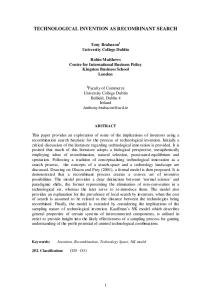
Mortality at three months was 29 percent in the placebo group, as compared with 18 percent in the three treatment groups combined — a relative reduction of 38 percent (P=0.02 by the chi-square test) (Fig. 1 and Table 3). All four outcome scales showed a more favorable global outcome for rFVIIatreated patients than for those who received placebo, in a dose–response fashion (Fig. 2). The results were significantly different from those in the patients who received placebo for all three doses of rFVIIa, as measured by the modified Rankin Scale and the NIHSS, and for the 80 and 160 µg per kilogram doses, according to the Barthel Index (Table 3). Treatment with rFVIIa more than doubled the odds of improving by one level on the modified Rankin Scale at 90 days and decreased the proportion of patients who died or were severely disabled from 69 percent in the placebo group to 53 percent in the three treatment groups combined, for an absolute reduction of 16 percentage points (95 percent confidence interval, 5 to 27; P=0.004). The E-GOS also showed a favorable effect of rFVIIa treatment on outcome, but this effect was not significant, owing to a large floor effect; overall, approximately three quarters of patients had died or were classified as severely disabled at three months.
Thromboembolic serious adverse events (Table 3) occurred in 2 percent of the placebo-treated patients, as compared with 7 percent of the rFVIIatreated patients overall (P=0.12 by Fisher’s exact test). There were no arterial thromboembolic serious adverse events in the placebo group; the overall frequency of such events was 5 percent among the rFVIIa-treated patients (P=0.01 by Fisher’s exact test). There were seven myocardial ischemic events and nine cases of cerebral infarction; all but four of these occurred within three days of the administration of rFVIIa. Two of the cases of cerebral infarction were massive and fatal, five were moderate in severity and disabling (two of these occurred 26 and 54 days after treatment and were not considered to be related to treatment), and two were asymptomatic. With the exception of one patient with an anterior-wall myocardial infarction who recovered with sequelae, the cardiac events that occurred in the rFVIIa-treated patients were characterized by small troponin I elevations, non–ST-segment elevation electrocardiographic abnormalities, and good recovery. Thromboembolic serious adverse events that were possibly or probably related to treatment (as opposed to those that were unlikely to be related to treatment) and that were fatal or disabling occurred in 2 percent of rFVIIa-treated patients and in 2 percent of the placebo group.
1.0
discussion
0.9
Proportion Surviving
0.8 0.7 0.6 0.5 0.4 0.3
Placebo rFVIIa, 40 µg/kg rFVIIa, 80 µg/kg rFVIIa, 160 µg/kg
0.2 0.1 0.0 0
15
30
45
60
75
90
Days
Figure 1. Survival at 90 Days According to Study Group. Mortality was reduced by approximately 35 percent in each rFVIIa group, as compared with that in the placebo group (P=0.10 by the log-rank test comparing all four groups; P=0.02 by the chi-square test for the comparison of the three rFVIIa groups combined with placebo).
782
n engl j med 352;8
In this study, rFVIIa given within four hours after the onset of intracerebral hemorrhage significantly reduced subsequent growth of the hemorrhage and improved the clinical outcome, despite a small increase in the frequency of thromboembolic adverse events. Treatment with rFVIIa resulted in reduced growth in the volume of intracerebral hemorrhage, as compared with placebo, by approximately 5 ml at 24 hours, which translated into an 11-ml reduction in total lesion volume at 72 hours, as compared with placebo. This difference was associated with an absolute reduction of 16 percentage points in the risk of death or severe disability (as measured by the modified Rankin Scale) at three months — consistent with the number needed to treat to prevent one unfavorable outcome of slightly more than six. The patients we treated were typical of those with spontaneous hypertensive intracerebral hemorrhage, but they had slightly smaller hemorrhages and better Glasgow Coma Scale scores than those
www.nejm.org
february 24 , 2005
Downloaded from www.nejm.org at EASTERN VIRGINIA MEDICAL SCHOOL on October 23, 2007 . Copyright © 2005 Massachusetts Medical Society. All rights reserved.
recombinant factor vii a for acute intracerebral hemorrhage
Table 3. Clinical Outcomes and Thromboembolic Severe Adverse Events at 90 Days According to Study Group.* Placebo (N=96)
Variable
rFVIIa 40 µg/kg (N=108)
80 µg/kg (N=92)
160 µg/kg (N=103)
Combined (N=303)
Survival 28 (29)
Died — no. (%) Odds ratio for survival (95% CI)
19 (18)
—
1.9 (1.0–3.8)
P value
17 (18) 1.8 (0.9–3.6)
20 (19) 1.7 (0.9–3.3)
56 (18) 1.8 (1.1–3.0)
0.05
0.10
0.11
0.02
59 (55)
45 (49)
56 (54)
160 (53)
Modified Rankin Scale† Unfavorable outcome — no. (%)
66 (69)
Odds ratio for improvement (95% CI)
—
2.2 (1.1–4.0)
2.4 (1.3–4.6)
2.1 (1.1–4.1)
2.2 (1.3–3.8)
P value
—
0.02
0.008
0.02
0.004
78 (72)
66 (72)
77 (75)
221 (73)
Extended Glasgow Outcome Scale‡ Unfavorable outcome — no. (%)
78 (81)
Odds ratio for improvement (95% CI)
—
1.9 (0.9–3.8)
P value
—
0.09
1.5 (0.7–3.2) 0.28
1.4 (0.7–3.0) 0.36
1.6 (0.9–3.0) 0.14
Barthel Index§ 25.0
Median score P value
55.0
—
67.5
55.0
60.0
0.07
0.01
0.02
0.006
6.0
5.0
7.0
6.0
0.03
0.004
0.02
0.008
National Institutes of Health Stroke Scale¶ 12.5
Median score P value
—
Thromboembolic serious adverse events — no. (%) 2 (2)
7 (6)
4 (4)
10 (10)
21 (7)
Arterial
0
6 (6)
2 (2)
8 (8)
16 (5)
Venous
2 (2)
1 (1)
2 (2)
2 (2)
5 (2)
Total
* Odds ratios are for survival as compared with the placebo group. CI denotes confidence interval. Outcome scores at day 15 were used according to the principle of the last observation carried forward when scores at day 90 were missing. Scores on the modified Rankin Scale and the Extended Glasgow Outcome Scale were not available for one patient in the group given 80 µg of rFVIIa per kilogram; otherwise, outcome scores were available for all patients. The numbers of patients who had one or more outcome scores carried forward from day 15 were 7, 6, 5, and 2 in the placebo, 40 µg, 80 µg, and 160 µg per kilogram groups, respectively. Odds ratio for improvement is the likelihood of improving by one scale level, as compared with that in the placebo group, with control for age, baseline intracerebral-hemorrhage volume, and location of the intracerebral hemorrhage. Modified Rankin Scale scores of 4 to 6 and Extended Glasgow Outcome Scale scores of 1 to 4 were pooled as a single category indicating an unfavorable outcome (death or severe disability). † Scores of 4 to 6 (defined as an unfavorable outcome) indicate death or survival with severe disability (bedbound and incontinent) or moderate-to-severe disability (unable to walk without assistance). ‡ Scores of 1 to 4 (defined as an unfavorable outcome) indicate death or inability to follow commands, care for oneself at home, or shop or travel locally without assistance. § A score of 100 indicates complete independence in activities of daily living, and 0 indicates total dependence or death. The treatment groups were compared with the placebo group with use of the Wilcoxon rank-sum test. ¶ A score of 0 indicates no neurologic deficit, and a score of 42 coma and quadriplegia or death. The treatment groups were compared with the placebo group with use of the Wilcoxon rank-sum test.
in other series, owing to our exclusion criteria.3,4 Accordingly, the 29 percent mortality rate in our placebo group was slightly lower than what is typically found in hospital registries and population-based studies.1,3,4 Thirty-two percent of placebo-treated patients had substantial growth in the volume of
n engl j med 352;8
intracerebral hemorrhage, defined as an increase of more than 33 percent or more than 12.5 ml from baseline (data not shown); this rate is similar to those in previous studies.5-8 The doses of rFVIIa that we studied in this doseranging proof-of-concept trial ranged from approx-
www.nejm.org
february 24, 2005
Downloaded from www.nejm.org at EASTERN VIRGINIA MEDICAL SCHOOL on October 23, 2007 . Copyright © 2005 Massachusetts Medical Society. All rights reserved.
783
The
Modified Rankin Scale
rFVIIa
0–1 24
160 µg/kg 80 µg/kg 40 µg/kg Placebo
2–3 21
21
29
17 8
4–5 35
29 23
40
new england journal
Barthel Index
Dead 19
30
18
37
18 29
95–100 55–90 37 15 36
19
27 22
0–50 Dead 29 19
24 17
27
18
31
18
32
29
percent of group
rFVIIa
Extended Glasgow Outcome Scale
7–8 5–6 160 µg/kg 15 11
2–4 55
0–1 18 21
80 µg/kg
13
15
53
18
40 µg/kg
11 16
55
18
Placebo
6 12
52
NIH Stroke Scale
Dead 19
29
2–8 40 40
10 13
≥9 22
51 29
Dead 19
20
18
22
18
29
29
percent of group
Figure 2. Outcome at 90 Days According to Study Group. Scores of 0 to 1 on the modified Rankin Scale, 7 to 8 on the Extended Glasgow Outcome Scale, 95 to 100 on the Barthel Index, and 0 to 1 on the National Institutes of Health (NIH) Stroke Scale indicate a favorable outcome. Percentages may not total 100 because of rounding. Twenty patients (5.0 percent) were alive but lacked complete outcome data at 90 days, and thus had some or all scores at day 15 carried forward.
imately half to twice the currently labeled dose of 90 µg per kilogram for bleeding related to hemophilia. Treatment with rFVIIa resulted in a relative reduction of approximately 50 percent in the growth of hemorrhage, and a dose–response effect was evident, with the smallest effect at 40 µg per kilogram and the strongest at 160 µg per kilogram. The timing of treatment also had a powerful effect on the capacity of rFVIIa to limit the growth of intracerebral hemorrhage; the best results were seen when patients were treated within three hours after the onset of symptoms. This suggests that active bleeding occurs in a large proportion of patients with intracerebral hemorrhage within the first few hours after onset and rapidly diminishes over time. Our subgroup analysis of patients treated more than three hours after onset was underpowered; accordingly, our data permit us to conclude only that treatment with rFVIIa within four hours after onset is beneficial. The precise mechanism by which rFVIIa arrests bleeding in patients with acute intracerebral hemorrhage is not fully understood. After blood-vessel damage and local initiation of the coagulation cascade, the administration of rFVIIa enhances throm-
784
n engl j med 352;8
of
medicine
bin generation on the surface of activated platelets, leading to accelerated formation of a fibrin clot.18 It seems most likely that the administration of rFVIIa after intracerebral hemorrhage accelerates thrombosis within ruptured small penetrating arteries or arterioles. Although the half-life of rFVIIa is only 2.6 hours,19 a sustained hemostatic effect may occur after a single dose because the clot that forms is denser than normal and more resistant to fibrinolysis.20 Mortality was significantly reduced, by 38 percent, with rFVIIa treatment. Both the 80 µg and the 160 µg per kilogram doses of rFVIIa significantly improved global outcomes as measured on three of the four standard scales that we evaluated. Although we did not record when decisions were made to withhold or withdraw life support, our use of the combination of death and severe disability as a single category made it unlikely that decisions to withdraw life-sustaining treatment influenced the results. Arterial thromboembolic serious adverse events occurred significantly more frequently with rFVIIa treatment than with placebo, primarily in the form of myocardial ischemic events and cerebral infarction within three days after the study drug was given. The majority of patients recovered from these complications, and the overall frequency of fatal or disabling thromboembolic serious adverse events did not differ significantly between the rFVIIa and the placebo groups. In summary, ultra-early hemostatic therapy with rFVIIa limits the growth of hemorrhage, reduces mortality, and improves functional outcomes after intracerebral hemorrhage. Until additional data on safety are available, however, rFVIIa should be administered with caution to patients with intracerebral hemorrhage who have risk factors for thromboembolic disease. Additional research is needed to identify patients at high risk for thromboembolic complications, to define the optimal therapeutic window, and to test rFVIIa for anticoagulationinduced intracerebral hemorrhage.21 Supported by Novo Nordisk, Bagsvaerd, Denmark. Presented in part at the Fifth World Stroke Congress, Vancouver, B.C., Canada, June 26, 2004. Drs. Brun and Skolnick and Ms. Begtrup are employees of Novo Nordisk and report being stockholders in the company. Dr. Mayer reports having received research support from Novo Nordisk. Drs. Mayer, Broderick, Davis, Diringer, and Steiner report having received consulting fees from Novo Nordisk, and Drs. Mayer, Davis, Diringer, and Steiner report having received lecture fees from Novo Nordisk. We are indebted to the study coordinators, the nurses and physicians in the emergency departments and intensive care units, and the patients and families who supported this trial.
www.nejm.org
february 24 , 2005
Downloaded from www.nejm.org at EASTERN VIRGINIA MEDICAL SCHOOL on October 23, 2007 . Copyright © 2005 Massachusetts Medical Society. All rights reserved.
recombinant factor vii a for acute intracerebral hemorrhage
appendix The following participated in the Recombinant Activated Factor VII Intracerebral Hemorrhage Trial: Steering Committee — S.A. Mayer, New York (chair); J. Broderick, Cincinnati; N.C. Brun, Bagsvaerd, Denmark (nonvoting); S. Davis, Melbourne, Australia; M.N. Diringer, St. Louis; B.E. Skolnick, Princeton, N.J. (nonvoting); and T. Steiner, Heidelberg, Germany; Statistician — K. Begtrup, Bagsvaerd, Denmark; Data and Safety Monitoring Board — T.G. Brott, Jacksonville, Fla. (chair); K. Asplund, Stockholm; T.P. Bleck, Charlottesville, Va.; M. Escobar, Houston; and I. Scharrer, Frankfurt, Germany; Neuroradiologists — R. Zimmerman, New York; J. Maldjian, Winston-Salem, N.C; Contract Research Organization — Quintiles Transnational (A. Lopez, global project manager); Clinical Centers — Australia: S. Davis, Royal Melbourne Hospital, Parkville, Vic.; G. Donnan, Austin and Repatriation Medical Centre, Heidelberg, Vic.; D. Freilich, Western Hospital, Footscray, Vic.; R. Gerraty, St. Vincent’s Hospital, Fitzroy, Vic.; T. Kimber, Royal Adelaide Hospital, Adelaide, S.A.; D. Schultz, Flinders Medical Centre, Bedford Park, S.A.; Austria: F. Fazekas, Universitat Klinik Graz, Graz; Belgium: S. Blecic, Hospital Erasme, Brussels; P.P. De Deyn, Algemeen Ziekenhuis Middelheim, Antwerp; V. Thijs, U.Z. Gasthuisberg, Leuven; Canada: P. Bailey, Saint John Regional Hospital, Saint John, N.B.; M. Hill, Foothills Hospital, Calgary, Alta.; D. Selchen, Trillium Health Centre, Mississauga, Ont.; C. Voll, Royal University Hospital, Saskatoon, Sask.; A. Woolfenden, Vancouver General Hospital, Vancouver, B.C.; Croatia: V. Demarin, Clinical Hospital Sestre Milosrdnice, Zagreb; Denmark: G. Andersen, Aarhus University Hospital, Aarhus; G. Boysen, Bispebjerg Hospital, Copenhagen; Finland: M. Kaste, Helsingin Yliopistollienen, Haartmaninkatu; Germany: O. Busse, Klinikum Minden, Minden; A. Ferbert, Universitaetsklinikum Kassel, Kassel; M. A. Grond, Kreiskrankenhaus Siegen, Siegen; R. Haberl, Steadt. Krakenhaus, Munich; M. Hennerici, Klinikum Mannheim, Mannheim; D. Schneider, Universitaets-Klinikum der Universitaet Leipzig, Leipzig; T. Steiner, Universitaetsklinik Heidelberg, Heidelberg; Italy: C. Argentino, Universitá La Sapienza, Rome; V. Gallai, Universitá di Perugia, Perugia; D. Guidetti, Azienda Hospedaliera Santa Maria, Reggio Emilia; G. Miceli, Istituto di Ricovero e Cura a Carattere Scientifico, C. Mondino UC Malattie, Pavia; Malaysia: R.A. Adman Zurin, Universiti Kebangsaan Malaysia, Kuala Lumpur; the Netherlands: J.U.R. Niewold, Scheper Ziekenhuis, Emmen; M. Vermeulen, Academisch Medisch Centrum, Amsterdam; New Zealand: C. Anderson, Middlemore Hospital, Auckland; A. Barber, Auckland Hospital, Auckland; Norway: U. Waje-Andreassen, Haukeland Sykehus, Bergen; Singapore: I. Ng, National Neuroscience Institute; C. Ning, National University Hospital; Spain: A. Chamorro, Hospital Clinic I Provincial de Barcelona, Barcelona; A. Dávalos, Hospital Universitario de Girona Dr. Josep Trueta, Girona; J. Egido, Hospital Clínico San Carlos, Madrid; J.V. Osorio, Hospital General Universitario Gregorio Marañón, Madrid; J.A. Sabin, Hospital Vall d’Hebrón, Barcelona; Sweden: M. Callander, Universitetssjukhuset I, Linköping; T.-B. Käll, Södersjukhuset, Stockholm; N.G. Walgren, Karolinska Sjukhuset, Stockholm; Switzerland: J. Bogousslavsky, Centre Hospitalier Universitaire Vaudois Service de Neurologie, Lausanne; Taiwan: Y.-H. Chiang, Tri-Service General Hospital, Taipei; T.-K. Lin, Chang Gung Memorial Hospital, Taoyuan; Y.-K. Tu, College of Medicine and Hospital National Taiwan University, Taipei; United Kingdom: J. Barrett, Arrowe Park Hospital, Merseyside; G. Ford, University of Newcastle, Newcastle upon Tyne; M.-J. Macleod, Aberdeen Royal Infirmary, Aberdeen; K. Richardson Lees, Western Infirmary, Glasgow; United States: B.F. Fitzsimmons, Columbia University Medical Center, New York; C. Graffagnino, Duke University Medical Center, Durham, N.C.; D. Green, Queen’s Medical Center, Honolulu; J. Grotta, University of Texas Medical School, Houston; S.E. Kasner, Hospital of the University of Pennsylvania, Philadelphia; R. Libman, Long Island Jewish Medical Center, New Hyde Park, N.Y.; T. Lowenkopf, Oregon Stroke Center, Portland; F. McGee, Neurological Associates, Richmond, Va.; B. Meyer, University of California–San Diego Stroke Center, San Diego; J. Rosand, Massachusetts General Hospital, Boston; C. Wijman, Stanford Stroke Center, Palo Alto, Calif. references 1. Counsell C, Boonyakarnkul S, Dennis M,
et al. Primary intracerebral hemorrhage in the Oxfordshire Community Stroke Project. 2. Prognosis. Cerebrovasc Dis 1995;5:26-34. 2. Broderick JP, Adams HP Jr, Barsan W, et al. Guidelines for the management of spontaneous intracerebral hemorrhage: a statement for healthcare professionals from a special writing group of the Stroke Council, American Heart Association. Stroke 1999; 30:905-15. 3. Broderick JP, Brott TG, Duldner JE, Tomsick T, Huster G. Volume of intracerebral hemorrhage: a powerful and easy-to-use predictor of 30-day mortality. Stroke 1993; 24:987-93. 4. Hemphill JC III, Bonovich DC, Besmertis L, Manley GT, Johnston SC. The ICH score: a simple, reliable grading scale for intracerebral hemorrhage. Stroke 2001;32:891-7. 5. Brott T, Broderick J, Kothari R, et al. Early hemorrhage growth in patients with intracerebral hemorrhage. Stroke 1997;28:1-5. 6. Fujii Y, Tanaka R, Takeuchi S, Koike T, Minakawa T, Sasaki O. Hematoma enlargement in spontaneous intracerebral hemorrhage. J Neurosurg 1994;80:51-7. 7. Fujitsu K, Muramoto M, Ikeda Y, Inada Y, Kim I, Kuwabara T. Indications for surgical treatment of putaminal hemorrhage: comparative study based on serial CT and time-
course analysis. J Neurosurg 1990;73:51825. 8. Kazui S, Naritomi H, Yamamoto H, Sawada T, Yamaguchi T. Enlargement of spontaneous intracerebral hemorrhage: incidence and time course. Stroke 1996;27:1783-7. 9. Mayer SA. Ultra-early hemostatic therapy for intracerebral hemorrhage. Stroke 2003;34:224-9. 10. Friederich PW, Henny CP, Messelink EJ, et al. Effect of recombinant activated factor VII on perioperative blood loss in patients undergoing retropubic prostatectomy: a double-blind placebo-controlled randomised trial. Lancet 2003;361:201-5. [Erratum, Lancet 2003;361:1138.] 11. Mayer SA, Brun N, Broderick J, et al. Safety and feasibility of recombinant factor VIIa for acute intracerebral hemorrhage. Stroke 2005;36:74-9. 12. Mayer SA, Brun N, Broderick J, Davis S, Diringer MN, Steiner T. Safety and preliminary efficacy of recombinant coagulation factor VIIa in acute intracerebral hemorrhage: U.S. phase 2A study. Stroke 2004; 35:332. abstract. 13. Teasdale G, Jennett B. Assessment of coma and impaired consciousness: a practical scale. Lancet 1974;2:81-4. 14. van Swieten JC, Koudstaal PJ, Visser MC, Schouten HJ, van Gijn J. Interobserver
n engl j med 352;8
www.nejm.org
agreement for the assessment of handicap in stroke patients. Stroke 1988;19:604-7. 15. Brott T, Adams HP Jr, Olinger CP, et al. Measurements of acute cerebral infarction: a clinical examination scale. Stroke 1989; 20:864-70. 16. Wilson JT, Pettigrew LE, Teasdale GM. Structured interviews for the Glasgow Outcome Scale and the extended Glasgow Outcome Scale: guidelines for their use. J Neurotrauma 1998;15:573-85. 17. Mahoney FI, Barthel DW. Functional evaluation: the Barthel index. Md State Med J 1965;14:61-5. 18. Monroe DM, Hoffman M, Oliver JA, Roberts HR. Platelet activity of high-dose factor VIIa is independent of tissue factor. Br J Haematol 1997;99:542-7. 19. Lindley CM, Sawyer WT, Macik BG, et al. Pharmacokinetics and pharmacodynamics of recombinant factor VIIa. Clin Pharmacol Ther 1994;55:638-48. 20. He S, Blombäck M, Jacobsson Ekman G, Hedner U. The role of recombinant factor VIIa (FVIIa) in fibrin structure in the absence of FVIII/FIX. J Thromb Haemost 2003;1: 1215-9. 21. Fewel ME, Park P. The emerging role of recombinant-activated factor VII in neurocritical care. Neurocrit Care 2004;1:19-30. Copyright © 2005 Massachusetts Medical Society.
february 24, 2005
Downloaded from www.nejm.org at EASTERN VIRGINIA MEDICAL SCHOOL on October 23, 2007 . Copyright © 2005 Massachusetts Medical Society. All rights reserved.
785