G.N
Pierce, M. Nagano, 1'! Zahradka, and NS. Dhalla (eds.).
ATHEROSCLEROSIS, HYPERTENSION Kluwer Academic Publishers. Boston. All rights reserved.
SELECTIVE
ATTENUATION
ENHANCED MEDIATED
Copyright ~ 2003.
II IN
STREPTOZOTOCIN
DIABETIC
THORACIC
BY
AORTA
DIABETES.
OF
ANGIOTENSIN RESPONSES
AND
THE RAT
TEMPOL
BETTADAPURA N. SRIKUMAR, SHAILESH SHASTRI, PODURI RAMARAO, and CHAMAN LAL KAUL 2 Department of Pharmacology and Toxicology, National and Research, Phase-X, Sector 67, S.A.S.Nagar
Summary.
Institute of Pharmaceutical Education
(Mohali}-160
062, Punjab, India
Previous studies suggest the role of superoxide anion radicals (02-)
as endothe-
lium derived contractile factor. However, their role in the enhanced responses to angiotensin II (Ang II) or phenylephrine (PE) and diminished relaxation responses to acetylcholine (ACh) in diabetes remain unclear. In the present study, we investigated the possible involvement 02- in the Ang II-, PE- and ACh-mediated tozotocin
(STZ)-diabetic
responses. Aortic
rats and contractile
of
rings were obtained from strep-
responses to Ang II and FE, and relaxation
responses to acetylcholine (ACh) were assessed in presence of tempol. The contractile responses to Ang II and PE were significantly increased and the ACh mediated responses were impaired. Ang II and PE mediated responses in control rat aorta were unaffected by the presence of tempol (100I.LM). The increase in responses to Ang II in diabetic aorta was decreased in presence of tempol (100 I.LM) but the PE mediated responses remained unchanged. Further, the impaired ACh mediated relaxation in diabetic aorta were restored by tempol. Removal of the endothelium led to an increase in the maximal response to Ang II and PE in both control and diabetic aorta. Tempol did not alter the responses to Ang II in endothelium-denuded aorta. The present findings indicate that 02- may be involved in the increased contractile responses to Ang II but not PE in the diabetic rat thoracic aorta and this is endothelium dependent. Key words: Streptozotocin-diabetic Endothelium
rat, Aorta, Angiotensin
II, Superoxide
radicals, Tempol,
I Deceasedbefore the completion of this work; 2Corresponding Author. C.L. Kaul, Director, National Institute of Pharmaceutical Education and Research, Phase-X, Sector 67, S.A.S. Nagar--160 062 (Pb), India. NIPER Communication No: -;Tel: +91-172-214682; Fax: +91-172-214692; e-mail:
[email protected]
328
III.
Diabetes
Mellitus
INTRODUCTION Vascular complications persist to be the main cause of morbidity and mortality in the diabetic population [1]. The mechanisms involved in the development of diabetic vascular diseaseremain poorly understood despite numerous investigations [2] . Reports regarding vascularreactivity to alpha-adrenergic agonistsand angiotensin II (Ang II) are inconsistent. Responseswere found to be increased [3], decreased[4] or unchanged [5] .However, there are consistent reports that endothelial dependent vasodilatation (EDV) responsesare impaired [6]. Reactive oxygen species (ROS), superoxides (O2-) in particular have been implicated to be responsible for the impaired vasorelaxation.In this context, increasedgeneration of O2- and other ROS [6] and decreasedplasma or tissue concentrations of superoxide dismutase (SOD), catalase,glutathione and ascorbic acid in both clinical and experimental diabetes are reported [7] .The role of ROS has been establishedin most of the vascularbeds like aorta [8], mesenteric artery [9], renal afferent arterioles [2] and basilar artery [10]. Amongst the ROS, O2- [11], hydoxyl radical (OH.-) [12] and hydrogen peroxide (H2O2) [13] have been found responsible for the impaired relaxation responsesto ACh. Therefore, the studies thus far have establishedthe role of ROS in the impaired vasorelaxation. However, there are no reports indicating the involvement of O2- in the evoked contractile responsesto agonists in diabetes. Ang II has been implicated in the diabetic vascular complications [14]. Ang II has been shown to produce O2- by vascular smooth cells [15]. Ang II mediated hypertension in rats has been shown to be increase O2- production in vascular smooth muscle and endothelium [16]. Increased levels ofAng II in rats [17] and decreasedlevels of Ang II in swine [18] and rats [19] have been found to cause oxidative stress.Recently, we have shown that the involvement of superoxidesin the contractile responseto acute addition of Ang II in aortic rings obtained from SHR [20]. Thus, although a link has been established for Ang II-stimulation of superoxide generation and superoxide involvement in the diabetic vascular complications, no systematic study has been undertaken to examine the involvement of O2- in the contractile responsest~ Ang II in diabetes.The likelihood of O2- role in the contractile response to Ang II may vary between the normal and diabetic rats. In this study, we have examined this possibility. Tempol, a membrane-permeable, water-soluble SOD mimetic [21], was used in this study.Tempol hasbeen shown to normalize mean arterial pressurein both shortterm infusion and 7-day intraperitoneal administration [22] .The same authors reported that long-term oral administration of tempol ameliorated hypertension and renal excretion of PGF2a, a marker of oxidative stressin vivo [23]. Furthermore, tempol has been found valuable in demonstrating the role of O2- in most models of hypertension like long-term infusion of Ang II [24], low levels of Ang II [19], renal hypertension [25], DOCA-salt hypertension [26], salt hypertensive Dahl rats [27] and lead-induced hypertension [28]. Hence, in order to evaluate the role of O2- in the contractile responsesto Ang II and PE in the diabetic rat aorta, we
Angiotensin
II Supersensitivity
in STZ-Diabetic
Rats
329
studied the Ang 11- and PE-mediated responsesin presence of tempol. ACh has been suggested to cause the release of 02- in afferent renal arterioles of diabetic rabbits [2] .Hence, to examine the role of 02- in the ACh mediated responsesin the rat aorta, we studied the Ach induced relaxation in pre-contracted aorta in presence of tempol. 2. MATERIALS AND METHODS All the experiments were approved by the Institutional
Animals Ethics Committee
(IAEC, NIPER).
2.1 Animals Experiments were performed on male Sprague-Dawley rats weighing 180-200 9 (Central Animal Facility, India), housed four per cage in a room with controlled ambient temperature (23 :t 1°C), humidity (50 :t 10%), 12h light/dark cycle and free accessto food and water through out the study. 2.2 Drugs STZ (Calbiochem, U.S.A.) L-PE, tempol (both from Sigma Chemical Co., St. Louis, MO, U.S.A.), glucose GOD-POD kit (Glaxo Qualigens, Mumbai, India) and Ang II (Bachem, Basel, Switzerland) were used in the study. All other chemicals were of reagent grade obtained from Loba chemicals, India. All the drugs except STZ were dissolved in distilled water.
2.3 Induction of diabetes Rats were made diabetic as described earlier [29]. Briefly, STZ (65mg/kg, i.p.; dissolved in cold 0.01 M citrate buffer, pH 4.4) was injected to rats. Age-matched control rats were injected with citrate buffer. After 48 h of the STZ injection, blood sampleswere taken from the retro-orbital plexus and the plasma was analyzed for glucose using the GOD-POD kit (Glaxo-Qualigens, Mumbai, India). Rats with a blood glucose level >250 mg/ dl were selected for the study. 2.4 Vascular tissue experiments 8 weeks after the STZ injection, rats were killed by cervical dislocation; the thoracic aorta was quickly excised and placed in cold oxygenated (95% 02-5% C02) Kreb's-Henseleit buffer (KHB). The aorta was cleaned of adhering fat and adventitial tissues and 5 mm segments were cut. Care was taken not to stretch the tissue or damage the endothelium. In some preparations, the endothelium was deliberately removed by passing a steel wire of suitable diameter through the lumen and gently rubbed using the finger. The composition (mM) of KHB was NaCl: 2.6, KCl: 4.7, MgCI2.2H2O: 1.2, CaCI2: 2.6, KH2P04: 1.2, NaHC03: 25 and glucose: 11.1 per liter for the depolarizing solution, 90 mM KCl was added with corresponding decreases in NaCl.
330
III.
Diabetes
2.5 Experimental
Mellitus
design
Aortic ring was suspended by a pair of stainless steel hooks in water-jacketed organ bath, filled with 10ml of oxygenated KHB maintained at 37°C. A resting tension of 2 g was applied to the aortic rings, which were then allowed to equilibrate for 2 h and the buffer was changed every 15 min. Isometric tension responses were measured on a student's physiograph chart recorder (BioDevices, Ambala, India) using an isometric force transducer (T-305, BioDevices, Ambala, India). The tissues were exposed to 90mM KCl buffer (depolarizing solution). After two such challenges, contractile responses to increasing concentrations of Ang II (10nM-30~M) were recorded. Further concentrations were not used because tissues responses underwent rapid desensitization. Similarly, PE (1 nM to 10~M) responses were obtained. In case of ACh, after the equilibration for 2 h, tissues were pre-contracted with 10 ~M PE and responses to increasing concentrations of ACh (1 nM 10 ~M) were obtained. 2.6 Effect of tempol
on vascular responses
To evaluate the role of 02- in the enhanced contraction to Ang II and PE or impaired relaxation to ACh, the responses were evaluated in presence of tempol (100 JlM). The tissues were incubated either in presence of tempol dissolved in Kreb's buffer (tempol treated) or Kreb's buffer alone (vehicle treated) for half an hour before the obtaining the responses. At the end of all the experiments, the rings were blotted dry, their lengths measured and weighed to calculate tension as normalized for cross-sectional area by the formula:
:ross -sectional
area (mm
=
wieght (fig) [Length (fifi) X density ]
with the density of vascular smooth muscle being Ang II responses are expressed as mg/mm2,
.05 mg/mm3.
The
KC1,
PE
and
2.7 Statistical analysis Cumulative concentration response curves were analyzed for pD2 value (the negative log concentration required to produce 50% of the maximal response) and Emax. Emaxis expressed as mg/mm2 for the tension responses for Ang II, PE and KCI. ACh responses are expressed as percentage of maximum PE responses. All experimental values are expressed as mean :1=S.E.M. Data analysis was done by Student's t test at 5% level of significance using GraphPad Prism version 3.00 for Windows, Pad Software, San Diego California
Graph-
USA.
3. RESULTS 3.1 Features of experimental animals All the STZ treated animals marked showed hyperglycemia (blood sugar levels >400 mg/ dl) and other characteristics like polyphagia, polydypsia and polyuria.
.9
-8 Log
-7 [MI
Ang
-6
"
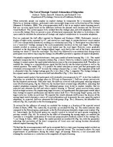
Figure 1. Cumulative Concentration Response Curves to Ang II in endothelium intact aortic ring preparations obtained from control and STZ-diabetic (STZ) rats in either absence (+ vehicle) or in presence (+ tempol) of tempol (l00~M). Tissues were incubated in presence of tempol (100IJ.M) or vehicle for half an hour prior to and during the exposure to Ang II. Each point is represented as mean :1:S.E.M., n = tissues obtained from 4-8 rats. *p < 0.05 vs respective control, #p < 0.01 vs respective control, @p < 0.001 vs respective control; bp < 0.01 vs respective + vehicle group.
3.2 Agonist induced contraction The log dose responsecurves for Ang II and PE are shown in Figs. 1 and 2, respectively. The maximal contractile responses(Ema,.)were significandy increased to Ang II (Fig. 1) and PE (Fig. 2) in STZ-rats as compared to age matched control rats. But there was no difference in the Emaxof the aorta from STZ diabetic and control rats to 90mM KCl (KCl Emaxfor control and diabetic rats were 2,538.7 :f: 209.8 and 2,782 :f: 65.3, respectively).There was no change in the sensitivity (pDV of the aortas from STZ diabetic rats to KCl and Ang II. The increasein the Ang II response was blocked by tempol (100~M). 3.3 ACh induced relaxation The maximum relaxation response to ACh in pre-contracted aorta was decreased in STZ diabetic rats (Fig. 3). There was no change in the pD2 in the ACh responses between control and diabetic rats. Tempol reversed the impaired ACh mediated responsesto near control values (Fig. 3). 3.4 Effect of endothelium denudation on tempol effect Removal of the endothelium led to an increase in the maximal responsesto Ang II (Fig.4) in both normal and STZ rats.There was no difference in the pD2 values
1500 N E,... E I "CJ1n 9 II 1000 c 0 .-~ (/)
I!. A
STZ + vehicle STZ + tempol
D
Control
.Control
+ vehicle
~
+ tempOIT/
c .
#
. Cw 0) . -(1) c +1
.0:: C IV
*
*
# 500-
#
~
-9
-8 Log
[M]
-5
-6
-7 PE
Figure 2. Cumulative Concentration Response Curves to FE in endothelium intact aortic ring preparations obtained from control and STZ-diabetic (STZ) rats in either absence (+ vehicle) or in presence (+ tempol) of tempol (100~M). Tissues were incubated in presence of tempol (100~M) or vehicle for half an hour prior to and during the exposure to FE. Each point is represented as mean :f: S.E.M., n = tissues obtained from 5-7 rats. *F < 0.05 vs respective control, #F < 0.01 vs respective control.
.9
-8
-6
-7 Jog
[M]
-5
ACh
Figure 3. Cumulative Concentration Response Curves to ACh in endothelium intact aortic ring preparations obtained from control and STZ diabetic (STZ) rats in either absence (+ vehicle) or in presence (+ tempol) of tempol (100~). Rings were pre-contracted with 10-sM PE. Tissues were incubated in presence of tempol (l00J.lM) or vehicle for half an hour prior to and during the exposure to FE and ACh Each point is represented as mean :t S.E.M., n = tissuesobtained from 4-8 rats. #F < 0 .01 vs respective control, 'F < 0.05 vs respective + vehicle group, bp < 0.01 vs respective + vehicle group, cp < 0.001 vs respective + vehicle group.
Angiotensin
1250, N
E
<0 1000I ~ ~ " ~ -
E
C O c: "in ~ C w aI - (,)
750-
.5 +I
500-
II Supersensitivity
in STZ-Diabetic
Rats
333
~ STZ + Vehicle ..STZ + tempol o Control + vehicle .Control + tempol
/)
250.
-9
.8 Log
.7 [MJ
Ang
-6
"
Figure 4. Cumulative Concentration Response Curves to Ang II in endothelium denuded aortic ring preparations obtained from control rats and STZ diabetic (STZ) rats in either absence (+ vehicle) or in presence (+ tempol) of tempol (100~). Tissues were incubated in presence of tempol (100~) or vehicle for half an hour prior to and during the exposure to Ang II. Each point is represented as mean :t S.E.M., n = tissues obtained from 4-{} rats.
between endothelium intact and endothelium denuded aortic rings. The KCl responseswere unaffected by the removal of the endothelium. The Emaxof KCl are 2,018 ::t 117 and 2,372 ::t 321.4, in intact and denuded, respectively).To evaluate the role of endothelium in the tempol-mediated effects, the vascular responsesof Ang II were studied in endothelium-denuded preparations. Tempol did not affect the vascular responsesto Ang II in the endothelium denuded aortic preparations (Fig. 4). 4. DISCUSSION In this study, we report that the probable involvement of Oi- in the enhanced contractile responsesto Ang II in diabetic rat aorta. In addition, O2- may not be involved in the enhanced responsesto PE in STZ diabetic aorta. Furthermore ACh mediated responsesmay recruit O2-, which may be escalated in the STZ diabetic rat aorta. Our data also indicates that the vascular endothelium might have a role in the superoxide-mediated responses. Studies indicate that the responsesto Ang II are elevated in STZ-diabetes [30,31]. Our studies show that contractile responsesto Ang II are elevated in STZ-diabetic rat aorta also. In good accordance with earlier reports [3,32], contractile responses to PE were increased in the STZ diabetic rat aorta. Further, the ACh mediated responseswere impaired in the STZ diabetic rat thoracic aorta, which supports
334
III.
Diabetes
Mellitus
results from other studies [33,34]. This indicates that the vascular complications in diabetes may be having a dual component, which includes enhanced contraction to vasoconstrictor agonists and impaired relaxation to vasodilators. The mechanisms, which contribute to these alterations in the responses,are multifarious. They may be changes in the receptor status or change in the postreceptor signal transduction mechanisms. Some of the mechanismsmay be changes in PLC activity and altered subcellular calcium [35]. The present study demonstrates that 02- might contribute to the exaggerated responsesto Ang II. How 02- cause the increase in the contractile response has been a investigated by several authors. 02- is known to stimulate IP3-induced Ca2+release by inhibiting the degradation of IP3 [36]. Changes in the carrier-mediated transport, passivediffusion and ligandreceptor interaction may occur as a consequence of 02- reaction with the lipids in the cell membrane [37]. Most importandy, 02- cause the degradation of NO which has a modulatory effect on the contractile responses of the blood vessels [38]. Tempol, which is a SOD mimetic, scavengesthe superoxide radicals and restoresthe enhanced vascular responsesto Ang II. Tempol, in addition to restoring the enhanced Ang II mediated responses to control values, brought it beyond it. This may not be due to the concentration of tempol used becausethe vascular responsesto Ang II were unaffected by lower concentrations «100~M) of tempol. Hence other mechanisms may be involved. H2O2 has been shown to cause direct vasorelaxation [39] .Studies have shown that this H202-induced relaxation is more in the diabetic aorta than in control vessels[13]. Superoxide radical levels are increased in diabetes [6] and in this study, we have shown that Ang II might induce the production of superoxide radicals in diabetic aorta. Tempol, a SOD mimetic causesthe dismutation of 02- and leads to the formation of H2O2. The H2O2, thus produced might cause vasorelaxation.This could possibly explain the tempol induced decreasein the Ang II induced contraction in diabetic aorta beyond the control response.Whether catalaseprevents this decrease remains to be investigated. Further, spontaneous release of EDRF might be more in diabetic vessels[40]. But due to the presence or generation of superoxide radicals, such release might not be obvious. Scavenging of 02- by tempol could cause more vasorelaxation or decreasedcontractile responseto agonistslike Ang II. Tempol at concentrations lower than 100J1M failed to produce any change in the responses to Ang II. There are many enzymes,which act as the source of superoxides in the vasculature, including xanthine oxidase, NADH/NADPH oxidase and eNOS. Ang II is thought to cause the generation of superoxides through the NADH/NADPH oxidase [16]. Whether Ang II produces superoxides in diabetes through this enzyme needs further investigation. Kawazoe and others [41] have demonstrated in rat aortic rings that superoxide radicals are involved in the contractile response to Ang II but not norepinephrine and epinephrine. Further, it has been shown that superoxide anions are involved in Ang II induced hypertension but not in catecholamine induced hypertension [17]. To determine whether superoxide radicals are involved in the enhanced contractile
Angiotensin
II Supersensitivity
in STZ-Diabetic
Rats
335
response to PE in diabetes, responsesto PE were studied in presence of tempol. Similar to the report [41] , tempol failed to alter the PE responsesin diabetic rat aorta implicating that superoxide radicals are not involved in the enhanced vascular responsesto PE. But in contrast to the same study, tempol did not alter Ang II induced contraction in control rat aorta. This may be attributed to the speciesdifference as they usedWKY rats and we in our study used SD rats. Investigators have shown that SOD reduces norepinephrine-induced contraction in the diabetic rat aorta [12]. Norepinephrine, a dihydroxyphenyl derivative that is subject to autooxidation in oxygenated buffer might be a source of superoxide radicals.Where as phenylephrine, which we used in our study is a synthetic monohydroxyphenyl derivative and is not prone to autooxidation. The reduction in the norepinephrineinduced contraction with pretreatment of SOD might have been in reality due to an improved EDV [12]. Superoxide radicals may be present both intracellularly and extracellularly implied by the physiological presence of different isoforrns of SOD like Cu/ZnSOD, MnSOD and ecSOD [42]. Exogenously administered SOD is presumably confined to the extracellular spacebecauseof its large molecular weight. In the present study, tempol, a water soluble, membrane permeable, SOD mimetic [21] was used which is capable of scavenging both intracellular and extracellular Oi-. There are numerous studies, which emphasize the role of superoxide radicals in the impaired EDV in diabetes. Recently, Schnackenberg and Wilcox proposed that ACh per se can lead to the generation of superoxide radicals in the diabetic rabbit afferent renal arterioles [2] .Whether this is true in the aorta is not known. In this study, we have demonstrated that tempol improves the impaired EDV in response to ACh in diabetic rat aorta. How ACh causesthe releaseof superoxidesis not yet clear. As studies have shown, eNOS could be a possible source of superoxides [43]. Superoxides are found to be produced by both VSMCs and endothelial cells [44]. Many studies have implicated the endothelium as the source of superoxide radicals in the vasculature in diabetes. SOD pretreatment failed to alter the endothelium independent relaxation [13,12]. Hence, we evaluated the responsesto Ang II in endothelium denuded aortic preparations. Similar to other reports [32], the removal of endothelium led to a drastic increase in the maximal responsesto Ang II and PE [45]. Tempol did not alter the vascular responsesto Ang II in endothelium denuded diabetic rat aorta. This indicates that the source of Ang II induced superoxide production may be the endothelium. NO is known to migrate to the VSMC from the endothelium, where it is produced. It is also possible that the superoxides released from the VSMC by Ang II may modulate the vascular tone by scavenging the NO and that modulation is lost on removal of endothelium. If endothelium is a source of superoxide radicals, how Ang II produces the superoxides in the endothelium is not known. eNOS has been shown to be a source of superoxide radicals and Ang II stimulates this to produce superoxide radicals in some vascular beds [44]. In conclusion, this study demonstrates the role of Oi- in the enhanced contractile responsesto Ang II and impaired relaxation responsesto ACh in the STZdiabetic rat aorta. Vascular complication associated with diabetes mellitus is of
336
III.
Diabetes
Mellitu
multifactorial origin and it is very unlikely that only one classof drug will produce tremendous change in the course of the disease.The results of this study lead to the speculation that SOD mimetics may prove to be a valuable therapeutic option in the treatment of diabetic vasculopathy in the future. ACKNOWLEDGEMENTS BNS was supported by a NIPER providing
funding. The authors acknowledgc
NIPER
for
the necessary facilities.
REFERENCES 1. Cooper ME, Bonnet F, Oldfleld M,Jandeleit-Dahm K. 2001. Mechanisms of diabetic vasculopathy: an overview. Am J Hypertens 14:475-486. 2. Schnackenberg CG, Wilcox CS. 2001. The SOD mimetic tempol restores vasodilation in afferent arterioles of experimental diabetes.Kidney Int 59:1859-1864. 3. Chang KS, StevensWC. 1992. Endothelium-dependent increase in vascular sensitivity to phenylephrine in long-term streptozotocin diabetic rat aorta. Br J Pharmacol 107:983-990. 4. Head RJ, Longhurst PA, Panek RL, Stitzel RE. 1987. A contrasting effect of the diabetic state upon the contractile responsesof aortic preparations from the rat and rabbit. Br J Pharmacol 91:275-286. 5. MacLeod KM, McNeill JH. 1985. The influence of chronic experimental diabetes on contractile responsesof rat isolated blood vessels.Can J Physiol Pharmacol 63:52-57. 6. De Vriese AS, Verbeuren TJ, Van de Voorde J, Lameire NH, Vanhoutte PM. 2000. Endothelial dysfunction in diabetes.Br J Pharmacol 130:963-974. 7. Giugliano D, Ceriello A, Paolisso G. 1996. Oxidative stress and diabetic vascular complications. Diabetes Care 19:257-267.
8. Chang KC, Chung SY, Chong WS, Suh JS, Kim SH, Noh HK, Seong BW, Ko HJ, Chun KW. 1993. Possible superoxide
radical-induced
alteration
of vascular reactivity
in aortas from
streptozotocin-
treated rats.J Pharmacol Exp Ther 266:992-1000. 9. Diederich D, Skopec J, Diederich A, Dai FX. 1994. Endothelial dysfunction in mesenteric resistance arteries of diabetic rats: role of free radicals.Am J Physiol 266:HI153-HI161. 10. Mayhan WG. 1997. Superoxide dismutase partially restores impaired dilatation of the basilar artery during diabetes mellitus. Brain Res 760:204-209. 11. Hat tori Y, Kawasaki H, Abe K, Kanno M. 1991. Superoxide dismutaserecovers altered endotheliumdependent relaxation in diabetic rat aorta. Am J Physiol 261:HI086-HI094. 12. Pieper GM, Langenstroer P, Siebeneich w: 1997. Diabetic-induced endothelial dysfunction in rat aorta: role of hydroxyl radicals. Cardiovasc Res 34:145-156. 13. Pieper GM, Gross GJ. 1988. Oxygen free radicals abolish endothelium-dependent relaxation in diabetic rat aorta. Am J Physiol 255:H825-H833. 14. Komers R, Komersova K. 2000. Therapeutic potential of ACE inhibitors for the treatment of hypertension in Type 2 diabetes.Expert Opin Investig Drugs 9:2601-2617. 15. Griendling KK, Minieri CA, Ollerenshaw JD, Alexander RW: 1994.Angiotensin II stimulates NADH and NADPH oxidase activity in cultured vascular smooth muscle cells. Circ Res 74:1141-1148. 16. Rajagopalan S, Kurz S, Munzel T, Tarpey M, Freeman BA, Griendling KK, Harrison DG. 1996. Angiotensin lI-mediated hypertension in the rat increasesvascular superoxide production via membrane NADH/NADPH oxidase activation. Contribution to alterations of vasomotor tone. J Clin 97:1916-1923. 17. Invest Laursen JB, Rajagopalan S, Galis Z, Tarpey M, Freeman BA, Harrison DG. 1997. Role of superoxide in angiotensin II-induced but not catecholamine-induced hypertension. Circulation 95:588-593. 18. Haas JA, Krier JD, Bolterman RJ, Juncos LA, Romero JC. 1999. Low-dose angiotensin II increases free isoprostane levels in plasma. Hypertension 34:983-986. 19. Ortiz MC, Sanabria E, Manriquez MC, Romero JC, Juncos LA. 2001. Role of endothelin and isoprostanesin slow pressor responsesto angiotensin II. Hypertension 37:505-510. 20. Shastri S, Ramarao P, Kaul CL, McNeill JR, Gopalakrishnan \I: Tempol selectively attenuates vasoconstrictor responsesto Ang II in aorta and perfused mesenteric vascularbed of spontaneously hypertensive rats (SHR). Annual Conferenceof Ind. Pharmacol.Soc.,2000. Gandhinagar, India; 2000.
~
Angiotensin
II Supersensitivity
in STZ-Diabetic
Rats
337