Psychological Medicine, 2003, 33, 1007–1018. f 2003 Cambridge University Press DOI : 10.1017/S0033291703007864 Printed in the United Kingdom
The functional neuroanatomy of schizophrenic subsyndromes G. D. H O N E Y, T. S H A R M A, J. S U C K L I N G, V. G I A M P I E T R O, W. S O N I, S. C. R. W I L L I A M S A N D E. T. B U L L M O R E1 From the University of Cambridge, Department of Psychiatry, Brain Mapping Unit, Addenbrooke’s Hospital, Cambridge ; Section of Cognitive Psychopharmacology, Department of Computing and Biostatistics, Department of Clinical Neurosciences, Institute of Psychiatry, King’s College, University of London
ABSTRACT Background. There is considerable variability between patients in their expression of the diverse range of symptoms encompassed by the syndrome of schizophrenia, which may modulate functional activation to cognitive processing. Method. Here we investigate associations between schizophrenic subsyndrome scores, identified by factor analysis, and experimentally controlled brain activation. Five factors were defined by rotated principal components analysis of PANSS rating scale measurements in 100 patients with schizophrenia. A subsample of 30 patients and a group of 27 comparison subjects were studied using functional magnetic resonance imaging (fMRI) during the performance of two periodically designed cognitive activation experiments : verbal working memory and psychomotor sequencing. Results. Factor analysis replicated the five dimensions consistently reported. Within the patient group, power of activation by working memory was negatively associated with global symptom severity in left lingual and temporo-parietal cortices ; negatively associated with positive subsyndrome scores in left inferior frontal and superior temporal cortices and basal ganglia ; and positively associated with negative subsyndrome scores in lateral and medial premotor cortex. No relationship was observed between subsyndrome scores and functional activation during the motor task. Between-group comparisons demonstrated reduced power of response to the working memory task by patients in bilateral dorsolateral prefrontal and left pre- and post-central cortices. Conclusions. In this study we observed task-specific modulation of functional response associated with symptom expression in schizophrenia. Our findings are compatible with previous empirical findings and theoretical conceptualization of human brain function, in terms of capacity constraints on activation in the face of competing demands from pathological and task-related cognitive activity.
INTRODUCTION Dimensional models of schizophrenia have attempted to address its phenomenological heterogeneity by identifying subsyndromes of cooccurring symptoms. Using factor analysis of symptom ratings, Liddle (1987 a) identified three subsyndromes of schizophrenia, labelled 1 Address for correspondence: Professor Edward T. Bullmore, University of Cambridge, Department of Psychiatry, Addenbrooke’s Hospital, Cambridge CB2 2QQ.
psychomotor poverty, reality distortion and disorganization, which have been widely replicated. Functional deficits in left dorsolateral and right ventrolateral prefrontal cortex, were hypothetically associated with psychomotor poverty and disorganization, respectively ; deficits in temporal lobe function were predicted in the reality distortion syndrome (Liddle, 1987 b). Using PET, Liddle et al. (1992) provided the first direct evidence of distinct neurobiological substrates for these three dimensions of psychopathology
1007
1008
G. D. Honey and others
in schizophrenia. Replication of these pioneering studies has provided partial corroboration (Ebmeier et al. 1993 ; Kaplan et al. 1993 ; Schroder et al. 1996) ; there are several methodological reasons that may explain some of the inconsistencies in these latter studies. As suggested by Liddle et al. (1994), ‘ The studies that have found three syndromes have not included the entire gamut of schizophrenic symptoms ’. Clinical rating scales that omit some of the full range of symptoms may yield a factor structure that distorts the underlying pattern of relationships between symptoms, given that the factor structure is generated on the strength of relationships between items that load on different factors, as well as relationships between items within a factor (Peralta & Cuesta, 1998). Factor analyses of the Positive and Negative Syndrome Scale (PANSS) (Kay et al. 1987), which provides a more comprehensive symptomatic assessment, have reliably reported a five factor model (Lindstrom & von Knorring, 1994 ; White et al. 1997). It should be noted, however, that while there is general agreement across factor analytic assessments of symptoms identified using the PANSS, the precise item composition of these factors varies to some extent across studies, and more ambivalent items that tend to load across multiple dimensions are occasionally excluded from the extracted factor structure. The cognitive dimension, in particular, shows a variable loading of items. Another potential source of variability is the use of resting or baseline measures of cortical physiology, which is highly variable across individual subjects. Variations in rCBF measures may, therefore, simply reflect the subjective experience of the procedure itself, rather than underlying cerebral pathology (Andreasen et al. 1997). It has also been suggested that there is no reason a priori why low resting rCBF should relate to a lack of functional activation under task-related demand ; indeed, it is conceivable that with a relatively lower starting point there is potential for greater increases in activity (Frith et al. 1995). Analogous to the use of cardiac stress tests in clinical medicine, cerebral functional deficits may only become apparent under conditions of increased physiological load (Berman, 1987). Hypofrontality, for example, has been demonstrated during the performance of cognitive tasks in patients who did not
demonstrate hypofrontality in measurement of resting perfusion levels (Volkow et al. 1987; Zemishlany et al. 1996). Here, we have addressed these two issues by applying factor analysis to PANSS ratings on a sample of 100 schizophrenic patients, and using their scores from each of the five factors to predict power of cerebral activation by two experimentally controlled tasks in a subsample of 30 patients. We predicted : (i) replication of established five symptom dimensional structure by factor analysis ; (ii ) consistent with previous imaging studies of working memory, schizophrenic patients would exhibit a hypofrontal response, as a group, compared to healthy volunteers (Fletcher et al. 1998; Bullmore et al. 1999 a); and (iii ) within the schizophrenic group, symptom dimensions would be differentially associated with power of functional response to the working memory task, given previous associations between working memory and psychotic symptomatology. METHOD Clinical and demographic data A group of 100 patients with a diagnosis of schizophrenia according to DSM-IV criteria was recruited from the patient population of the Bethlem Royal and Maudsley NHS Trust for the assessment of clinical symptomatology using the PANSS. This was assessed by two clinicians trained on the use of these scales, and having established >0.8 inter-rater reliability. The mean age of the group was 35 (¡S.D. 9.2) years. Mean duration of illness was 10.24 (¡10) years, with a mean age of onset of 24.9 (¡6.5) years. Mean scores of the PANSS positive, negative and general psychopathology subscales were 15.8 (¡7.2), 16.6 (¡7.2) and 32.7 (11.2). There were 77/100 males in the group. A subset of 30 patients was randomly sampled for fMRI scanning. For this subset of patients, mean duration of illness was 13 (¡9.8) years, with a mean age of onset at 23.9 (¡6.3) years, and mean dose of typical antipsychotic drugs was 327.3 (¡205.3) chlorpromazineequivalent mg/day. Mean scores on the PANSS positive, negative and general psychopathology subscales (rated within 1 week of the fMRI scan) were 11.7 (¡6.1), 14.6 (¡7.1) and 26.3 (¡10.3) respectively.
The functional neuroanatomy of schizophrenic subsyndromes
1009
A group of 27 healthy volunteers without personal or familial neurological or psychiatric history also participated in the fMRI aspect of the study. There was no significant difference in the mean age of the scanned patient and comparison groups (36.9 (¡9.2) and 35.1 (¡9.9) years respectively). There were 27/30 male subjects in the patient group and 21/27 males in the comparison group. All subjects were righthanded. After comprehensive description of the study, written informed consent was obtained from all participants. Scanning procedures were in accordance with the guidelines of the Bethlem Royal & Maudsley Hospital (Research) Ethical Committee.
(Talairach & Tournoux, 1988). The fundamental power quotient (fundamental power divided by its standard error) was estimated at each voxel. If the median fundamental power quotient exceeded the critical value of the median fundamental power quotient obtained by randomization, that voxel was considered generically activated with a probability of false positive activation of a<0.001. To estimate the difference in mean power of response between the groups of patients and comparison subjects, we fitted the following one-way ANCOVA model at each intracerebral voxel : (1) P=m+b1 G+b2 Age+e:
Factor analysis Orthogonal rotation of principal components with eigenvalues >1 by the equamax procedure was used to identify five subsyndromal factors in the standardized PANSS measurements made on the full sample of 100 patients (see Nunnally, 1994) for general introduction to factor analysis). Factor scores were derived from this analysis for each of the 30 patients in the fMRI subsample. Global symptom severity was measured by summing the five subsyndrome scores for each subject.
Here, m is the overall mean power, G is a vector coding group membership, Age is the subject’s age, and e is an error. The null hypothesis that the coefficient b1=0 was tested by permutation at cluster level, as described in detail elsewhere (Bullmore et al. 1999 c). Briefly, the procedure was as follows : a map of the standardized coefficient b1/SE( b1) was thresholded so that if | b1/SE( b1)|>2, the voxel value was set to | b1/SE( b1)|x2. This resulted in a set of suprathreshold voxel clusters, spatially contiguous in two dimensions, each of which can be described in terms of its mass or the sum of suprathreshold voxel statistics it comprises. The 2D mass of each cluster was tested against a null distribution ascertained by repeated random permutation of the vector coding group membership in Eq (1), followed by thresholding of the standardized voxel statistic map and generation of 2D clusters under the null hypothesis (Bullmore et al. 1999 c). To estimate the association between power of response and subsyndromal scores within the group of patients, we fitted the following multiple regression model at each intracerebral voxel : (2) P=m+b1 S1+b2 Age+e:
Image acquisition Gradient-echo echoplanar MR images were acquired using a 1.5 Tesla GE Signa System at the Maudsley Hospital, UK. In each of 14 non-contiguous planes parallel to the inter-commissural (AC–PC) line, 100 T2*weighted MR images depicting BOLD contrast were acquired : TE=40 ms, TR=3 s, in-plane resolution=3.1 mm, slice thickness=7 mm, slice skip=0.7 mm. Image analysis After estimation and correction of head motion during scanning (Bullmore et al. 1999b), experimentally determined power was estimated in each fMRI time series by sinusoidal regression, described in detail elsewhere (Bullmore et al. 1996 ; Brammer et al. 1997). Briefly, the power of periodic signal change at the (fundamental) frequency of stimulation was estimated by the iterated least squares fitting of a sinusoidal regression model at each voxel in standard space
Here, S1 indicates the vector of scores on the first of the five subsyndromes separately modelled in this way ; the other abbreviations are as before. The methods of inference used in the between-group comparison were identically applied to identify voxel clusters where the association between subsyndrome scores and power of response was significant.
1010
G. D. Honey and others
For the voxel-wise hypothesis tests conducted for generic brain activation mapping, a onetailed probability of type 1 error a<0.001 was adopted as the threshold for significance. For the cluster-wise hypothesis tests conducted for within- and between-group comparisons, a twotailed probability of type 1 error a<0.01 was adopted as the threshold for significance. This more lenient threshold for cluster-level testing is justified by the much smaller number of tests entailed at cluster level. Thus, for each of the maps reported here, the expected number of false positive clusters (number of clusters tested multiplied by a) was less than or equal to 1 over the whole map. Cognitive paradigms Five blocked periodic cycles of two contrasting conditions were presented in 30 s epochs, for 5 min for two experiments (order of presentation pseudo-randomized). Accuracy and reaction time were monitored by button press. Verbal working memory Baseline condition : the participants viewed a series of 11 letters, presented individually (interstimulus interval (ISI)=2.3 s), and were required to press a button in response to the letter ‘ X ’. Activation condition : the participants viewed a series of 11 letters and were required to press the button if the currently presented letter was the same as that presented two trials previously. Psychomotor task Baseline condition : the subject viewed the word ‘ REST ’ visually presented for 0.5 s (ISI 2.5 s). Activation condition : the subject viewed the word ‘ MOVE ’, paced identically to the stimulus in the control condition; in response to the visual cue, subjects were required to move a joystick using the right hand, which was secured in the left hand, in one of four possible directions (up, down, left or right) ; subjects were required to make the sequence of movements as random as possible, based on preliminary findings from pilot data indicating that motor response selection elicited more robust activation of medial and lateral motor cortical areas than repetitive movements, consistent with previous findings (Deiber et al. 1991; Touge et al. 1995).
RESULTS Subsyndromal factor structure Five principal components, with eigenvalues >1, accounted for 67.5 % of the total variance. After rotation these factors were identified as negative, positive, excited, cognitive and depressed subsyndromes based on the symptom loadings (Table 1) and reference to the prior literature (Lindstrom & von Knorring, 1994; White et al. 1997). A close correspondence was observed between the loading of items of the five dimensions in this study, and that reported in previous studies. However, it is notable that the conceptual disorganization item (P2), generally observed to load on the ‘ cognitive ’ dimension, loaded on the first dimension, labelled the negative factor in this study. There were no significant correlations between any of the five factors and any demographic variables (two-tailed probability a<0.05). Behavioural data Patients correctly identified 80% of targets in the working memory task on average ; however, this was significantly less than the mean accuracy of the control group (96 %). Patients also had significantly slower reaction times for both the working memory task, the working memory control task and the psychomotor task ; see Table 2 for details. There were no significant correlations between any of the five subsyndromal factors and any of the behavioural measures of task performance (two-tailed probability a<0.05). fMRI data Generic brain activation maps Verbal working memory Both groups demonstrated significant median power of activation in three main brain regions : (i) supplementary motor area (SMA) extending inferiorly to cingulate gyrus ; (ii ) dorsolateral prefrontal cortex (DLPFC) extending inferiorly to ventrolateral prefrontal cortex and superiorly to lateral premotor cortex ; and (iii ) parietal cortex, extending from the angular and supramarginal gyri to precuneus and dorsal occipital gyri medially and superiorly. Frontal and parietal regions were activated bilaterally (see Table 3).
1011
The functional neuroanatomy of schizophrenic subsyndromes
Table 1. Rotated component loadings : according to the recommendations of Nunnally (1994), and given the relatively small number of patients included relative to the number of PANSS items, only those items whose communality was >0.5 are reported Component Factor label
Description
Item
1
Negative
Passive/apathetic social withdrawal Lack of spontaneity and flow of conversation Emotional withdrawal Blunted affect Active social withdrawal Poor rapport Conceptual disorganization Disturbance of volition
N4 N6 N2 N1 G16 N3 P2 G13
0.804 0.775 0.706 0.698 0.682 0.560 0.506 0.501
Positive
Grandiosity Unusual thought content Delusions Preoccupation Hallucinatory behaviour
P5 G9 P1 G15 P3
Excited
Hostility Poor impulse control Poor attention
P7 G14 G11
Cognitive
Somatic concern/delusions Mannerisms and posturing Lack of judgement and insight Difficulty in abstract thinking Stereotyped thinking Disorientation
G1 G5 G12 N5 N7 G10
Anxiety Tension Guilt feelings Depression
G2 G4 G3 G6
Depressive
Table 2.
Group Patient Control t statistics
2
3
4
5
0.787 0.704 0.654 0.527 0.510 0.787 0.779 0.526
0.515
0.519
0.706 0.616 0.583 0.515 0.513 0.508 0.834 0.742 0.731 0.652
% Total variance
Cronbach’s alpha
41.2
0.91
9.6
0.86
8.0
0.84
5.1
0.79
3.7
0.81
Behavioural data for the verbal working memory and psychomotor tasks in patient and control groups (S.D. in parentheses) Reaction time (s)
Working memory condition targets identified (maximum=11)
Working memory control condition
Working memory condition
8.8 (2.5) 10.58 (0.78) t=3.5, df=52***
0.61 (0.18) 0.49 (0.11) t=4.02, df=52****
0.72 (0.12) 0.57 (0.08) t=3.8, df=52****
Psychomotor task 0.56 (0.2) 0.42 (0.08) t=3.07, df=50**
** P<0.01; *** P<0.001 ; **** P<0.0001.
Psychomotor task Both groups demonstrated significant median power of activation in the cerebellum bilaterally, the supplementary motor area and preand post-central gyrus bilaterally, extending caudally into adjacent parietal cortex. The patients additionally showed activation of inferior and middle frontal gyri bilaterally (see Table 3).
Between group comparisons Verbal working memory In comparison to the control group, schizophrenic patients demonstrated significant reduction of mean power in the dorsolateral prefrontal cortex bilaterally (Brodmann Area (BA) 9/46 ; Talairach co-ordinates (x, y, z mm) : 30, 42, 28 and x41, 26, 32) and left pre-central gyrus (BA4 ; x43,x11, 50) (see Fig. 1).
1012
G. D. Honey and others
Table 3.
Main regional foci of generic activation in the working memory and psychomotor tasks in patients and controls Controls
Patients
Talairach co-ordinates Task
Region
Working memory
L/R
No. of voxels
x
y
z
No. of voxels
x
y
z
40/39/7/19
L/R*
412
x42
x55
42
376
49
x31
42
9/44 46/10 45 45 6/32 6 18 18 —
L R L R Med L L R L R
74 24 11 10 90 43 56 23 3 5
x43 38 x32 52 3 x43 x38 32 x12 29
3 42 19 14 8 x3 x69 x64 x78 x64
37 15 4 20 48 42 x13 x13 x18 x18
3 26 12 7 20 14 7 4 26 10
x38 55 x43 46 6 x32 x38 38 x6 23
14 8 14 8 17 0 x59 x64 x72 x58
31 26 9 9 42 48 x13 x13 x13 x13
Cerebellum Post-central gyrus/posterior parietal cortex SMA Pre-central gyrus
— 1/2/3/40
397 287 226 77
17 x38 43 3
x53 x28 x36 x8
x13 48 48 48
320 1198
23 x35
x56 x39
x13 48
Inferior frontal gyrus
44
Middle frontal gyrus
9
L/R L(L/R)*# R Med R L R L L R
376 103 19 78 14 35 7
43 46 x38 52 x49 x20 40
0 3 6 8 8 35 31
37 9 9 26 31 37 26
Posterior parietal cortex/ precuneus Dorsolateral prefrontal cortex Inferior frontal gyrus SMA/anterior cingulate Lateral premotor cortex Extrastriate cortex Cerebellum
Psychomotor
BA
Talairach coordinates
6 6
* Coordinates represent the centroid of a spatially dispersed three dimensional cluster, covering several brain regions. # Laterality differs across groups ; patients laterality in brackets.
Psychomotor task The patient group demonstrated significant enhancement of mean power in right lateral premotor area (BA6 ; 42, 0, 45) in comparison to the control group. Associations between brain function, total symptom scores and subsyndrome scores Verbal working memory Global symptom severity scores were negatively associated with the power of functional response in the left lingual gyrus and left superior temporal gyrus, extending superiorly into inferior parietal cortex (see Figs. 1 and 2 and Table 4). Negative subsyndrome scores were positively associated with power of response in lateral premotor cortex bilaterally, SMA and right posterior parietal cortex (see Figs. 1 and 2 and Table 4). Positive subsyndrome scores were negatively associated with power of response in several left hemisphere regions including inferior
frontal gyrus, middle and superior temporal gyri, inferior parietal cortex, lateral premotor cortex, and the caudate nucleus. There was also a negative association in right middle temporal gyrus extending inferiorly to superior occipital gyrus (see Figs. 1 and 2 and Table 4). Excited subsyndrome scores were negatively associated with power of response in left middle and superior temporal gyrus, posterior parietal cortex and precuneus, and in left middle frontal gyrus, SMA and right angular gyrus ; (see Table 4). Cognitive subsyndrome scores were negatively associated with functional response in left temporal, right inferior frontal gyrus, right cingulate gyrus, supramarginal gyrus bilaterally, and left lateral premotor cortex ; (see Table 4). Depressive subsyndromes scores were not significantly associated with functional response. Psychomotor task There were no clusters of significant association between functional response to this task
1013
The functional neuroanatomy of schizophrenic subsyndromes
50 40 30 20 –4 –3 –2 –1 0 1 2 3 4 Summed subsyndrome scores
Negative subsyndrome Supplementary motor area 100 80 60 40 20 –4 –3 –2 –1 0 1 2 3 4 Negative subsyndrome scores
Regional power of activation
60
Severity of psychosis Temporo-parietal area
Regional power of activation
Regional power of activation
FIG. 1. Maps of significant between-group differences in power of response to a verbal working memory task (top row) and of significant associations between functional response to working memory and global and subsyndrome scores. Top row, red voxels indicate clusters of significantly reduced power in the group of patients with schizophrenia (N=30) relative to a group of comparison subjects (N=27) ; second row, blue voxels indicate clusters of significant negative association between global severity of psychosis (summed factor scores) and functional response ; third row, yellow voxels indicate clusters of significant positive association between severity of negative subsyndrome and functional response ; fourth row, blue voxels indicate clusters of significant positive association between severity of positive subsyndrome and functional response. The distance of each map above the intercommissural line in the standard space of Talairach & Tournoux (1988) is given in millimeters below. The right side of the brain is shown on the left side of each map.
Positive subsyndrome Inferior frontal gyrus 180 160 140 120 100 80 60 40 20 –4 –3 –2 –1 0 1 2 3 4 Positive subsyndrome scores
FIG. 2. Scatter plots of regional activation versus global, negative and positive subsyndrome scores. Left, negative association between global severity and power of response in left temporo-parietal cortex (R2=0.58) ; middle, positive association between negative subsyndrome and power of response in supplementary motor area (R2=0.54); right, positive association between positive subsyndrome and left inferior frontal gyrus (R2=0.28). Note that the y axes for these plots are differently scaled, as a result of the differences in the amplitude of response across these regions.
1014
G. D. Honey and others
Table 4. Talairach coordinates of clusters showing significant correlation between power of functional response during the working memory task and subsyndrome scores Negative correlations
Positive correlations Talairach coordinates
Subsyndrome
Region
Negative
Positive
Excited
Cognitive
L/R
BA
x
y
Talairach coordinates z
No correlation
Inferior frontal gyrus Middle/superior temporal gyrus Inferior parietal lobe/superior temporal gyrus Middle temporal/occipital gyrus Caudate nucleus Lateral premotor area Middle/superior temporal gyrus into parietal cortex and precuneus* Middle frontal gyrus SMA Angular gyrus Superior/middle temporal gyrus Inferior frontal gyrus Cingulate gyrus Supramarginal gyrus Lateral premotor cortex
Depressed
Lateral premotor area Posterior parietal cortex SMA
L L L
44/45 21/22 40/42
x40 13 x50 x53 x57 x33
20 12 28
R L L
39/19 — 6/4
x57 x33 x10 13 x41 x9
28 16 55
L
21/22/42/ x40 x65 40/7/19
32
L 9 Med 6 R 39
x45 10 3 x3 41 x64
40 55 32
L R R L R L
x42 50 23 x43 36 x33
x52 6 x33 x32 x69 4
12 20 20 32 32 55
26
34
22/39 44 30 39/40 39/19 6
No correlation
Symptom severity Superior temporal gyrus into posterior parietal cortex* Lingual gyrus
Region
L/R
BA
R L R
6 6 40
Med
6
x
y
z
37 x4 55 x42 x4 55 41 x33 40 6
0 50
No correlation
No correlation
No correlation
No correlation
L
22/40
x47
L
18
x13 x67 x4
No correlation
Coordinates are from Talairach & Tournoux (1988). * Coordinates represent the centroid of a spatially dispersed three dimensional cluster, covering several brain regions.
and any of the global or subsyndromal factor scores. DISCUSSION This study demonstrated anatomically distinct psychophysiological associations between global and subsyndromal symptom scores and power of fMRI activation by a verbal working memory task in 30 patients with schizophrenia. No relationship was observed between symptom expression and cerebral response to the psychomotor task, indicating a degree of specificity to the relationship observed between symptomatic status and cognitive activation during the working memory task. These data therefore provide complementary information to previous
studies of associations between symptoms of schizophrenia, which have focused on resting cerebral activity, by demonstrating the nature of symptomatic modulation of brain function under cognitive challenge. The regional specificity of the psychophysiological associations observed between symptom dimension/severity and fMRI-measured cognitive activation is compatible with previous studies of resting state neurophysiology in schizophrenia. Global symptom severity was associated with functional activation in the left superior temporal gyrus and left lingual gyrus. The first of these results is comparable to previous associations reported between summed subsyndromal scores and resting cerebral perfusion demonstrated in left medial temporal
lobe (Friston et al. 1992) and also between total BPRS scores and resting glucose metabolism in left superior temporal gyrus (DeLisi et al. 1989). These studies provide convergent evidence that left temporal lobe function seems especially sensitive to the overall severity of schizophrenic psychosis. The association observed in this study between negative subsyndrome scores and power of functional response in bilateral and medial premotor cortex is consistent with several studies examining resting CBF/metabolism, which have also reported an association between negative symptoms and frontal resting activity (Volkow et al. 1987; Liddle et al. 1992; Wolkin et al. 1992 ; Ebmeier et al. 1993 ; Kawasaki et al. 1996; Schroder et al. 1996). Positive subsyndrome scores were associated with the power of functional response in several left hemispheric brain regions including inferior frontal gyrus, middle/superior temporal gyrus, inferior parietal cortex, premotor cortex and caudate nucleus. Previous studies which have examined the relationship between co-occurring positive symptoms and resting cerebral state have inconsistently found association with temporal lobe activity (Liddle et al. 1992; Ebmeier et al. 1993 ; Kaplan et al. 1993; Kawasaki et al. 1996 ; Schroder et al. 1996). However, auditory hallucinations have been consistently associated with abnormal blood flow in interconnected left hemispheric language areas, such as inferior frontal and superior temporal gyri, and/or SMA (DeLisi et al. 1989 ; McGuire et al. 1993 ; Suzuki et al. 1993). We also observed significant association between functional response to the working memory task and patients’ scores on both the excited and cognitive subsyndromes, but not the depressive subsyndrome. There is similarity between these observed patterns of regional associations, and the findings reported by the only previous study to investigate the neurobiological basis of the PANSS, which measured resting state perfusion (Kawasaki et al. 1996). However, given the small amount of variance accounted for by these subsyndromes in this sample, and the lack of previous research to constrain interpretation, these latter findings should be regarded cautiously, and require replication. Although the regional localizations of the psychophysiological correlations observed in this study generally correspond with the prior
1015
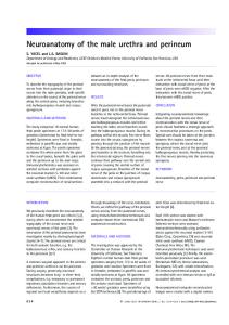
Functional activation
The functional neuroanatomy of schizophrenic subsyndromes
Normal function
Symptom severity
Positive Negative syndrome syndrome
FIG. 3. Schematic representation of a simple capacity model of cortical function. The model assumes a fixed capacity for cognitive processing in a given cortical region. Endogenous processing demands due to ‘resting ’ brain activity can compete for capacity-limited neural resources with exogenous, experimentally-controlled processing demands. The power of functional activation, as measured here by fMRI, is inversely proportional to the amount of total capacity consumed by endogenous processes. Thus, patients with positive symptoms may have increased endogenous activity in left perisylvian cortex and concomitantly attenuated power of functional activation by exogenous tasks, such as verbal working memory, which make competitive demands on these regions. Likewise, patients with negative symptoms may have reduced endogenous activity in premotor cortex and concomitantly increased power of functional activation of this region by a competitive experimental task. ( , Functional reserve ; , exogenous task-related activity ; &, endogenous activity.)
literature on resting state perfusion/metabolism in schizophrenia, the direction of associations in our study is reversed relative to the resting state literature. We suggest that these apparent discrepancies may be resolved within the theoretical context of a capacity model for cortical function (Just & Carpenter, 1992; Just et al. 1996) (schematically illustrated in Fig. 3). Briefly, this model suggests that different cognitive tasks may compete for common, capacity-limited neural resources. As the demands on processing resources increase, there is increasing functional activation of the brain regions specialized to perform the relevant tasks until a capacity limit is reached, at which point activation by one or more of the competing tasks is attenuated. Cognitive activation studies incorporating parametric modulation of working memory load (Callicott et al. 1999) and dual-task performance interference (Klingberg, 1998) provide supporting evidence for this model of brain function. Perturbations of endogenous or baseline activity may also compete for neural resources with exogenous or experimentally administered cognitive tasks ; for example, a negative correlation between focal activation during visual stimulation and baseline rCBF has recently been
1016
G. D. Honey and others
demonstrated in healthy volunteers (Kastrup et al. 1999). In extending this model to the psychopathological literature, we propose that psychotic symptoms represent an endogenous demand on cortical processing resources which may be competitive with exogenous or experimental task demands. Thus, it is clear from studies of ‘resting ’ brain activity in schizophrenic patients that positive symptoms are associated with a regionally-specific increase in endogenous activity of superior temporal and inferior frontal cortex and SMA (DeLisi et al. 1989 ; McGuire et al. 1993; Suzuki et al. 1993). The capacity model predicts attenuated activation of these regions by competitive tasks, i.e., tasks which would normally activate these regions strongly, in patients with positive psychotic symptoms. Indeed it has previously been shown that patients with a history of auditory hallucinations have attenuated activation of frontal and temporal regions in response to a verbal self-monitoring task (McGuire et al. 1995), and attenuated activation of the auditory cortex in response to external auditory stimulation (David et al. 1996; Woodruff et al. 1997). Our finding of a negative association between positive subsyndrome scores and power of activation by a working memory task in frontal and temporal cortex is likewise compatible with the predictions of the capacity model. Applying the same logic to interpretation of our results on negative subsyndrome scores, we conclude that endogenous processing demands on lateral promoter cortex may be reduced in patients with severe negative symptoms, so that the capacity available to process an exogenously presented working memory task is increased, and power of activation by the experimental task is also increased. Similarly, our results on global symptom severity would suggest that schizophrenic symptoms generally impose an endogenous processing demand on left superior temporal and ventral occipital cortices. It should be noted that in this study we did not directly measure baseline activity, and therefore these predictions, based on a simple capacity model of cortical function, are preliminary but potentially testable in future studies combining variable measures of exogenous task load, and resting baseline activity. The implication of our current findings, with reference to previous
studies of the effect of schizophrenia on resting activity, is that symptom expression likely involves an interactive effect on both resting and activated states, the former compromising the latter, and that this is dependent on the nature of the neurocognitive requirements and neural resources engaged. Finally, it is noteworthy that the significant between-group differences in activation of frontal cortex by the working memory task were not located in the same regions of frontal cortex that demonstrated significant psychophysiological associations with subsyndrome scores. One possible explanation is that relatively reduced power of activation in dorsolateral prefrontal cortex, or ‘ hypofrontality ’, in these data may be related to the relatively poor behavioural performance of the patients on the verbal working memory task, as reported previously (Fletcher et al. 1998). The differences in location of hypofrontality and psychophysiological associations are likewise compatible with the lack of correlation between subsyndrome scores and behavioural measures of task performance, suggesting that cognitive impairment and psychotic symptom severity are not strongly linked and may have anatomically dissociable neural correlates (Honey et al. 2002). The possibility of an interaction between symptom status, cognitive performance and neurophysiological response was not tested in these data, and remains an intriguing possibility for further study. In summary, we have reported anatomically distinct psychophysiological associations between global and subsyndromal symptom scores and power of fMRI activation by a verbal working memory task in a sample of 30 patients with schizophrenia. We have generally interpreted these associations, in the light of complementary prior studies on ‘ resting ’ blood flow or metabolism, as indicating competition for limited cortical capacity between exogenous (experimental) and endogenous (psychopathological) processes in patients with schizophrenia.
REFERENCES Andreasen, N. C., Arndt, S., Alliger, R., Miller, D. & Flaum, M. (1997). Symptoms of schizophrenia : methods, meanings and mechanisms. Archives of General Psychiatry 52, 341–351. Berman, K. F. (1987). Cortical ‘stress tests ’ in schizophrenia : regional cerebral blood flow studies. Biological Psychiatry 22, 1304–1326.
The functional neuroanatomy of schizophrenic subsyndromes Brammer, M. J., Bullmore, E. T., Simmons, A., Williams, S. C., Grasby, P. M., Howard, R. J., Woodruff, P. W. & Rabe-Hesketh, S. (1997). Generic brain activation mapping in functional magnetic resonance imaging : a nonparametric approach. Magnetic Resonance Imaging 15, 763–770. Bullmore, E. T., Brammer, M., Williams, S. C., Rabe-Hesketh, S., Janot, N., David, A., Mellers, J., Howard, R. & Sham, P. (1996). Statistical methods of estimation and inference for functional MR image analysis. Magnetic Resonance in Medicine 35, 261–277. Bullmore, E. T., Brammer, M., Williams, S. C., Curtis, V., McGuire, P., Morris, R., Murray, R. & Sharma, T. (1999 a). Functional MR imaging of confounded hypofrontality. Human Brain Mapping 8, 86–91. Bullmore, E. T., Brammer, M. J., Rabe-Hesketh, S., Curtis, V. A., Morris, R. G., Williams, S. C., Sharma, T. & McGuire, P. K. (1999 b). Methods for diagnosis and treatment of stimuluscorrelated motion in generic brain activation studies using fMRI. Human Brain Mapping 7, 38– 48. Bullmore, E. T., Suckling, J., Overmeyer, S., Rabe-Hesketh, S., Taylor, E. & Brammer, M. J. (1999 c). Global, voxel, and cluster tests, by theory and permutation, for a difference between two groups of structural MR images of the brain. IEEE Transactions in Medical Imaging 18, 32–42. Callicott, J. H., Mattay, V. S., Bertolino, A., Finn, K., Coppola, R., Frank, J. A., Goldberg, T. E. & Weinberger, D. R. (1999). Physiological characteristics of capacity constraints in working memory as revealed by functional MRI. Cerebral Cortex 9, 20–26. David, A. S., Woodruff, P. W., Howard, R., Mellers, J. D., Brammer, M., Bullmore, E., Wright, I., Andrew, C. & Williams, S. C. (1996). Auditory hallucinations inhibit exogenous activation of auditory association cortex. Neuroreport 7, 932–936. Deiber, M. P., Passingham, R. E., Colebatch, J. G., Friston, K. J., Nixon, P. D. & Frackowiak, R. S. (1991). Cortical areas and the selection of movement : a study with positron emission tomography. Experimental Brain Research 84, 393–402. DeLisi, L. E., Buchsbaum, M. S., Holcomb, H. H., Langston, K. C., King, A. C., Kessler, R., Pickar, D., Carpenter, W. T., Morihisa, J. M. & Margolin, R. (1989). Increased temporal lobe glucose use in chronic schizophrenic patients. Biological Psychiatry 25, 835–851. Ebmeier, K. P., Blackwood, D. H., Murray, C., Souza, V., Walker, M., Dougall, N., Moffoot, A. P., O’Carroll, R. E. & Goodwin, G. M. (1993). Single-photon emission computed tomography with 99m TC-exametazime in unmedicated schizophrenic patients. Biological Psychiatry 33, 487–495. Fletcher, P. C., McKenna, P. J., Frith, C. D., Grasby, P. M., Friston, K. J. & Dolan, R. J. (1998). Brain activations in schizophrenia during a graded memory task studied with functional neuroimaging. Archives of General Psychiatry 55, 1001–1008. Friston, K. J., Liddle, P. F., Frith, C. D., Hirsch, S. R. & Frackowiak, R. S. (1992). The left medial temporal region and schizophrenia. A PET study. Brain 115, 367–382. Frith, C. D., Friston, K. J., Herold, S., Silbersweig, D., Fletcher, P., Cahill, C., Dolan, R. J., Frackowiak, R. S. & Liddle, P. F. (1995). Regional brain activity in chronic schizophrenic patients during the performance of a verbal fluency task. British Journal of Psychiatry 167, 343–349. Honey, G. D., Bullmore, E. T. & Sharma, T. (2002). De-coupling of cognitive performance and cerebral functional response during working memory in schizophrenia. Schizophrenia Research 53, 45–56. Just, M. A. & Carpenter, P. A. (1992). A capacity theory of comprehension : individual differences in working memory. Psychological Review 99, 122–149. Just, M. A., Carpenter, P. A. & Keller, T. A. (1996). The capacity theory of comprehension : new frontiers of evidence and arguments. Psychological Review 103, 773–780. Kaplan, R. D., Szechtman, H., Franco, S., Szechtman, B., Nahmias, C., Garnett, E. S., List, S. & Cleghorn, J. M. (1993). Three clinical
1017
syndromes of schizophrenia in untreated subjects : relation to brain glucose activity measured by positron emission tomography (PET). Schizophrenia Research 11, 47–54. Kastrup, A., Li, T.-Q., Kruger, G., Glover, G. & Moseley, M. (1999). Relationship between cerebral blood flow changes during visual stimulation and baseline flow levels investigated with functional MRI. Neuroreport 10, 1751–1756. Kawasaki, Y., Maeda, Y., Sakai, N., Higashima, M., Yamaguchi, N., Koshino, Y., Hisada, K., Suzuki, K. & Matsuda, H. (1996). Regional cerebral blood flow in patients with schizophrenia: relevance to symptom structures. Psychiatry Research 67, 49–58. Kay, S. R., Fiszbein, A. & Opler, L. A. (1987). The positive and negative syndrome scale (PANSS) for schizophrenia. Schizophrenia Bulletin 13, 261–276. Klingberg, T. (1998). Concurrent performance of two working memory tasks : potential mechanisms of interference. Cerebral Cortex 8, 593–601. Liddle, P., Carpenter, W. T. & Crow, T. (1994). Syndromes of schizophrenia. Classic literature. British Journal of Psychiatry 165, 721–727. Liddle, P. F. (1987 a). The symptoms of chronic schizophrenia. A re-examination of the positive-negative dichotomy. British Journal of Psychiatry 151, 145–151. Liddle, P. F. (1987b). Schizophrenic syndromes, cognitive performance and neurological dysfunction. Psychological Medicine 17, 49–57. Liddle, P. F., Friston, K. J., Frith, C. D., Hirsch, S. R., Jones, T. & Frackowiak, R. S. (1992). Patterns of cerebral blood flow in schizophrenia. British Journal of Psychiatry 160, 179–186. Lindstrom, E. & von Knorring, L. (1994). Symptoms in schizophrenic syndromes in relation to age, sex, duration of illness and number of previous hospitalizations. Acta Psychiatrica Scandinavica 89, 274–278. 443 (erratum). McGuire, P. K., Shah, G. M. & Murray, R. M. (1993). Increased blood flow in Broca’s area during auditory hallucinations in schizophrenia. Lancet 342, 703–706. McGuire, P. K., Silbersweig, D. A., Wright, I., Murray, R. M., David, A. S., Frackowiak, R. S. & Frith, C. D. (1995). Abnormal monitoring of inner speech : a physiological basis for auditory hallucinations. Lancet 346, 596–600. Nunnally, J. C. (1994). Psychometric Theory, 2nd edn. McGraw Hill Book Company : New York. Peralta, V. & Cuesta, M. J. (1998). Factor structure and clinical validity of competing models of positive symptoms in schizophrenia. Biological Psychiatry 44, 107–114. Schroder, J., Buchsbaum, M. S., Siegel, B. V., Geider, F. J., Lohr, J., Tang, C., Wu, J. & Potkin, S. G. (1996). Cerebral metabolic activity correlates of subsyndromes in chronic schizophrenia. Schizophrenia Research 19, 41–53. Suzuki, M., Yuasa, S., Minabe, Y., Murata, M. & Kurachi, M. (1993). Left superior temporal blood flow increases in schizophrenic and schizophreniform patients with auditory hallucination : a longitudinal case study using 1231-IMP SPECT. European Archives of Psychiatry and Clinical Neuroscience 242, 257–261. Talairach, J. & Tournoux, P. (1988). A Co-planar Stereotaxic – Atlas of a Human Brain. Theime Verlag : Stuttgart. Touge, T., Werhahn, K. J., Rothwell, J. C. & Marsden, C. D. (1995). Movement-related cortical potentials preceding repetitive and random-choice hand movements in Parkinson’s disease. Annals of Neurology 37, 791–799. Volkow, N. D., Wolf, A. P., Van Gelder, P., Brodie, J. D., Overall, J. E., Cancro, R. & Gomez-Mont, F. (1987). Phenomenological correlates of metabolic activity in 18 patients with chronic schizophrenia. American Journal of Psychiatry 144, 151–158. White, L., Harvey, P. D., Opler, L. & Lindenmayer, J. P. (1997). Empirical assessment of the factorial structure of clinical symptoms in schizophrenia. A multisite, multimodel evaluation of the factorial structure of the Positive and Negative Syndrome Scale. The PANSS Study Group. Psychopathology 30, 263–274.
1018
G. D. Honey and others
Wolkin, A., Sanfilipo, M., Wolf, A. P., Angrist, B., Brodie, J. D. & Rotrosen, J. (1992). Negative symptoms and hypofrontality in chronic schizophrenia. Archives of General Psychiatry 49, 959–965. Woodruff, P. W., Wright, I. C., Bullmore, E. T., Brammer, M., Howard, R. J., Williams, S. C., Shapleske, J., Rossell, S., David, A. S., McGuire, P. K. & Murray, R. M. (1997). Auditory
hallucinations and the temporal cortical response to speech in schizophrenia: a functional magnetic resonance imaging study. American Journal of Psychiatry 154, 1676–1682. Zemishlany, Z., Alexander, G. E., Prohovnik, I., Goldman, R. G., Mukherjee, S. & Sackeim, H. (1996). Cortical blood flow and negative symptoms in schizophrenia. Neuropsychobiology 33, 127–131.