The Ne w E n g l a nd Jo u r n a l o f Me d ic i ne
Brief Report
The abnormal findings in muscle-biopsy specimens from both thighs and the finding of severely impaired oxygen extraction when the forearm muscles were repeatedly contracted8 suggested generalized muscular involvement.
METHODS
P ATERNAL I NHERITANCE OF M ITOCHONDRIAL DNA MARIANNE SCHWARTZ, PH.D., JOHN VISSING, M.D., PH.D.
AND
M
AMMALIAN mitochondrial DNA (mtDNA) is thought to be strictly maternally inherited.1,2 Sperm mitochondria disappear in early embryogenesis by selective destruction, inactivation, or simple dilution by the vast surplus of oocyte mitochondria.3 Very small amounts of paternally inherited mtDNA have been detected by the polymerase chain reaction (PCR) in mice after several generations of interspecific backcrosses.4 Studies of such hybrids and of mouse oocytes microinjected with sperm support the hypothesis that sperm mitochondria are targeted for destruction by nuclear-encoded proteins.5-7 We report the case of a 28-year-old man with mitochondrial myopathy due to a novel 2-bp mtDNA deletion in the ND2 gene (also known as MTND2), which encodes a subunit of the enzyme complex I of the mitochondrial respiratory chain. We determined that the mtDNA harboring the mutation was paternal in origin and accounted for 90 percent of the patient’s muscle mtDNA. CASE REPORT The patient was a 28-year-old man with severe, lifelong exercise intolerance. He had never been able to run more than a few steps. His cardiac and pulmonary functions were normal, and he was otherwise well. Both parents and a 23-year-old sister were healthy and had normal exercise tolerance. The myopathic symptoms were associated with severe lactic acidosis induced by minor physical exertion. His plasma lactate level after walking 100 m at a slow pace was 6 to 8 mmol per liter (the normal level is below 2.5 mmol per liter). His creatine kinase levels were marginally elevated in periods of no physical exertion. Biopsies of the right and left quadriceps muscle revealed that 15 percent of the fibers were of the ragged-red type, a result consistent with the accumulation of abnormal mitochondria with impaired respiratory function. Biochemical analysis demonstrated an isolated deficiency of the mitochondrial enzyme complex I of the respiratory chain in muscle. There were no signs of muscular atrophy or weakness.
From the Department of Clinical Genetics (M.S.) and the Department of Neurology and the Copenhagen Muscle Research Center (J.V.), University Hospital Rigshospitalet, Copenhagen, Denmark. Address reprint requests to Dr. Schwartz at the Department of Clinical Genetics, Rigshospitalet 4062, Blegdamsvej 9, Copenhagen DK 2100, Denmark, or at
[email protected].
DNA was isolated from the patient’s blood, muscle, hair roots, and fibroblasts (derived from a skin biopsy) by standard methods. DNA was also isolated from the blood of the patient’s parents and paternal uncle, and from the blood and the quadriceps muscle of the patient’s sister. The mtDNA was amplified into two products with the primers OLA (5756–5781) + D1B (282–255) and D1A (336–363) + OLB (5745–5721),9 and the products were purified. We sequenced most of the mtDNA, including all transfer RNA (tRNA) genes, CYTB, and all seven genes encoding subunits of enzyme complex I, using a genetic analyzer (ABI PRISM 310, Applied Biosystems) and a terminator cycle-sequencing ready-reaction kit (ABI PRISM BigDye, Applied Biosystems). The sequences obtained were compared with the revised Cambridge reference sequence10,11 (AC J01415) with use of the DNAsis program (Hitachi Software Engineering Europe). Two different mtDNA haplotypes were found in the patient; presumably, one came from the father and the other from the mother. Solid-phase minisequencing12 was performed to establish the ratios of the mtDNA haplotypes in blood and muscle. The target was nucleotide position 3197, which, among others, distinguished the maternal haplotype (3197T) from the paternal one (3197C). PCR products spanning the position in question were generated by the 5'-biotinylated forward primer (3014–3034) and the reverse primer (3376–3356). PCR products were immobilized on a streptavidin-coated solid support (96-well plate) and denatured by sodium hydroxide. A sequencing primer (3220–3198) was designed to anneal adjacent to (upstream from) nucleotide 3197. The nucleotide at position 3197 was analyzed by the primer extension reaction, in which a tritium-labeled deoxynucleoside triphosphate corresponding to either the maternal nucleotide (deoxyadenosine triphosphate) or the paternal nucleotide (deoxyguanosine triphosphate) was added to two parallel reactions. After washing, the elongated primers were eluted by sodium hydroxide, and the amount of incorporated [3H]deoxynucleoside monophosphate was determined with a liquid scintillation counter. The ratios of adenine to guanine incorporated into each sequencing primer were determined and compared with the values on a standard curve constructed on the basis of known proportions of cloned segments of mtDNA harboring 3197T and 3197C, respectively. The ratio of the 2-bp deletion to wild-type mtDNA in tissues (the level of heteroplasmy) was determined by PCR fragment analysis. The mtDNA was amplified by the 5'-fluorochrome–labeled forward primer (5041–5060) and the reverse primer (5196–5177). The PCR products were analyzed on a genetic analyzer with a GeneScan standard (PE Applied Biosystems) as a size marker. The areas of the mutant (2-bp deletion) and wild-type peaks were used to calculate the percentage of mutant (paternal) mtDNA in the patient’s muscle. The nuclear genotypes of the patient, his parents, and his sister were determined for the highly polymorphic markers (microsatellites) D7S2212, D7S817, D19S219, D19S559, and TNFB. PCR products were analyzed on a genetic analyzer with GeneScan software (Applied Biosystems) and a GeneScan standard as a size marker. The patient and his family provided oral consent for testing after receiving counseling. Written consent was not required by the institutional review board because the investigation was considered part of clinical care.
RESULTS
Sequencing the mitochondrial genome from a specimen from a biopsy of the quadriceps muscle re-
576 · N Engl J Med, Vol. 347, No. 8 · August 22, 2002 · www.nejm.org Downloaded from www.nejm.org on January 1, 2008 . For personal use only. No other uses without permission. Copyright © 2002 Massachusetts Medical Society. All rights reserved.
BR IEF REPOR T
vealed a deletion of 2 bp, 5132delAA, in the ND2 gene. The 2-bp deletion causes a frame shift, introducing a stop codon downstream from the deletion. Furthermore, a novel variant, 1303G→A, was detected in the gene encoding 12S ribosomal RNA (rRNA). The 1303G→A variant was not found in 50 normal controls, but a conservation analysis showed that this position is not conserved through evolution (Table 1). The patient was apparently homoplasmic (only one type of mtDNA was present) for both the 2-bp deletion and 1303G→A. The patient also harbored several known mtDNA polymorphisms. To evaluate the ratio of normal to mutant mtDNA (heteroplasmy) in other tissues, we analyzed mtDNA from the patient’s blood, hair roots, and cultured fibroblasts. Neither the 1303G→A variant nor the 2-bp deletion was present in mtDNA from these tissues. In fact, the blood and muscle mtDNA sequences differed at 18 positions, some of which allowed assignment of the two sequences to separate European mtDNA haplogroups, H and U5, respectively (Table 2 and Fig. 1). Mixing up of samples was ruled out by analyses of repeated blood and muscle samples. The repeated muscle biopsies were in the right and left vastus lateralis muscles. Furthermore, genotyping of all samples for five highly polymorphic nuclear markers (microsatellites) indicated that all samples came from the same person (Fig. 2). Sequencing of blood mtDNA from the patient’s healthy parents and from his paternal uncle demonstrated that the haplotype of the patient’s muscle mtDNA was identical to that of his father’s and uncle’s blood (with the exception of the 2-bp deletion, which was found only in the patient). The haplotype of the patient’s blood was identical to that of his mother. Analysis of mtDNA from both blood and muscle of the patient’s sister found only the maternal mtDNA haplotype (data not shown). The nuclear microsatellite genotypes of the patient, his parents, and his sister showed a distribution of alleles consistent with their biologic relationships (Fig. 2). According to direct sequencing of PCR-amplified mtDNA, muscle tissue from the patient was homoplasmic for the paternal haplotype and for the 2-bp deletion. However, solid-phase minisequencing12 and fragment analysis showed about 10 percent maternal (normal) mtDNA in both muscle-biopsy specimens (Fig. 1). The mtDNA from the patient’s blood, hair roots, and cultured fibroblasts showed only the maternal haplotype. DISCUSSION
We report the case of a patient with severe exercise intolerance caused by a 2-bp deletion in the ND2 gene of mtDNA. A striking finding was that the mutation occurred on a paternal mtDNA background. Because
TABLE 1. CONSERVATION ANALYSIS OF 1303G IN 12S RIBOSOMAL RNA ACROSS SPECIES.*
SPECIES
SEQUENCE HOMOLOGY
GENBANK ACCESSION NO.
Human Gorilla Chimpanzee Cow Pig Mouse Rat Elephant Rabbit
GTAAGCGCAAGT GTAAGCACAAGT GTAAGCACAAGT GTAAGCGTAATT GTGAGCGTAATT GTAAGCAAAAGA GTAAGCACAAGA GTGAGCTTAATC GTAAGCTCAATT
X62996 D38114 X93335 J01394 AJ222767 AJ279438 AJ428514 X93602 AJ001588
*Nucleotides 1297 to 1308 are shown, and nucleotide 1303 is shown in boldface.
TABLE 2. SEQUENCE DIFFERENCES BETWEEN THE TWO MITOCHONDRIAL DNA (mtDNA) HAPLOTYPES FOUND IN THE PATIENT’S BLOOD AND MUSCLE AND IN HIS PARENTS’ BLOOD. NUCLEOTIDE POSITION
PATIENT’S BLOOD*
PATIENT’S MUSCLE
MOTHER’S BLOOD
FATHER’S BLOOD
477 1303 3192 3197† 3591 4592 5132–5134 11296 11467† 11719‡ 11938 12308† 12372† 12618 13617† 14766‡ 14793 15218
C G C T G T AAA C A G C A G G T C A A
T A T C A C delAA T G A T G A A C T G G
C G C T G T AAA C A G C A G G T C A A
T A T C A C AAA T G A T G A A C T G G
*This is the Cambridge reference sequence.10,11 †The haplotype of the mtDNA from the patient’s muscle and his father’s blood, as defined by the nucleotides at these sites, is diagnostic of mtDNA haplogroup U5.13,14 ‡The haplotype of the mtDNA from the patient’s blood and that of his mother, as defined by the nucleotides at these two sites, and the presence of 4580G in all samples (data not shown) are diagnostic of mtDNA haplogroup H.13-15
N Engl J Med, Vol. 347, No. 8 · August 22, 2002 · www.nejm.org · 577
Downloaded from www.nejm.org on January 1, 2008 . For personal use only. No other uses without permission. Copyright © 2002 Massachusetts Medical Society. All rights reserved.
The Ne w E n g l a nd Jo u r n a l o f Me d ic i ne
Figure 1. Partial Sequence Analysis of Mitochondrial DNA from the Patient’s Blood and Muscle and from His Parents’ Blood. Panel A demonstrates part of the difference in haplotypes between the patient’s muscle (paternal) and his blood (maternal). The arrows indicate the polymorphic nucleotides 3192 and 3197. Panel B shows fragment analysis for the 2-bp deletion. Fragments of 152 bp (normal) and 150 bp (2-bp deletion) are shown in purple. The GeneScan size standard is shown in orange. The muscle is heteroplasmic for the deletion, with 10 percent wildtype and 90 percent mutant mitochondrial DNA.
A T
C A
A C
T
Patient’s Blood
Patient’s Muscle
the patient had an isolated myopathy due to a mutation found only in skeletal muscle, and because family members were unaffected and did not carry the mutation in blood or muscle, we conclude that the 2-bp deletion arose spontaneously in early embryogenesis or in the paternal germ line. However, we cannot rule out the possibility that the father harbored this mutation at a low level in other tissues. The origin of the mutation could be similar to that of sporadic, single, large-scale deletions, which so far have been thought to arise spontaneously in maternal mtDNA, either in the germ line or in early embryogenesis.16 Mutations of mtDNA cause symptoms only when high levels of mutant mtDNA are present: typically, 50 to 60 percent for single, large-scale deletions and 80 to 90 percent for point mutations.17,18 For a high percentage of mutant mtDNA to be present, a replicative advantage for the mutated mitochondria is probably required. However, not only single, large-scale deletions, but also mtDNA point mutations, can proliferate with time and outnumber wild-type mtDNA.19 It has been suggested that the replicative advantage of the largescale deletions is due to a faster completion of replication of the smaller mtDNA molecules, but the experimental evidence for this hypothesis is controversial.2 Complex I of the respiratory chain consists of 41 subunits, 7 of which are encoded by mtDNA. The stop codon introduced by the 2-bp deletion affects translation of the C-terminal portion of the gene product. It therefore inactivates the ND2 subunit and the catalytic function of complex I. In support of this concept, complex I activity was severely impaired in muscle from the patient we describe. The patient also harbored a novel variant (1303G→A) in the 12S rRNA gene. This variant could theoretically influence the patient’s phenotype. However, the variant is situated in a part of the 12S rRNA gene that is not conserved in evolution. Mouse and rat 12S rRNA genes have an adenine, and rabbit and elephant genes have a thymine in this position (Table 1), which suggests that 1303G→A is a harmless variant. Furthermore, the homoplasmic occurrence of
Father’s Blood
Mother’s Blood
T
A
G T A T 160
3192 C
T
A T
3197 T
T
C
T
C
C
T
B
Blood
Muscle
152 bp
150 bp
152 bp
578 · N Engl J Med, Vol. 347, No. 8 · August 22, 2002 · www.nejm.org Downloaded from www.nejm.org on January 1, 2008 . For personal use only. No other uses without permission. Copyright © 2002 Massachusetts Medical Society. All rights reserved.
A C
BR IEF REPOR T
D19S219 D19S559 D7S817 D7S2212 TNFB
I-1
I-2
161–173 173–181 155–163 192–200 107–117
171–173 169–181 151–159 196–196 99–99
II-1
II-2
171–173 169–173 159–163 192–196 99–117
171–173 173–181 151–163 196–200 99–107
eight-cell stage of some abnormal embryos.25 However, paternal mtDNA has not been detected in infants born after intracytoplasmic sperm injection.26 The underlying mechanism responsible for the elimination of sperm mtDNA in normal embryos is not well understood. We speculate that the process in some cases may be defective, allowing sperm mitochondria to survive and giving those with a selective advantage the possibility of prevailing in certain tissues. The present findings suggest that investigation of paternal mtDNA inheritance may be warranted in cases with sporadic mitochondrial mutations.
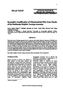
Figure 2. Pedigree of the Family with Genotypes of Five Microsatellite Nuclear DNA Markers. Allele sizes are given in base pairs. These results confirm that Subject I-1 is the biologic father of the patient (Subject II-1). The alleles found in nuclear DNA from the muscle of Subject II-1 were identical to those found in nuclear DNA from his blood.
1303G→A in the healthy father and uncle also indicates the nonpathogenic nature of this variant. Until now, pathogenic mtDNA has been assumed to be maternally inherited or to have arisen spontaneously on a maternal mtDNA background. However, paternal mtDNA inheritance may go unrecognized in cases with sporadic, single, large-scale deletions, because mitochondrial haplotypes are rarely investigated in diagnostic analyses. The same may be true of the rare cases of sporadic point mutations in mtDNA that give rise to a clinical picture similar to that in the present patient. Mutations in mtDNA that cannot be detected in the mother, are found in skeletal muscle only, and have no effects other than exercise intolerance have been reported for the respiratory-chain subunits ND4,20 cytochrome b,21 cytochrome c oxidase subunit I,22 and cytochrome c oxidase subunit III.23 Among these, mutations of the cytochrome b gene have been reported in more than one patient.21 However, sporadic cytochrome b mutations may also affect tissues other than muscle.24 The present case could be the result of the survival of one or a few sperm mitochondria that probably would have been diluted out and never have been recognized had the pathogenic mutation not conferred a selective proliferative advantage on the mitochondria. There is now strong evidence that highly effective processes exist for eliminating healthy sperm mitochondria in early mammalian embryogenesis.5-7 When a sperm, including the midpiece, which is rich in mitochondria, is injected directly into an oocyte, as in assisted reproductive techniques to treat human infertility, paternal mtDNA can be detected in the four-to-
Supported by an unrestricted grant from the Novo Nordic Foundation (to Dr. Schwartz) and a grant from the Danish National Research Foundation (504-14, to Dr. Vissing).
We are indebted to Dr. Søren Nørby for constructive discussions and advice and to Dorthe Munkløv for her excellent technical assistance.
REFERENCES 1. Birky CW Jr. Uniparental inheritance of mitochondrial and chloroplast genes: mechanisms and evolution. Proc Natl Acad Sci U S A 1995;92: 11331-8. 2. Idem. The inheritance of genes in mitochondria and chloroplasts: laws, mechanisms, and models. Annu Rev Genet 2001;35:125-48. 3. Cummins JM, Wakayama T, Yanagimachi R. Fate of microinjected sperm components in the mouse oocyte and embryo. Zygote 1997;5:301-8. 4. Gyllensten U, Wharton D, Josefsson A, Wilson AC. Paternal inheritance of mitochondrial DNA in mice. Nature 1991;352:255-7. 5. Cummins JM, Wakayama T, Yanagimachi R. Fate of microinjected spermatid mitochondria in the mouse oocyte and embryo. Zygote 1998;6:21322. 6. Shitara H, Hayashi JI, Takahama S, Kaneda H, Yonekawa H. Maternal inheritance of mouse mtDNA in interspecific hybrids: segregation of leaked paternal mtDNA followed by the prevention of subsequent paternal leakage. Genetics 1998;148:851-7. 7. Shitara H, Kaneda H, Sato A, et al. Selective and continuous elimination of mitochondria microinjected into mouse eggs from spermatids, but not from liver cells, occurs throughout embryogenesis. Genetics 2000;156: 1277-84. 8. Jensen TD, Kazemi-Esfarjani P, Skomorowska E, Vissing J. A forearm exercise screening test for mitochondrial myopathy. Neurology 2002;58: 1533-8. 9. Kleinle S, Wiesmann U, Superti-Furga A, et al. Detection and characterization of mitochondrial DNA rearrangements in Pearson and KearnsSayre syndromes by long PCR. Hum Genet 1997;100:643-50. 10. Anderson S, Bankier AT, Barrell BG, et al. Sequence and organisation of the human mitochondrial genome. Nature 1981;290:457-65. 11. Andrews RM, Kubacka I, Chinnery PF, Lightowlers RN, Turnbull DM, Howell N. Reanalysis and revision of the Cambridge reference sequence for human mitochondrial DNA. Nat Genet 1999;23:147. 12. Suomalainen A, Syvänen AC. Quantitative analysis of human DNA sequences by PCR and solid-phase minisequencing. Mol Biotechnol 2000; 15:123-31. 13. Macaulay V, Richards M, Hickey E, et al. The emerging tree of West Eurasian mtDNAs: a synthesis of control-region sequences and RFLPs. Am J Hum Genet 1999;64:232-49. 14. Finnilä S, Lehtonen MS, Majamaa K. Phylogenetic network for European mtDNA. Am J Hum Genet 2001;68:1475-84. 15. Saillard J, Magalhães PJ, Schwartz M, Rosenberg T, Nørby S. Mitochondrial DNA variant 11719G is a marker for the mtDNA haplogroup cluster HV. Hum Biol 2000;72:1065-8. 16. DiMauro S. Mitochondrial encephalopathies. In: Rosenberg RN, Prusiner SB, DiMauro S, Barchi RL, Kunkel LM, eds. The molecular and genetic basis of neurological disease. Stoneham, Mass.: Butterworth– Heinemann, 1993:665–94. 17. Hayashi JI, Ohta S, Kikuchi A, Takemitsu M, Goto Y-I, Nonaka I. Introduction of disease-related mitochondrial DNA deletions into HeLa cells
N Engl J Med, Vol. 347, No. 8 · August 22, 2002 · www.nejm.org · 579
Downloaded from www.nejm.org on January 1, 2008 . For personal use only. No other uses without permission. Copyright © 2002 Massachusetts Medical Society. All rights reserved.
The Ne w E n g l a nd Jo u r n a l o f Me d ic i ne
lacking mitochondrial DNA results in mitochondrial dysfunction. Proc Natl Acad Sci U S A 1991;88:10614-8. 18. Chomyn A, Martinuzzi A, Yoneda M, et al. MELAS mutation in mtDNA binding site for transcription termination factor causes defects in protein synthesis and in respiration but no change in levels of upstream and downstream mature transcripts. Proc Natl Acad Sci U S A 1992;89: 4221-5. 19. Yoneda M, Chomyn A, Martinuzzi A, Hurko O, Attardi G. Marked replicative advantage of human mtDNA carrying a point mutation that causes the MELAS encephalomyopathy. Proc Natl Acad Sci U S A 1992; 89:11164-8. 20. Andreu AL, Tanji K, Bruno C, et al. Exercise intolerance due to a nonsense mutation in the mtDNA ND4 gene. Ann Neurol 1999;45:820-3. 21. Andreu AL, Hanna MG, Reichmann H, et al. Exercise intolerance due to mutations in the cytochrome b gene of mitochondrial DNA. N Engl J Med 1999;341:1037-44.
22. Karadimas CL, Greenstein P, Sue CM, et al. Recurrent myoglobinuria due to a nonsense mutation in the COX I gene of mitochondrial DNA. Neurology 2000;55:644-9. 23. Keightley JA, Hoffbuhr KC, Burton MD, et al. A microdeletion in cytochrome c oxidase (COX) subunit III associated with COX deficiency and recurrent myoglobinuria. Nat Genet 1996;12:410-6. 24. Wibrand F, Ravn K, Schwartz M, Rosenberg T, Horn N, Vissing J. Multisystem disorder associated with a missense mutation in the mitochondrial cytochrome b gene. Ann Neurol 2001;50:540-3. 25. St John J, Sakkas D, Dimitriadi K, et al. Failure of elimination of paternal mitochondrial DNA in abnormal embryos. Lancet 2000;355:200. 26. Houshmand M, Holme E, Hanson C, Wennerholm UB, Hamberger L. Is paternal mitochondrial DNA transferred to the offspring following intracytoplasmic sperm injection? J Assist Reprod Genet 1997;14:223-7. Copyright © 2002 Massachusetts Medical Society.
FULL TEXT OF ALL JOURNAL ARTICLES ON THE WORLD WIDE WEB Access to the complete text of the Journal on the Internet is free to all subscribers. To use this Web site, subscribers should go to the Journal’s home page (http://www.nejm.org) and register by entering their names and subscriber numbers as they appear on their mailing labels. After this one-time registration, subscribers can use their passwords to log on for electronic access to the entire Journal from any computer that is connected to the Internet. Features include a library of all issues since January 1993 and abstracts since January 1975, a full-text search capacity, and a personal archive for saving articles and search results of interest. All articles can be printed in a format that is virtually identical to that of the typeset pages. Beginning six months after publication the full text of all original articles and special articles is available free to nonsubscribers who have completed a brief registration.
580 · N Engl J Med, Vol. 347, No. 8 · August 22, 2002 · www.nejm.org Downloaded from www.nejm.org on January 1, 2008 . For personal use only. No other uses without permission. Copyright © 2002 Massachusetts Medical Society. All rights reserved.