MILITARY MEDICINE, 173, 2:t67, 2008
Acute Effects of MK63 Stun Device Discharges in Miniature Swine Daniel J. Valentino, MD*f; Robert J. Walter, PhD*f; Andrew J. Dennis, DO*t; Kimberly Nagy, MD*f; Michele M. Loor, MD*f; Jerry Winners, BS*; Faran Bokhari, MD*t; Dorion Wiley, MD"t, Azher Merchant, BSf; Kimberly Joseph, MD*f; Roxanne Roberts, MD f ABSTRACT Objective: Electronitiscular incapacitation (EMI) devices are beitig used and evaluated by both military and law enforcement agencies. Although Ihe gross muscular response is obvious, pbysiologica! respotises to these devices are poorly understotxl. We hypothesized that the intense, repetitive, muscle contractions evoked by EMI devices would cause dose-dependent metabolic acido.sis, accompanied by netiromuscular or cardiac injury. Methods: Using ati approved protocol. 26 Yucatan mini-pigs (22 experimenlal atiimals and 4 control animals) were anesthetized with ketaniine and xylazine. Expetimental animals were exposed to MK63 (Aegis Industries, Bellevue, Idaho) discharges over the left anterior hind limb for 10, 20.40, (tr 80 seconds. Electrocardiognims, electromyograms, troponin I levels, blotxl gas values, and eleetrolyte levels were recorded before imd 5. 15, 30, and 60 minutes and 24,48, and 72 hours after discharge. Skin, muscle, and nerve biopsies were taken frotn the sht)cked and contralateral sides. Results: Core body temperature significantly decreased (1.0-1.5°C) in all shocked animals but not in sham-treated control animals. No cardiac dysrhythmias or deaths were seen, and heart rate was unaffected. No clinically significant changes wet^ seen in troponin I, myoglobin, orcttatine kinase-MB levels. Central venous blood pH decreased. wherea.s carbon dioxide pressure and lactate levels increased for 60 minutes after discharge. All values retumed to normal by 24 hours after discharge, and no significant histological or electromyographic changes were found. Conclusions: Changes in blood chemistry were observed but were of little clinical significance, and tio neuRimuscular damage was detected. Therefore, within the limitations of this model, it appears that EMI can safely be achieved by using this device, even for lengthy peri(xls, without eausing significant injury.
INTRODUCTION Electromuscular incapacitation (EMI) devices use direct current (DC) of bigb voltage (1.5-900 kV), low frequency (10-100 Hz), and time-varying amperage (up to 18 A) to produce pain and strong muscle contractions, resulting in the incapacitation of volitional movement. The discharge times of stun devices, as used in the field, may vary greatly. Short bursts (—5 seconds) are sufficient to subdue most subjects, but these devices are capable of delivering very prolonged continuous discharges. The only physical litnit on the discharge duration is the amount of battery pt)wer available; continuous discharges can be administered for >iO minutes, and instances of discharges exceeding 90 .seconds have occurred with the Taser X26 (Taser Intematiotial, ScotLsdale, Arizona).' EMI devices have been shown to be effective when used to incapacitate combative individuals, while reducing risks to officers, suspects, and bystanders.^ The utility of these devices in both military and law enforcement operations has led to their proliferation and widespread use as an alternative to lethal force. *Cook County Trauma Unit, John Stroger Hospital of Cook County, Chicago. IL 60612. tDepartment of General Surgery, Rush University Medical Center. Chicago, IL 60612. Presented iit the Annual Eastern Association for the Surgery of Trauma Meeting, January 16-20. 2007, Fon Myers, FL. This manuscript was received for review in April 2007. The revised manuscript was accepted for puhlication in September 2{X)7. Reprint & Copyright © hy Association of Military Surgeons of U.S.,
MILITARY MEDICINE, Vol. 173, Febtiiary 2008
All EMI devices generate time-varying DC. with waveforms that are similar but distinctive for each specific device. The immediate effects and safety profiles of these discharges on animals and humans are poorly understood.''^ However, well over 200 fatalities involving subjects who had been exposed to EMI discharges have been reported in tbe United States and Canada.-•^•'^ Tbis list of fatalities is growing at an alarming rate, drawing a great deal of public attention and raising questions about tbe safety of EMI devices and tbeir potential complications, particularly fatal ventricular dysrhythmia.^'^ Eatalities temporally associated witb EMI discharges are generally ascribed to one or more of the following by medical examiners'''^": (1) "excited delirium," a controversial condition"'^ characterized by irrational, often violent behavior, higb core body temperature, pain insensitivity, and severe cardiovascular stress'"; (2) preexisting medical conditions, typically cardiac or psychiatric conditions"''^; or (3) bigh concentrations of illicit drugs, usually methampbetamine. pbencyclidine, or cocaine.'-'^ In addition, fatalities occur almost itntnediately after tbe discharge(s) or up to 24 hours later, while suspects are in custody. Finally, fatalities tnay be associated with exposure to repeated or prolonged stun device discharges.' Despite the increasing use of EMI devices, there is no consensus in the medical community regarding the safety of or range of injuries produced by these devices, Early studies on stun devices performed before 1999 used much lesspowerful, first- or .second-generation devices." Our initial understanding of the pre.sent fourth-generation EMI devices
Effects ofMK63 Stun Device Discharges came from studies performed by the Department of Defense (Human Effects Center of Excellence and the Joint NonLethal Weapons Program).''^~"' The Department of Justice and the Department of Defense continue to pursue studies in this area. Peer-reviewed literature on these devices is also emerging, but findings are contradictory. Some studies show no evidence of acute dysrhythmia' '^ in swine and no acidosis or hyperkalemia in healthy human volunteers.'^'" Others indicate the potential for the development of significant acidosis''' or dysrhythmia.-"-' Such conflicting results have made it difficult to arrive at a consensus regarding the need for treatment or monitoring of exposed individuals. As a result of the increasing usage and deployment of EMi devices, a growing number of individuals are presenting with injuries related to their use. We have developed a tnodel system to study the effects of EMI devices in anesthetized miniature swine. We used this model to test the hypothesis that the time-varying DC used by one type of BMI device, the MK63 stun baton, may produce significant myocardial or other injury associated with acute dysrhythmia, acidosis, and electrolyte or biochemical abnormalities. METHODS
Animals and Groups Three- to 4-month-old Yucatan mini-pigs (Sinclair Research. Columbia, Missouri), weighing 15 to 33 kg (22 ± 4.6 kg; mean ± SD). were used. Animals in the experimental groups received 10-second (n = 4), 20-second (n = 6), 40-second (n = 6), or 80-second (n = 6) discharges over the anterior thigh. The dedicated control group was composed of four additional animals that were exposed to the same monitoring and anesthesia procedures as the experimental groups but did not receive any discharges. All aspects of this project were reviewed and approved by the institutional animal care and use committee for the Hektoen Institute for Medical Research.
Anesthesia and Monitoring of Vitai Signs Animals were sedated with intramuscularly administered ketanfiine (Ketaset; Fort Dodge Animal Health, Fort Dodge, Iowa) and xylazine (Anased; Lloyd, Shenandoah. Iowa), at 30 mg/kg and 3.0 mg/kg. respectively. During EMI discharge and for all subsequent monitoring, animals were anesthetized with ketamine and xylazine (5.6 mg/mL and 0.8 mg/mL. respectively) in sterile saline solution, instilled intravenously by using an infusion pump (Flogard 6200; Travenol, Deerfield, Illinois). Animals were intubated with cuffed endotracheal tubes (5.0-6.5 mm; Rusch. Kemen, Germany). Breathing was controlled (15 breaths per minute; tidal volume, 10 mL/kg; minute volume, 150 mL/kg). Animals were maintained in dorsal recumbence for all electrical discharge and monitoring procedures. Vital signs were monitored and respiration was controlled by using a Narkomed 3 anesthesia machine (Drager, Telford. Pennsylvania) and a pulse oxime-
168
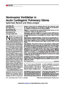
E-Pod nith Elect roiic Cap Attached
E-Pod Electrode Cap Lah Simulator with E-Pod Inserted FIGURE 1. E-Pod and MK63 laboratory simulator. The distance between the outer electrodes on the MK63 was 2.1 inches, and the minimum distance between the inner electrodes was O..35 inches. The total mass of the experimental device was 1.5 kg. and the device moved freely on the vertical axis, to produce constant pressure on the target area despite movements resulting from muscular contractions.
ter (Palco Mediaid, Torrance, California), using either an oral or rectal probe to monitor heart rate, tissue oxygen saturation, and body temperature.
Test Device The MK63 stun baton (Aegis Industries. Bellevue, Idaho), specifically the component containing the electronic circuitry for generating EMI discharges (e-pod), was studied. This device is representative of the class that evokes EMI by using similar principles of high-voltage and time-varying DC. The e-pod was incorporated into a custom-made laboratory apparatus fashioned from 1.25-inch- and 2-inch-diameter polyvinyl chloride pipe and supplemental weights (Fig. I). The smaller pipe with the e-pod could slide freely up and down within the larger pipe while maintaining uniform downward force resulting from a final total mass of 1.5 kg. A 12.0-VDC, 800-mA power supply was used as the power source.
Experimentai Configuration and EMi Discharges In dorsal recumbence, all four limbs of the animal were restrained to the table in a position of moderate extension. The e-pod was placed over the left anterior thigh just inferior to the inguinal ligament, and discharges were administered with the electrodes oriented parallel to the femur and thus to the muscle fibers of the underlying quadratus muscles. The contralateral thigh was used as an intra-animal control. The e-pod was discharged continuously for the 10-. 20-. and 40-second groups but was discharged in two 40-second periods separated by a 10-second rest for the 80-second group. The ventilator was shut off during the discharges, but spontaneous breathing was permitted.
Cardiac Monitoring and Biood Sampies Cardiac rhythtn was tnonitored continuously during anesthesia by using a five-lead electrocardiogram and monitor (Datex Instruments. Helsinki. Finland), and electrocardiograms were recorded during the discharge. There were eight time points
MILITARY MEDICINE, Vol. 173. February 2008
Effects of MK63 Stun Device Discharges
(before discharge [time 0] and 5, 10, 15, 30, and 60 minutes and 24, 48, and 72 hours after discharge) at which central venous blood was drawn from the precaval venous complex, vital signs (tissue oxygen saturation, heart rate, and blood pressure) were recorded, and electrocardiogram strips were printed. Animals were humanely euthanized according to American Veterinary Medical Association standards after the 72 hours of monitoring. Immediately after drawing, each blood sample was placed in beparinized and plain vacuum tubes. The heparinized blood was tested by using an iSTAT analyzer (Abbott Pointof-Care, Abbott Park, Illinois) with CG8 + , CG4+, creatinine, and troponin I (Tnl) cartridges. These cartridges return data on a variety of parameters, including pH, carbon dioxide pressure (PCO2), and bicarbonate, lactate, potassium, Tnl. and creatinine levels. Blood samples were centrifuged (3.000 X g for \5 minutes at 4°C). plasma and serum were aliquotted into 400-/LIL microcentrifuge tubes, and samples were stored at -85°C until use. Serum samples from each time point were thawed once and assayed for creatine kinase (CK)-MB and myoglobin by using microplate enzyme-linked immunosorbent assays, according to the manufacturer's protocol (Diagnostic Automation, Calabasas, California).-^
Tissue Histological Analyses and Electromyography hnmediately after eutbanasia, tissue biopsies were obtained from the skin at the discharge site, from skeletal muscle underlying this site, and from the fetnoral nerve. Biopsies were also obtained from these tissues in the contralateral unshocked leg. Tissue samples were immersed in 10% buffered formalin, embedded in paraffin, sectioned, and stained with bematoxylin and eosin. Sections were evaluated for possible pathological changes. Compound muscle action potentials were studied by using electromyography (Dantec Instruments, Skovlunde, Denmark) with Ag/AgCl surface electrodes (Neotrode; Conmed, Utica, New York).-' These were placed over the rectus femoris belly and tendon, and stimulatory pulses were delivered cutaneously over the femoral nerve,
rants of the body. These contractions continued tbrougbout tbe discharge but became less intense after 20 to 25 seconds of discharge.
Core Body Temperature Decreased after EMI Discharges As shown in Figure 2, a 1.0°C to 1.5°C decrease in body temperature was observed in the 60-minute postdischarge period. This decrease was significant for all groups except the 20-second discharge group (j) < 0.01 or p < 0.05; ANOVA with Tukey's post hoc tests or linear regression). Body temperature normalized in 24 to 72 hours. These changes in temperature occurred despite the use of heating pads, heat lamps, and warm water blankets but were not seen in sbamtreated control animals tbat were exposed to the satne regimen of sedation, anesthesia, intubation, and monitoring.
Vital Signs Were Not Affected by EMI Discharges Heart rate, blood pressure, and pulse oximetry were not significantly affected by MK63 discharges. These data indicated no changes that might suggest pain perception.
Wo Evidence of Significant Acute Dysrhythmia or Myocardial Injury Was Found Electrocardiogram strips taken before, during, and after discbarge of the MK63 on the thigh showed no acute changes in cardiac rhythm at any time (Fig. 3). Despite electronic and motion artifacts seen during the discharges, electrocardiograms showed continued regular ventricular contractions during all discharges, and no dysrhythmias were observed. Mean CK-MB levels were not significantly affected in the experimental groups, compared with control or baseline values (Fig. 4). Mean Tnl values increased at the 24-hour time point in the control group (0.023 ± 0.019 ng/mL) and in the 40-second (0.040 ± 0.030 ng/mL) and 80-second (0.023 ± 0.015 ng/ mL) discharge groups (Fig. 5). These increases in experimen-
Body Temperature - • - Control •-O- 80 sec - V- 40 sec •O-- 20 sec
O
Statisticai Analyses
£ 37
All data points represent means ± SEMs. Reference or normal values for each parameter were drawn from published data.-"" -'' One-way or two-way analysis of variance (ANOVA) followed by Tukey's post hoc tests, paired or unpaired t tests, or regression analyses were used to compare parametric data. Groups were compared by using Prism and InStat software (GraphPad Software, San Diego, California).
(0
RESULTS Discharges from the MK63 caused strong, tetanic, muscle contractions pritnarily in extensor muscle groups in all quad-
MILITARY MEDICINE, Vol. 173, February 2008
--D -10 sec
3635-
34 0 5 1015
30
60
1
2
3
Time Post-Shock (min // days) FIGURE 2. Body temperatures before and after EMI discharge. Body tenipenitures decreased at u steady rate after discharge, such that 1.0°C to L5°C decreases were evident at 60 minutes for the 10-. 40-, and 8()-second discharge groups. No change in temperature was seen in the sham-treated control group {p < O.OI or p < 0,05; ANOVA with Tukey's post hoc tests and linear regression).
169
Effects of MK63 Stun Device Discharges
Troponin I Concentration
Sham Control
0.09-)
Contro 80 sec - y - 40 sec - 0 - 20 sec -a- 10 sec —•—
0.07-
10 Sec Discharge 0 05"c 0.03I0.01-
20 Sec Discharge
-to Sec Discharge
-0.01 1 2 30 60 0 5 15 Time Post-Shock (min // days)
80 Sec Discharge FIGURE 3. Representative electrocardiograms from ail groups, as seen 5 minutes after discharge or after sham discharge for control animals. Electrocardiograms show sinus rhythm and no change in rate for all groups. No dysrhythmias were observed for any animal at any time.
3
FIGURE 5. Tnl values during the 72-hour period after EMI discharge. At 24 hours, increases in Tnl levels occurred in Ihe dedicated control group (0.023 ± 0.019 ng/mL) and in the 40-second discharge (0.040 ± 0.0.10 ng/mL) and 80-second discharge (0.023 ± 0.015 ng/mL) groups. These increases in experimental groups were not significant, compared with the control group (p = 0.59).
Central Venous pH 7.70 n
CK-MB Concentration ^ ^ Control o 80 sec - ^ - 40 sec -<^ - 20 sec -D -10 sec
18-
—•— Control •o- 80 sec
7.60-
- V- 40 sec - 0- - 20 sec -Q--10 sec
7.50-
7,40-
7.30
15 30 60 1 2 Time Post-Shock (min // days)
3
FIGURE 4. CK-MB concentrations during the 72-hour period after EMI discharge. CK-MB concentrations (mean ± SEM) did not change significantly at any time in any group. There was some variability with time in all groups, but the differences between the group values and their own baseline values were not significant (one-way ANOVA).
tal groups were not statistically significant, compared with the control group {p = 0.59). Tnl values of 0.040 ng/mL represent the upper limit of normal in humans."*^ Tnl levels in all animals returned to baseline at 48 hours.
Miid Acidosis Was Seen after Lengthy EMi Discharges Central venous blood pH decreased at the 5-minute postdischarge time for the 40-second (7.49 ± 0.03 to 7.47 ± 0.03) and 80-second (7.48 ± 0.03 to 7.42 ± 0.04) groups (Fig. 6). The latter change was statistically significant (p < 0.03), although all values were still within nortnal ranges. Blood pH returned to baseline after 60 minutes. Control animals had a significantly higher pH (p < 0.01) over the initial 60-minute time period. Central venous pCOj was not significantly changed by EMI discharges. A small increase in pCO^ (36.6 ± 2.6 mm Hg) was seen at 5 minutes after discharge in the 80-second
170
30 0 5 15 60 1 2 Time Post-Shock (min // days)
3
FIGURE 6. Central venous pH over time for control and experimental groups. Experimental animals in the 40-second and 80-second groups showed small pH decreases 5 minutes after EMI discharge. The latter change was statistically significant ip < 0.05), although the observed values for all groups were within normal ranges. Central venous blood pH returned to baseline by 60 mitiutes after discharge.
group, but this was not significant in comparison with the baseline value of 34.4 ± 2.7 mm Hg. All bicarbonate levels were within the normal reference range. Baseline lactate values showed small (p < 0.05) increases at the 5-minute time point for the 40-second and 80-second groups (Fig. 7). Experimental anitnals showed significantly higher lactate levels, compared with control animals, over the initial 60-minute monitoring period (p < 0.05; one-way ANOVA). Lactate levels returned toward baseline values at 24 hours.
EMi Discharges Had Minor Effects on Eiectrolyte Leveis Sodium, potassium, and creatinine levels were not affected by EMI discharges. Sodium levels increased slightly in the 20second discharge group, from a baseline value of 139 mmol/L to 146 mmol/L at 30 minutes after discharge (p < 0.05; paired (test). However, values remained within normal physiological limits at all time points in all animals.
MILITARY MEDICINE, Vol. 173. February 2008
Effects of MK63 Stun Device Discharges
Lactate Concentration 5-,
-•— •o - V-o-
Control 80 sec 40 sec 20 sec
- • • • 10 sec
0 5
15
30
60
1
2
3
Time Post-Shock (min // days) FIGURE 7. Lactate levels over time for control and experimenlal gniups. Lactate levels showed smdl increases at 5 minules in the 40-second and SO-sectind discharge groups. These increases were not statistically significant (j? > 0.05). compared with baseline values. As a group, the experimental animals showed signiticantly higher lactate levels, compared with control animals, over Ihe inilial AO-minute monitoring period (p < 0.05; one-way ANOVAI. These values returned to baseline al 24 hours.
EMI Discharges Had Limited Effects on Serum Myogiobin Levels Mean myogiobin levels in the 20-, 40-, and 80-second experimental groups did not change relative to the baseline value tor each group. In the 10-second group, mean myoglobin levels increased at the 30- and 60-minute time points (65 ng/mL and 61 ng/mL, respectively), compared with the baseline value for this group (32 ng/mL). These differences were signilicant (p < 0.05; paired / test). Elevated myoglobin values in this group returned to baseline levels at 24 hours and remained there subsequently.
baseline values. In contrast, studies with the Taser X26 in anesthetized swine reported prolonged tachycardia after dischaige.''^ This effect was also reported in studies of healthy hutnan volunteers, where the response was ascribed to intense pain associated with tbe discharge.'^'" Because tachycardia was nearly absent in our model, either a deep plane of anesthesia completely suppressed pain or the MK63 did not cause enough metabolic acidosis to result in compensatory elevation of the heart rate. Discbarges frotn the MK63 did not appear to interfere with or capture cardiac rhythm. Sudden deaths associated with Taser discharges in hutnans may restilt from direct or indirect injuries to the myocardium, which then lead to dysrhythmia.'^ Two cardiac markers, CK-MB and Tnl, were assayed here to assess myocardial injury. There were no elevations in CK-MB levels after 80-second MK63 discharges, and Tnl levels showed small but insignificant increases. Tnl is released frotn cardiac myocytes^' when their cell membranes are damaged. Release of Tnl from both human and swine cardiac myocytes peaks 18 to 24 hours after tbe injury and tben gradually decreases to normal over the course of the next several days.-**-'' The time-related pattern and magnitude of Tnl increases seen here were very different from those seen in humans or swine with myocardial inj
Blood Gas Data
Cardiac Effects
Sudden or delayed deaths associated with EMI discbarges may result from cardiac instability attributable to EMl-induced lactic acidosis,*^ exacerbated by greatly elevated body temperature associated with a posttilated condition known as excited delirium.'" No evidence of severe acidosis was seen in the present study. There were minor fluctuations in acidbase status, but none of the changes was statistically or clinically significant. Similarly, pCO,. lactate levels, and bicarbonate levels showed either no changes or small transient increases that correlated with the obsen'ed changes in blood pH. Furthermore, a signilicant decrease in body temperature was observed here during the 6()-minute postdischarge period. This short-tenn thermal dysregulation may bear some relationship to the excited delirium phenomenon but suggests that MK63 exposure may actually promote short-term hypH)thermia and not hyperthermia. Although this temperature decrease did not seem to have any lasting effect in our model, the causes and consequences of such dysregulation deserve further study with this and other stun devices. In particular, time points between 60 minutes and 24 hours after discharge should be examined.
Case repotts, autt)psies. and retrospective analyses indicate that EMI discbarges can be associated with fatal ventricular fibrillation in humans, altbough the frequency of this complication is extremely low.^^ In the present study, no changes in cardiac rhythm were seen even after lengthy MK63 exposures, and no animals died during the 72-hour monitoring period. Experimental animals maintained a mean heart rate that increased slightly after discharge but returned rapidly to
Tbe results of our study were largely at variance with those of a recent Air Force study by Jauchem et al.'"* that examined the effects of discharges from a Taser X26 in swine. In that study, severe acidosis (pH of <7.0) was seen immediately after EMI exposure and was accompanied by dramatic hypercapnia (pCO, of >1()() mm Hg) and lactate level elevation (>I5 mmol/L). There were, however, a number of methodological differences between our study and that
EMI Discharges Had Limited Effects on Electromyographic Responses and Tissue Histological Findings No effect on tissue structure was observed for skeletal muscle or peripheral nerve. In some skin biopsies, minor, highly localized, inflammatory changes, including some interstitial edema, intracellular vacuolization, or inflammatory cell inflltration, were seen at tbe discharge site. M-wave latency and amplitude were unaffected in tbe shocked limb and the contralateral control limb. M-wave area showed small increases during the 60-minute postdischarge period and retumed to baseline subsequently, DISCUSSION
MILITARY MEDICINE, Vol. 173, February 2008
171
Effects of MK63 Stun Device Discharges
of Jauchem et al.." including the anesthetic agents used and methods of physiological support, which may account for these disparities. Most importantly, the devices used in each study generate different waveforms, although both ptoduce an EMI response. The Taser X26 used by Jauchem et al.''' delivers DC pulses at a voltage of -^50 kV, with a pulse duration of 140 microseconds, a frequency of 19 Hz, and power of 0.36 J per pulse.*" The MK63 device used here also delivers DC pulses but at a lower voltage (—19 kV), with pulse dutations of 15 microsecotids at 65 Hz and power of 0.08 J per pulse (C. Hathcock, personal communication). Differences in the device designs or the discharge patterns tnight account for discrepancies between these two studies, Electrical injuries can cause severe muscle and deep-tissue injury through a combination of joule heating and membrane electn)potalion.^' Tissue biopsies taken at 72 hours after discharge showed no evidence of skeletal muscle or peripheral nerve datnage. Altht)Ugh a transient increase in myoglobiti levels was observed in the 10-second discharge group during the 60-minute postdischarge period, this resolved at later times and no other elevations in myoglobin levels occurred in other groups. Electromyographic data gave no indication of serious muscle injury; instead, only minor transitory effects were seen. Our model for studying the MK63 using anterior thigh discharges has some inherent limitations. (1) For ethical reasons, anesthesia was used. Anesthesia precludes pain perception, which is one of the two principal effects of stun device discharges in conscious humans. Pain perception would undoubtedly alter some of the responses reported here. (2) In the field, stun devices are used to subdue cotnbative individuals, who are usually in a state of elevated sympathetic activity and often are under the influence of alcohol or other drugs that may alter the thresholds for dysrhythmia and pain. We have not yet studied the effects of stimulants in this system, although others'^-' have reported the effects of cocaine and epinephrine in swine exposed to Taser discharges, with conflicting results. (3) Thigh discharges were used instead of thoracic discharges over the heart. The importance of the di.scharge vector and proximity to the heart is not completely understood for the MK63. Recently, however, we showed that even lengthy discharges from the MK63 administered to the chest wall directly over the heart did not cause dysrhythmia in swine.-*If we speculate on the relevance of these data obtained in anesthetized resting swine, relative to effects in conscious agitated humans, we must do so with the understanding that there are implicit limitations (see above). With this in mitid, several fairly direct extrapolations are possible. First, the MK63 did not have any significant effect on cardiac function despite the administration of lengthy (80-second) discharges, whereas others'' '^ reported that Tasers can have significant effects on cardiac rhythm in swine, even to the point of triggering ventricular fibrillation. Second. MK63 discharges caused sotne acidosis in swine but this did not generate
172
cardiac arrhythmias. This finding and the findings of Jauchem et al.'^ suggest that stun device-induced acidosis, which can be profound, may not be related to cardiac arrhythmias occurring in healthy individuals with initially normal acid-base status. Third, the decrease in core body temperature seen here after MK63 discharges may actually serve to counter-balance the temperature elevations often seen in agitated individuals. In sutTimary. contrary to our hypothesis, no evidence of acute dysrhythmias, myocardial damage, severe acidosis, skeletal tiiuscle or nerve injury, or electrolyte and biochemical abnormalities were seen in the present study. Because this differs from findings with the Taser X26 in swine,'^ our findings may be attributable to the unique waveform and pulse power generated by the MK63. dilfcrences in the electrode spacing for the MK63 compared with the Taser X26, or differences between the model systems. Further studies are needed to distinguish amotig these possibilities and to develop guidelines and treatment protocols for the growing nutnbcr of individuals exposed to EMI devices. We conclude that, within the limitations of this study, discharges from the MK63 administered on the lower extremity do not appear to cause any measurable neuromuscular or cardiac injury in this swine model.
ACKNOWLEDGMENTS We thank the animal facility staff members for their valuable assistance. This work was suppotied by Aviicet Polymer Technologies (Plainfield, Illinois).
REFERENCES 1. Blackwell V: Chief questions use of Taser on disabled woman. First Coast News, February- 12. 2007. Available at http://www.firstcoastnews. coin/news/news-article.aspx'?storyid=757y5. 2. Battershill P. Naiighton B. Laur D. Panton K. Massine M. Anthony R: Taser Technology Review: Final Report. OPCC File 2474. Vancouver. Canada. Pathlinder Forum, 2(X)5. Available at www.cprc.org/docs/ bcopcc_linal.pdf. 3. McDaniol WC. Stratbucker RA. Nerheim M. Brewer JE: Cardiac safety of neuromuscular incapacitating defensive devices. Pacing Clin Electrophysiol 2(X)5; 2K{Suppl 1): S2S4-7. 4. Bleetman A. Steyn R: The advanced Taser: a medical review. Ta.ser International 2003. Available at http://www.taser.com/research/Science/ Documents/Bleetman%20TASER%2(Kafety.pdf. 5. Amnesty International: Canada: inappropriate and excessive use of tasers. Available at http://web.amnesty.org/en/alfresco_asset/6c6342eO-a2bf-11 dc8d74-6l45!'3y9S4c5/amr2(KX)22()()7en.pdf. 6. Ward C: Taser death update. Available at http://www.cameronward. com/tasers. 7. Amnesty International: United States of America: excessive and lethal force? Amnesty International's concerns about deaths and ill-treatmeni involving police use of tasers. Available at http://www.amnestyusa.org/ countries/u sa/Ta se r_re po rt. pd f. 8. Sullivan L: Death by excited delirium: diagnosis or coverup'.' Available at http://www,npr.org/tcmplates/stury/storv.php?storyld = 7608386. 9. Stratfon SJ, Rogers C. Brickett K. Gnj/.inski G: Factors associated with sudden death of individuals requiring restraint for excited delirium. Am J Emerg Med 2001: 19: 187-91.
MILITARY MEDICINE, Vol. 173, Febtuary 2008
Effects ofMK63 Stun Device Discharges 10. Liiur D: Excited Deltriiini iind Iis Corrckitioii to Sudden and Unexpected Death Proximal lo ResLmim. Ottawa. C:in!i(J:i. Canadian Police Research Centre. 2(H)4, Available al http://www.cprc.orgAr/tr-2(K)5-()2_e.pdf. 11. Ordog GJ. Wasserberger J. Schlater T. Bala.subramanium S; Electronic gun iTaser) injuries, Ann Emerg Mcd 1987; 16: 73-8. 12, KDrnbliim RN. Reddy SK: Effects of the Ta.ser in fatalitie,'; Involving police contronlalion, J Forensic Sci 1991; 36: 434-8. 13, Lakkireddy D. Wallick D. Ryschon K, et al: Effects of cocaine intoxication on Ihe threshold for stun gun induction of* ventricular fibrillation, J Am Coll Cardiol 2(H)6: 48: 803-11, 14, Maici- A. Nance P. Price P. et al: Human effectiveness and risk characterization ot electromuscular incapacitation devices. Available at http:// www.taser.com/documents/HECOE_Report_Summary_l01804.pdf; accessed April 9. 2007. 15. Maier A. Nance P. Price P. et al: Human effectiveness and risk characterization of the electromuscular incapacitation device: a limited analysis of the Taser. Available at http://www.iacp.org/re.search/Cuttinglidge/ HECOEReport.pdt"; accessed September 7. 2007. 16. Sherry C. Brown C. Beason C. et ai: An evaluation of the electrical properties and bio-bchavioral effects of four commercially available TASERs and ilie Jaycor Sticky ShiKker, Available at http://slinet.dtic.niii/ cgi-bin/GetTRDoc7AD-ADA4l6553&Location=U2&dcx;=GetTRDoc, pdf: ac-cessed April 9. 2007, 17, Ho JD, Miner JR. Lakkireddy D. Bultman LL, Heegaard WG: Cardiovascular and physiologic effects of conducted electrical weapon discharge in resting adults, Acad Emerg Med 2(MKx 13: 589-9S. 18, Levine SD. Sloane CM. Chan TC. Dunford JV, Vilke GM: Cardiac monitoring of subjects exposed to the taser. J Emerg Med 2007; 33: II.V7. 19. Jaucheni JR. Sherry CJ. Fines DA. Cook MC: Acidosis. lactate. electrolytes, muscle enzymes, and other factors in the blood of 5M,V scrofa following repeated Taser exposures. Forensic Sci !nt 2(H)6: 161: 20-30. 20. Webster JG. Will JA. Sun H. et al: Can Tasers directly cause ventricular tibrillation? IFBMF Proc 2006: 14: 3307-10. 21, Nanthakumar K. Billingsley IM. Masse S, et al: Cardiac electrophysio-
MILITARY MEDICINE, Vol, 173. February 2008
22.
23. 24. 23, 26.
27,
28,
29.
30,
31. 32. 33,
logical consequences of neiiroinuscular incapacitating device discharges. J Am Coll Cardiol 2(K)6: 48: 798-804, Gibler WB, Runyon JP, Levy RC, et al: A rapid diagnostic and treatment center for patients with chest pain in the emergency depanment, Ann Emerg Med 199S: 25: 1-8, Jabre JF, Hackett ER: EMG Manual. Springfield. IL. Charles C. Thomas. 1983, Swindle MM: Surgery. Anesthesia, and Experimental Techniques in Swine, Ames. lA. Iowa State University Press. 1998, Sinclair Research: Clinical Chemistry Values of Sinclair Pigs. Columbia. MO. Sinclair Research. 1991, Apple FS. Ler R. Chung AY, Berger MJ. Murakami MM: Point-of-care i-STAT cardiac troponin I for assessment of patients with symptoms suggestive of acute cortmary syndrome. Clin Chem 2006; 32: 322-5. Apple FS. Christensoii RH. Valdes R Jr. et al: Simultaneous rapid measurement of whole blood niyoglobin. creatine kinase MB, and cardiac troponin I by the triage cardiac panel for detection of niyocardial infarction, Clin Chem 1999; 45: 199-205, Zipes DP. Libby P. Bonow RO. Braunwald E: Braunwald's Hoan Disease: A Text Book of Cardiovascular Medicine, pp 1158-61. Philadelphia. PA. Saunders. 200S, Feng YJ. Chen C. Fallon JT. et al: Comparison of cardiac troponin I, creatine kinase-MB. and myoglobin for detection of acute ischemic niyocardial injury in a swine model. Am J Clin Pathol 1998: 110: 70-7. TASER International: Basii; Electrical Principles. March 12. 2007, Available at www,2,t;Lser,coni/research/science/pages/basicelectricprinciples. aspx. Lee RC: Injury by electrical forces: pathophysiology. manifestations. and therapy. Curr ProbI Surg 1997: 34: bll-lbA. Valentino DJ. Walter RJ. Nagy K. et al: Repeated thoracic discharges from a stun device. J Trauma 2007: 62: 1134-42, Wu J-Y. Sun H. 0 Rourke AP. Huebncr S, Will JA. W^ebster JG: Taser dan-to-hean distance that causes ventricular librillation in pigs. Available at http://www.engr,wisc,edu/bme/faculty/Webster.john/EB20()6Final, pdf; accessed June 21, 2007,
173