The
new england journal
of
medicine
review article
current concepts
Use of the Electrocardiogram in Acute Myocardial Infarction Peter J. Zimetbaum, M.D., and Mark E. Josephson, M.D.
t
he electrocardiogram remains a crucial tool in the identification and management of acute myocardial infarction. A detailed analysis of patterns of ST-segment elevation may influence decisions regarding the use of reperfusion therapy. The early and accurate identification of the infarct-related artery on the electrocardiogram can help predict the amount of myocardium at risk and guide decisions regarding the urgency of revascularization. Electrocardiographic signs of reperfusion represent an important marker of microvascular blood flow and consequent prognosis. The electrocardiogram is also crucial for identifying new conduction abnormalities and arrhythmias that influence both short- and long-term outcome. In this review, we discuss approaches to the interpretation of the electrocardiogram in the clinical management of patients during the first 24 hours after a myocardial infarction.
From the Cardiovascular Division, Department of Medicine, Beth Israel Deaconess Medical Center, Boston. Address reprint requests to Dr. Zimetbaum at the Division of Cardiology, Beth Israel Deaconess Medical Center, 1 Deaconess Rd., Boston, MA 02215, or at
[email protected]. N Engl J Med 2003;348:933-40. Copyright © 2003 Massachusetts Medical Society.
identification of the infarct-related artery The specificity of the electrocardiogram in acute myocardial infarction is limited by large individual variations in coronary anatomy as well as by the presence of preexisting coronary artery disease, particularly in patients with a previous myocardial infarction, collateral circulation, or previous coronary-artery bypass surgery. The electrocardiogram is also limited by its inadequate representation of the posterior, lateral, and apical walls of the left ventricle. Despite these limitations, the electrocardiogram can help in identifying proximal occlusion of the coronary arteries, which results in the most extensive and most severe myocardial infarctions. inferior myocardial infarction
The culprit vessel in inferior myocardial infarction may be either the right coronary artery (in 80 percent of the cases) or the left circumflex artery. Greater ST-segment elevation in lead III than in lead II and ST-segment depression of more than 1 mm in leads I and aVL suggest involvement of the right coronary artery rather than the left circumflex artery (Fig. 1).1 ST-segment elevation in lead III is greater than that in lead II in the presence of infarction involving the right coronary artery because the ST-segment vector is directed toward the right (lead III). The added finding of ST-segment elevation in lead V1 suggests proximal occlusion of the right coronary artery with associated right ventricular infarction.2 Conversely, infarction involving the left circumflex artery produces an ST-segment vector directed toward the left (lead II). In this case, ST-segment elevation in lead III is not greater than that in lead II, and there is an isoelectric or elevated ST segment in lead aVL.3 ST-segment depression in leads V1 and V2 with ST-segment elevation in the inferior leads also suggests involvement of the left circumflex vessel, but this pattern may also be seen in infarction caused by occlusion of a dominant right coronary artery.4 In either circumstance, ST-segment depression in leads V1 and V2 suggest con-
n engl j med 348;10
www.nejm.org
march 6, 2003
933
The
new england journal
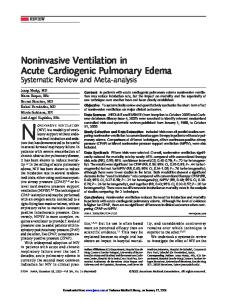
ST-segment elevation in III > ST elevation in II and ST-segment depression in I, aVL, or both (>1 mm)
Yes
No
Right coronary artery Sensitivity 90% Specificity 71% Positive predictive value 94% Negative predictive value 70%
ST-segment elevation in I, aVL, V5, and V6 and ST-segment depression in V1, V2, and V3
In addition, ST-segment elevation in V1, V4R, or both
Left circumflex coronary artery Sensitivity 83% Specificity 96% Positive predictive value 91% Negative predictive value 93%
Proximal right coronary artery with right ventricular infarction Sensitivity 79% Specificity 100% Positive predictive value 100% Negative predictive value 88%
of
medicine
occlusion of the left anterior descending coronary artery. ST-segment elevation in these three leads and in lead aVL in association with ST-segment depression of more than 1 mm in lead aVF indicates proximal occlusion of the left anterior descending artery (Fig. 2).6,7 In this case, the ST-segment vector is directed upward, toward leads V1, aVL, and aVR, and away from the inferior leads. ST-segment elevation in leads V1, V2, and V3 without significant inferior ST-segment depression suggests occlusion of the left anterior descending artery after the origin of the first diagonal branch.6 ST-segment elevation in leads V1, V2, and V3 with elevation in the inferior leads suggests occlusion of the left anterior descending artery distal to the origin of the first diagonal branch, in a vessel that wraps around to supply the inferoapical region of the left ventricle.6 New right bundle-branch block with a Q wave preceding the R wave in lead V1 is a specific but insensitive marker of proximal occlusion of the left anterior descending artery in association with anteroseptal myocardial infarction6 (Fig. 2). left bundle-branch block
Figure 1. Algorithm for Electrocardiographic Identification of the Infarct-Related Artery in Inferior Myocardial Infarction. Data on sensitivity, specificity, positive predictive value, and negative predictive value are from Zimetbaum et al.,1 Herz et al.,2 Bairey et al.,3 Hasdai et al.,4 and Lopez-Sendon et al.5
comitant infarction of the posterior wall of the left ventricle. right ventricular myocardial infarction
Right ventricular myocardial infarction is always associated with occlusion of the proximal segment of the right coronary artery. The most sensitive electrocardiographic sign of right ventricular infarction is ST-segment elevation of more than 1 mm in lead V4R with an upright T wave in that lead. This sign is rarely present more than 12 hours after the infarction. As discussed above, ST-segment elevation in lead V1 in association with ST-segment elevation in leads II, III, and aVF (with greater elevation in lead III than in lead II) is highly correlated with the presence of right ventricular infarction1,5 (Fig. 1).
Spontaneous or pacing-induced left bundle-branch block can obscure the electrocardiographic diagnosis of acute myocardial infarction. In the presence of left bundle-branch block or a right ventricular paced rhythm, right ventricular activation precedes left ventricular activation; this activation of the infarcted left ventricle occurs later and is obscured within the QRS complex. Thus, Q waves cannot be used to diagnose infarction. An indicator of myocardial infarction in the presence of left bundle-branch block is primary ST change — that is, ST deviation in the same (concordant) direction as the major QRS vector. Concordant ST changes in the presence of left bundle-branch block include ST-segment depression of at least 1 mm in lead V1, V2, or V3 or in lead II, III, or aVF and elevation of at least 1 mm in lead V5. Extremely discordant ST deviation (>5 mm) is also suggestive of myocardial infarction in the presence of left bundle-branch block.8
electrocardiographic predictors of reperfusion
The management of acute myocardial infarction is targeted toward restoration of blood flow in the inmyocardial infarction of the anterior wall farct-related artery. There is increasing evidence that In myocardial infarction of the anterior wall, ST- the presence of normal epicardial blood flow does segment elevation in leads V1, V2, and V3 indicates not always correlate with microvascular perfusion
934
n engl j med 348;10
www.nejm.org
march 6 , 2003
current concepts
ST-segment elevation in V1, V2, and V3
ST-segment elevation in V1 (>2.5 mm) or right bundle-branch block with Q wave or both
ST-segment depression (>1 mm) in II, III, and aVF
ST-segment depression (≤1 mm) or ST-segment elevation in II, III, and aVF
Proximal left anterior descending artery Sensitivity 12% Specificity 100% Positive predictive value 100% Negative predictive value 61%
Proximal left anterior descending artery Sensitivity 34% Specificity 98% Positive predictive value 93% Negative predictive value 68%
Distal left anterior descending artery Sensitivity 66% Specificity 73% Positive predictive value 78% Negative predictive value 62%
Figure 2. Algorithm for Electrocardiographic Identification of the Infarct-Related Artery in Anterior Myocardial Infarction. Data on sensitivity, specificity, positive predictive value, and negative predictive value are from Engelen et al. 6
of the myocardial tissue.9 The absence of tissue perfusion is the most potent predictor of impaired ventricular function and the risk of death after myocardial infarction.9 Conversely, resolution of ST-segment elevation is believed to be an excellent marker of tissue perfusion, and the degree of resolution has proved to be a powerful indicator of shortterm (30-day) and long-term (1-year) prognosis.10,11 Assessment of ST-segment resolution is also useful for guiding reperfusion therapy: the absence of ST-segment resolution during the first 90 minutes after the administration of fibrinolytic medications should prompt consideration of rescue angioplasty. A reduction in ST-segment elevation by more than 70 percent in the leads with maximal elevation is associated with the most favorable outcomes.12-14 In the future, therapies that promote microvascular blood flow after restoration of blood flow in the infarct-related artery may become available. The simplicity of assessing ST-segment resolution will probably make this step an important component of the decision to administer such therapies. Other electrocardiographic markers of reperfusion include T-wave inversion within four hours after myocardial infarction. T-wave inversion that occurs during the first few hours of reperfusion therapy is a highly specific sign of reperfusion.15 T-wave inversion that develops more than four hours after the start of reperfusion therapy is consistent with the normal electrocardiographic evo-
n engl j med 348;10
lution of myocardial infarction and does not indicate that reperfusion has occurred. An accelerated idioventricular rhythm (defined as a heart rate of 60 to 120 beats per minute initiated by a late, coupled, ventricular premature depolarization) is a highly specific marker of reperfusion.16,17 This rhythm is benign and should not be suppressed with medication. Isolated ventricular premature depolarizations may also be seen with reperfusion. Polymorphic ventricular tachycardia and ventricular fibrillation may be seen with reperfusion but are rare and should raise the suspicion of ongoing arterial occlusion.
arrhythmias and conduction disease in acute myocardial infarction Conduction abnormalities, including bundle-branch block or varying forms of heart block during acute myocardial infarction, may be associated with a poor prognosis.18-23 The incidence of conduction abnormalities associated with acute myocardial infarction has diminished in the era of early revascularization therapy, but the mortality and morbidity associated with these abnormalities remain unchanged.23 The presence and clinical significance of different types of bradyarrhythmias and conduction disease depend on the location of the infarct and the mass of the involved myocardium. An understanding of the bradyarrhythmias and
www.nejm.org
march 6, 2003
935
The
new england journal
conduction disease that may be associated with acute myocardial infarction requires a review of the anatomy and blood supply of the conduction system. The sinus node is supplied by the right coronary artery in 60 percent of people and by the left circumflex artery in 40 percent. The atrioventricular node is supplied by the right coronary artery in 90 percent of people and by the left circumflex artery in 10 percent. The bundle of His is supplied by the atrioventricular nodal branch of the right coronary artery, with a small contribution from the septal perforators of the left anterior descending artery.11 The bundle of His divides into the right and left bundle branches in the interventricular septum. The right bundle branch receives most of its blood from septal perforators of the left anterior descending artery. There may also be collateral blood supply from the right coronary artery or left circumflex artery. The proximal left bundle branch divides into the left anterior fascicle and the left posterior fascicle. The left anterior fascicle is supplied by septal perforators from the left anterior descending artery and is particularly susceptible to ischemia or infarction. The proximal portion of the left posterior fascicle is supplied by the atrioventricular nodal artery (i.e., the right coronary artery) and by septal perforators of the left anterior descending artery. The distal por-
of
medicine
tion of the posterior fascicle has a dual blood supply from the anterior and posterior septal perforating arteries.
inferior my ocardial infarction Conduction abnormalities in association with inferior myocardial infarction can occur immediately or hours or days after infarction. Sinus bradycardia or varying degrees of atrioventricular block (including complete heart block) can occur within the first two hours after an acute inferior myocardial infarction as a result of heightened vagal tone. Such conditions often resolve within 24 hours,24 and they are very responsive to atropine. Later in the course of inferior myocardial infarction, progressive conduction delay and block may occur. Their spontaneous resolution is regressive, as third-degree atrioventricular block becomes second-degree and then first-degree block and finally resolves, with normal conduction ensuing. This phase of atrioventricular conduction problems appears to be related to edema and local accumulation of adenosine.25 It is less responsive to atropine than the acute phase and may respond to aminophylline.25 The atrioventricular node is the site of conduc-
I
aVR
V1
V4
II
aVL
V2
V5
III
aVF
V3
V6
II
Figure 3. Electrocardiogram Showing Inferior Myocardial Infarction Associated with Complete Heart Block with a Narrow Escape Rhythm. There is ST-segment elevation in lead III that is greater than the ST-segment elevation in lead II, marked ST-segment depression in leads I and aVL, and ST-segment elevation in lead V1 — all consistent with the occurrence of proximal occlusion of the right coronary artery in association with right ventricular infarction. If the patient is in hemodynamically stable condition, a temporary pacemaker is not required.
936
n engl j med 348;10
www.nejm.org
march 6 , 2003
current concepts
tion disturbances in inferior myocardial infarction; therefore, complete atrioventricular block is generally associated with a narrow complex escape rhythm of between 40 and 60 beats per minute (Fig. 3). It is usually asymptomatic but may be associated with hemodynamic instability due to loss of atrioventricular synchrony. It is generally transient and resolves within five to seven days but may persist for up to two weeks. A ventricular escape rhythm with a widened QRS complex may signify the presence of block below the atrioventricular node and impaired collateral circulation to an occluded left anterior descending artery. The management of conduction abnormalities associated with myocardial infarction depends on the associated symptoms. As noted above, bradyarrhythmias during the first few hours after an acute inferior myocardial infarction are responsive to atropine; conduction disease that begins or persists after the first 24 hours of myocardial infarction is not responsive to atropine. If the patient has hemo-
dynamic instability, worsening ischemia, or ventricular arrhythmias, a temporary pacemaker should be used. Placement of temporary pacing wires in the right atrium (or coronary sinus) and right ventricle allows restoration of atrioventricular synchrony and improves hemodynamic function. In most instances, conduction abnormalities associated with acute inferior myocardial infarction resolve within two weeks, and permanent pacing is not required (Table 1). anterior myocardial infarction
As opposed to inferior myocardial infarction, conduction disease associated with anterior myocardial infarction is not related to heightened vagal tone but instead to necrosis of the intramyocardial conduction system. This condition occurs almost exclusively in the presence of proximal occlusion of the left anterior descending artery and septal necrosis. PR prolongation in acute anterior myocardial infarction is rarely due to atrioventricular nodal ischemia, since in most people the atrioventricular node is
Table 1. Guidelines of the American College of Cardiology and the American Heart Association for Temporary or Permanent Implantation of Pacemakers in Patients with Acute Myocardial Infarction.* Class†
Indications for Temporary Pacing
Indications for Permanent Pacing
I
Asystole Symptomatic bradycardia (including sinus bradycardia or Mobitz type I block with hypotension) Bilateral BBB (alternating BBB or right BBB alternating with LAFB or LPFB) Bifascicular block that is new or of indeterminate age (right BBB with LAFB or LPFB or left BBB) with a prolonged PR interval Mobitz type II second-degree AV block
Persistent second-degree AV block in the His– Purkinje system, with bilateral BBB or thirddegree AV block within or below the His– Purkinje system after myocardial infarction Transient advanced (second- or third-degree) infranodal AV block and associated BBB‡ Persistent and symptomatic second- or thirddegree AV block
IIa
Right BBB and LAFB or LPFB that is new or of indeterminate age Right BBB with a prolonged PR interval Left BBB that is new or of indeterminate age Recurring sinus pauses not responsive to atropine
None
IIb
Bifascicular block of indeterminate age Isolated right BBB that is new or of indeterminate age
Persistent second- or third-degree AV block at the level of the AV node
III
Prolonged PR interval Type 1 second-degree AV block with normal hemodynamics Accelerated idioventricular rhythm BBB or fascicular block known to exist before acute myocardial infarction
Transient AV conduction disturbances in the absence of intraventricular conduction defects Transient, isolated AV block in the presence of isolated LAFB Acquired LAFB in the absence of AV block Persistent first-degree AV block in the presence of BBB that is old or of indeterminate age
* The information is adapted from Ryan et al.26 and Gregoratos et al.27 AV denotes atrioventricular, BBB bundle-branch block, LAFB left anterior fascicular block, and LPFB left posterior fascicular block. † Class designations refer to the level of evidence supporting the effectiveness of the procedure or treatment, where class I indicates that the evidence is very strong and class III that it is absent or that the procedure is not useful and may be harmful. ‡ An electrophysiological study may be useful to determine the site of the block.
n engl j med 348;10
www.nejm.org
march 6, 2003
937
The
new england journal
supplied by the right coronary artery. More commonly, necrosis of the septum is associated with slight PR prolongation (usually <0.25 second) due to involvement of the conducting system below the atrioventricular node. In this situation, PR prolongation is often associated with a wide QRS complex (>0.12 second) with a right bundle-branch block pattern. Second-degree atrioventricular block with anterior myocardial infarction is usually Mobitz type II block secondary to block in the His–Purkinje system. Complete heart block results from extensive necrosis of the ventricular septum. It usually occurs abruptly during the first 24 hours after myocardial infarction and is almost always preceded by the development of right bundle-branch block with rightor left-axis deviation (Fig. 4). In right bundle-branch block associated with anterior myocardial infarction, a Q wave precedes the R wave in lead V1 (QR in lead V1). Both the left anterior fascicle and the right bundle branch are supplied by the septal branches of the proximal left anterior descending artery. Anteroseptal infarctions may be associated with the development of new right bundle-branch block, with left anterior (or, less commonly, left posterior) fascicular block. The development of bifascicular
of
medicine
block with anteroseptal infarction is associated with as much as a 30 percent excess risk of complete heart block.18,21 The addition of PR prolongation to bifascicular block further increases this risk of complete heart block. The mortality associated with complete heart block in anterior myocardial infarction, with or without preceding right bundle-branch block and left fascicular block, may be as high as 80 percent.18 This mortality rate is largely related to progressive pump failure as a result of extensive myocardial necrosis. A temporary pacemaker should be placed in patients with anterior infarction and new right bundle-branch block (QR in lead V1) with left anterior or left posterior fascicular block if there is associated PR prolongation (Table 1). Another indication for temporary pacing during anterior myocardial infarction is alternating right and left bundle-branch block.
tachy arrhythmias Tachyarrhythmias that occur during acute myocardial infarction may result from reperfusion, altered autonomic tone, or hemodynamic instability. Sinus tachycardia generally results from heightened adrenergic tone and is usually a manifestation of he-
I
aVR
V1
V4
II
aVL
V2
V5
III
aVF
V3
V6
II
II
II
II
Figure 4. Electrocardiogram Showing Anterior Myocardial Infarction Associated with Right Bundle-Branch Block with a QR Pattern and ST-Segment Elevation. Left anterior fascicular block and slight PR prolongation are also present. Together, the findings (which are new in comparison with an electrocardiogram obtained before myocardial infarction) suggest proximal occlusion of the left anterior descending artery. The patient has a substantial risk of complete heart block, and implantation of a temporary pacemaker is indicated.
938
n engl j med 348;10
www.nejm.org
march 6 , 2003
current concepts
modynamic failure. Atrial fibrillation may also occur in acute infarction and may result from increased vagal tone, increased left atrial pressure, atrial infarction, or pericarditis. Atrial fibrillation is associated with a worsened prognosis, regardless of the site of infarction.28 Ventricular premature depolarizations are common during acute myocardial infarction but do not predict the subsequent development of sustained ventricular arrhythmias and should not be suppressed.29 Sustained monomorphic ventricular tachycardia (with a heart rate above 150 beats per minute) is not generally associated with acute myocardial infarction unless there is a preexisting scar in the region that has become ischemic or a very large myocardial infarction.30 Initially, ventricular fibrillation may be seen as an acute manifestation of ischemia; later (at two or three weeks), it may be seen as a consequence of progressive pump dysfunction. The presence of anterior myocardial infarction with right bundle-branch block and an ejection fraction of 35 percent or less is associated with recurrent ventricular tachycardia or fibrillation in the second and third weeks after myocardial infarction. In most instances, ventricu-
lar fibrillation in acute myocardial infarction is associated with lack of reperfusion of the infarct-related artery and should prompt evaluation for cardiac catheterization.31
conclusions Important information to guide management and determine prognosis can be derived from the electrocardiogram in patients with acute myocardial infarction. Electrocardiographic markers of proximal coronary-artery occlusion identify relatively large myocardial infarctions that benefit most from early and complete revascularization strategies, such as primary angioplasty. The degree of ST-segment resolution is a simple and powerful predictor of ventricular function and prognosis after myocardial infarction. Finally, the recognition of abnormalities of conduction that result from different types of myocardial infarction is essential to the proper management of these conditions. We are indebted to William C. Quist, M.D., for his review of the anatomy of the cardiac conduction system.
references 1. Zimetbaum P, Krishnan S, Gold A, Car-
rozza JP II, Josephson M. Usefulness of STsegment elevation in lead III exceeding that of lead II for identifying the location of the totally occluded coronary artery in inferior wall myocardial infarction. Am J Cardiol 1998;81:918-9. 2. Herz I, Assali AR, Adler Y, Solodky A, Sclarovsky S. New electrocardiographic criteria for predicting either the right or left circumflex artery as the culprit coronary artery in inferior wall acute myocardial infarction. Am J Cardiol 1997;80:1343-5. 3. Bairey CN, Shah K, Lew AS, Hulse S. Electrocardiographic differentiation of occlusion of the left circumflex versus the right coronary artery as a cause of inferior acute myocardial infarction. Am J Cardiol 1987;60: 456-9. 4. Hasdai D, Birnbaum Y, Herz I, Sclarovsky S, Mazur A, Solodky A. ST segment depression in lateral limb leads in inferior wall acute myocardial infarction: implications regarding the culprit artery and the site of obstruction. Eur Heart J 1995;16:1549-53. 5. Lopez-Sendon J, Coma-Canella I, Alcasena S, Seoane J, Gamallo C. Electrocardiographic findings in acute right ventricular infarction: sensitivity and specificity of electrocardiographic alterations in right precordial leads V4R, V3R, V1, V2, and V3. J Am Coll Cardiol 1985;6:1273-9. 6. Engelen DJ, Gorgels AP, Cheriex EC, et al. Value of the electrocardiogram in local-
izing the occlusion site in the left anterior descending coronary artery in acute anterior myocardial infarction. J Am Coll Cardiol 1999; 34:389-95. 7. Tamura A, Kataoka H, Mikuriya Y, Nasu M. Inferior ST segment depression as a useful marker for identifying proximal left anterior descending artery occlusion during acute anterior myocardial infarction. Eur Heart J 1995;16:1795-9. 8. Sgarbossa EB, Pinski SL, Barbagelata A, et al. Electrocardiographic diagnosis of evolving acute myocardial infarction in the presence of left bundle-branch block. N Engl J Med 1996;334:481-7. [Erratum, N Engl J Med 1996;334:931.] 9. Ito H, Maruyama A, Iwakura K, et al. Clinical implications of the ‘no reflow’ phenomenon: a predictor of complications and left ventricular remodeling in reperfused anterior wall myocardial infarction. Circulation 1996; 93:223-8. 10. van ’t Hof AW, Liem A, Suryapranata H, Hoorntje JC, de Boer MJ, Zijlstra F. Angiographic assessment of myocardial reperfusion in patients treated with primary angioplasty for acute myocardial infarction: myocardial blush grade. Circulation 1998; 97:2302-6. 11. Matetzky S, Novikov M, Gruberg L, et al. The significance of persistent ST elevation versus early resolution of ST segment elevation after primary PTCA. J Am Coll Cardiol 1999;34:1932-8.
n engl j med 348;10
www.nejm.org
12. Schroder K, Wegscheider K, Zeymer U, Tebbe U, Schroder R. Extent of ST-segment deviation in a single electrocardiogram lead 90 min after thrombolysis as a predictor of medium-term mortality in acute myocardial infarction. Lancet 2001;358:1479-86. 13. Poli A, Fetiveau R, Vandoni P, et al. Integrated analysis of myocardial blush ST-segment elevation recovery after successful primary angioplasty: real-time grading of microvascular reperfusion and prediction of early and late recovery of left ventricular function. Circulation 2002;106:313-8. 14. Angeja BG, Gunda M, Murphy SA, et al. TIMI myocardial perfusion grade and ST segment resolution: association with infarct size as assessed by single photon emission computed tomography imaging. Circulation 2002;105:282-5. 15. Wehrens X, Doevendans P, Ophuis TJ, Wellens H. A comparison of electrocardiographic changes during reperfusion of acute myocardial infarction by thrombolysis or percutaneous transluminal coronary angioplasty. Am Heart J 2000;139:430-6. 16. Gressin V, Louvard Y, Pezzano M, Lardoux H. Holter recording of ventricular arrhythmias during intravenous thrombolysis for acute myocardial infarction. Am J Cardiol 1992;69:152-9. 17. Gorgels AP, Vos MA, Letsch IS, et al. Usefulness of the accelerated idioventricular rhythm as a marker for myocardial necrosis and reperfusion during thrombolytic therapy
march 6, 2003
939
The
new england journal
in acute myocardial infarction. Am J Cardiol 1988;61:231-5. 18. Hindman MC, Wagner GS, JaRo M, et al. The clinical significance of bundle branch block complicating acute myocardial infarction. 1. Clinical characteristics, hospital mortality, and one-year follow-up. Circulation 1978;58:679-88. 19. Idem. The clinical significance of bundle branch block complicating acute myocardial infarction. 2. Indications for temporary and permanent pacemaker insertion. Circulation 1978;58:689-99. 20. Harpaz D, Behar S, Gottleib S, Boyko V, Kishon Y, Eldar M. Complete atrioventricular block complicating acute myocardial infarction in the thrombolytic era. J Am Coll Cardiol 1999;34:1721-8. 21. Sgarbossa EB, Pinski SL, Topol EJ, et al. Acute myocardial infarction and complete bundle branch block at hospital admission: clinical characteristics and outcome in the thrombolytic era. J Am Coll Cardiol 1998;31: 105-10. 22. Go AS, Barron HV, Rundle AC, Ornato JP, Avins AL. Bundle-branch block and inhospital mortality in acute myocardial infarction. Ann Intern Med 1998;129:690-7. 23. Newby KH, Pisano E, Krucoff MW, Green
of
medicine
C, Natale A. Incidence and clinical relevance of the occurrence of bundle-branch block in patients treated with thrombolytic therapy. Circulation 1996;94:2424-8. 24. Feigl D, Ashkenazy J, Kishon Y. Early and late atrioventricular block in acute inferior myocardial infarction. J Am Coll Cardiol 1984;4:35-8. 25. Bertolet BD, McMurtrie EB, Hill JA, Belardinelli L. Theophylline for the treatment of atrioventricular block after myocardial infarction. Ann Intern Med 1995;123:509-11. 26. Ryan TJ, Antman EM, Brooks NH, et al. 1999 update: ACC/AHA guidelines for the management of patients with acute myocardial infarction: executive summary and recommendations: a report of the American College of Cardiology/American Heart Association Task Force on Practice Guidelines (Committee on Management of Acute Myocardial Infarction). Circulation 1999;100: 1016-30. 27. Gregoratos G, Abrams J, Epstein AE, et al. ACC/AHA/NASPE 2002 guideline update for implantation of cardiac pacemakers and antiarrhythmia devices: summary article: a report of the American College of Cardiology/American Heart Association Task Force on Practice Guidelines (ACC/AHA/NASPE
Committee to Update the 1998 Pacemaker Guidelines). Circulation 2002;106:2145-61. 28. Eldar M, Canetti M, Rotstein Z, et al. Significance of paroxysmal atrial fibrillation complicating acute myocardial infarction in the thrombolytic era. Circulation 1998;97: 965-70. 29. Antman EM, Berlin JA. Declining incidence of ventricular fibrillation in myocardial infarction: implications for the prophylactic use of lidocaine. Circulation 1992;86: 764-73. 30. Mont L, Cinca J, Blanch P, et al. Predisposing factors and prognostic value of sustained monomorphic ventricular tachycardia in the early phase of acute myocardial infarction. J Am Coll Cardiol 1996;28:1670-6. 31. Berger PB, Ruocco NA, Ryan TJ, Frederick MM, Podrid PJ. Incidence and significance of ventricular tachycardia and fibrillation in the absence of hypotension or heart failure in acute myocardial infarction treated with recombinant tissue-type plasminogen activator: results from the Thrombolysis in Myocardial Infarction (TIMI) Phase II trial. J Am Coll Cardiol 1993;22:1773-9. Copyright © 2003 Massachusetts Medical Society.
clinical problem-solving series The Journal welcomes submissions of manuscripts for the Clinical Problem-Solving series. This regular feature considers the step-by-step process of clinical decision making. For more information, please see http://www.nejm.org/hfa/articles.asp.
940
n engl j med 348;10
www.nejm.org
march 6 , 2003