Teratogenesis, Carcinogenesis, and Mutagenesis 21:97–106 (2001)
CD44 in Normal Human Pancreas and Pancreatic Carcinoma Cell Lines Jörg Ringel,1,2 Ralf Jesnowski,2 Christian Schmidt ,2 Jens Ringel,3 Hans J. Köhler,2 Joachim Rychly,2 Surinder K. Batra,1 and Matthias Löhr2,4* 1 Department of Biochemistry and Molecular Biology, University of Nebraska Medical Center, Omaha, Nebraska 2 Department of Medicine, University of Rostock, Germany 3 Department of Biochemistry, Friedrich Schiller University of Jena, Germany 4 Department of Medicine IV, Medical Faculty Mannheim, Germany CD44 is an integral cell-surface glycoprotein. Overexpression of the CD44 standard (CD44st) and its variants (CD44v) has been implicated in transformation and progression of many cancer types. Here, we investigated expression of CD44st, CD44v3-7, CD44v7/8, and v10 in five human pancreatic tumor cell lines and normal human pancreatic duct cells transfected with the SV40 large T antigen. CD44st and its variant proteins were quantified using immunocytochemistry and flow cytometry. CD44v7 was expressed at low levels, whereas CD44st, CD44v3, CD44 v4, CD44v, and CD44v6 were expressed at moderate levels in all pancreatic tumor cell lines. In contrast, CD44v7/8 and CD44v10 were expressed at very low levels in two out of the five pancreatic tumor cell lines. Overall, staining of CD44st and CD44 variants was significantly weaker compared to another surface molecule, ICAM-1, reported to be overexpressed in pancreatic cancer cells. Furthermore, the SV40 large T transfected duct cells showed only a weak staining for CD44st, CD44v5, and CD44v6. To determine a possible mechanism for the regulation of surface expression of CD44st, v5 and v6, we incubated Panc-1 cells with bFGF, TGF-β1, EGF, TNFα, and IFNγ. Only IFNγ affected the CD44 expression by downregulation of CD44v6. The constitutive expression of CD44 variants seems to be associated with the malignant state of invasive carcinoma. Teratogenesis Carcinog. Mutagen. 21:97–106, 2001. © 2001 Wiley-Liss, Inc. Key words: CD44; growth factors; pancreatic cancer cell lines; pancreatic adenocarcinoma
Contract grant sponsor: Deutsche Forschungsgemeinschaft (DFG); Contract grant number: Lo431/6-2; Contract grant sponsor: Minister of Science, State of Mecklenburg; Contract grant number: RVFF UR40; Contract grant sponsor: National Institutes of Health, USA; Contract grant number: P50 CA 7271. *Correspondence to: Dr. Matthias Löhr, Department of Medicine IV,Medical Faculty Mannheim, University of Heidelberg, Theodor Kutzer Ufer 1, D-68135 Mannheim, Germany. E-mail: matthias.loehr@ med4.ma.uni-heidelberg.de
© 2001 Wiley-Liss, Inc.
98
Ringel et al.
INTRODUCTION
Altered expression of cell-surface molecules and the overexpression of growth factors and their receptors may be involved in malignant progression and metastasis of human exocrine pancreatic cancer. It has been reported that such growth factors as basic fibroblast growth factor (bFGF), transforming growth factor (TGF)-β1, and epidermal growth factor (EGF) are abundantly expressed in pancreatic cancer [1– 3]. These growth factors may also be involved in the regulation of adhesion molecules supposed to be involved in invasion and metastasis. CD44 is a transmembrane glycoprotein that is implicated in wide variety of functional roles including cell-cell and cell-matrix interactions. It mediates cell differentiation, T-cell activation, cell migration, and metastasis [4,5]. The human CD44 gene is encoded by a gene containing 20 exons. The CD44st standard molecule expresses ten exons, whereas the remaining so-called variant (v) exons can be alternatively spliced in more than 30 different combinations. These variant exons are expressed in the CD44 variant isoforms [4,5]. The alternative splicing as well as different degrees of glycosylation result in a multitude of protein isoforms. Variant isoforms of CD44 were recently described to determine the metastatic potential of cancer cells. In a rat pancreatic cancer model, the transfection of CD44v6 led to metastatic behavior of the non-metastatic parental pancreatic cancer cell line [6]. CD44 expression has been analyzed in a broad of tissue and cancer types [7– 12]. Different CD44variant isoforms are considered to play a critical role in growth and metastatic behavior of various tumors [13]. In addition, in some cancer types, a correlation between CD44v expression and clinical prognosis was observed [7,12]. However, results in different cancer types are controversial [13,14]. The tissue- and cancer-specific expression pattern of CD44 molecules may be the result of specific splicing and regulation processes. The influence of cytokines like interferon (IFN) γ or tumor necrosis factor (TNF) α on the CD44 surface expression has been demonstrated in epithelial kidney and lung cancer cell systems [15]. The “standard“ form of CD44 [CD44st) has been found to be overexpressed in pancreatic tumors (16, 17). Other reported that CD44v5 and CD44v6 are newly expressed in human pancreatic carcinoma and may play role in tumor cell invasion and metastasis (18, 19, 20). However, the data concerning the expression of CD44st and CD44v isoforms in pancreatic cancer are somehow contradictory (20, 21, 22, 23). Little is known about the regulation of the CD44 expression in human pancreatic cancer cells. To clarify the CD44 protein expression, we investigated different human pancreatic cancer cell lines and two SV40 transfected human ductal cell lines using immunocytochemistry and flow cytometry. Besides the semi-quantitative protein expression analysis of CD44st and CD44v in these cell lines, we evaluated the effects of bFGF, TGF-β1, EGF, IFNγ, and TNFα on the expression of the CD44st molecule and the CD44 variant v5 and v6. MATERIALS AND METHODS Cell Lines
Two human SV40 transfected human pancreatic duct epithelial cell lines (PDEC) were used: M540 (provided by F. Real, Barcelona) [24] and E4 (established by our group) [25,26]. The establishment, culture conditions, and cell characterization of
CD44 in Pancreatic Cancer Cell Lines
99
the PDEC were published previously [25,26]. A panel of well-characterized human pancreatic cancer cell lines was used; BxPC-3 (well differentiated) and Panc-1 (poorly differentiated) were purchased from the American Type Culture Collection (ATCC; Rockville, MD). The cell line PaCa-44 (poorly differentiated) was a gift from Dr. J. Mollenhauer (Rush University, Chicago, IL) [27]. The cell lines SW850 and SW979 were obtained from Dr. Kalthoff (University of Kiel, Germany) [28]. All cancer cell lines were routinely grown in DMEM supplemented with 10% fetal calf serum, 100 U/ml penicillin, and 100 µg/ml streptomycin. We also analyzed Panc-1cells stable transfected with full-length cDNA of TGFβ1 under the control of a CMV early promoter (Löhr et al., submitted). This plasmid also codes for the neo resistance gene, such enabling selection of transfectants with the antibiotic G418. As control cells we used MOCK transfected Panc-1 cells. Immunocytochemistry
The following panel of mAbs were used: SFF-2 (CD44st) (Serva, Heidelberg, Germany), anti CD44v3 (R&D), VFF-8 (CD44v5), VFF-18 (CD44v6), VFF-9 (CD44v7), VFF-17 (CD44v7/8), VFF-16 (CD44v10) (all from Serva), anti-ICAM-1 (Roche, Mannheim, Germany), IgG-control antibodies, and FITC-conjugated antimouse Fab (Sigma Chemical Co., St. Louis, MO). Expression of CD44st, CD44v5 and CD44v6 was determined by immunocytochemistry performed by the three-step method with HRP-conjugated secondary and tertiary antibodies as described [26]. All primary antibodies and control antibodies were used in dilutions ranging from 1:10–1:100. FACS Analysis
For flow cytometric analysis of CD44st, CD44 variants v3, v4, v5, v6, v7, v7/8, v10, and ICAM-1 cells were detached with 5mM EDTA solution and washed with PBS; 105 cells were incubated with primary antibodies or with an isotype-matched control mAb for 30 min at 4°C. Then, the cells were washed once and incubated with 100 ml FITC-conjugated secondary antibody for 30 min at 4°C. The determination of the surface expression was performed by measuring 10,000 cells of each sample in a FACScan flow cytometer (Becton Dickinson, Mountain View, CA) by using FACScan Research software [29]. Growth Factor Stimulation
For stimulation experiments Panc-1 cells (106) were grown in culture dishes with DMEM supplemented with 10% FCS for 1 day. Thereafter, the cells were washed and refed with DMEM without FCS containing bFGF (1, 5, 10, and 100 ng/ml). After 24, 48, and 72 h, the cells were detached with 5 mM EDTA. Cells were incubated with specific mAbs and stained with a FITC-conjugated secondary antibody as described above. The effects of IFNγ, TNFα, and EGF were determined by incubation with IFNγ (250 and 500 U/ml), TNFα (100 and 200 U/ml), and EGF (10 and 20 ng/ml) for 24 and 48 h. For stimulation experiments with TGF-β1, native Panc-1, and TGF-β1 transfected Panc-1 cells (106) (transfected cells grown in the presence of 400 µg/ml G418) were grown in culture dishes with DMEM (without FCS) supplemented with 1 or 10 ng/ml TGF-β1 for 48 h. The CD44st and CD44 variant exon v5 and v6 expression was analyzed by FACScan flow cytometer [29]. All experiments were done three times independently.
100
Ringel et al.
RESULTS Immuncytochemical Staining and Flow Cytometric Analysis of CD44st and CD44v
CD44st, v5, and v6 expression may play a critical role in pancreatic cancer. Therefore, the expression patterns of CD44st and of the variant exons v5 and v6 were examined by immunocytochemistry in the pancreatic cancer cell lines BxPC-3 (well differentiated), Panc-1 (poorly differentiated), and the SV40 large T transfected human pancreatic duct cell lines M450 and E4. Both cancer cell lines displayed moderate staining for CD44st (mAb SFF-2), CD44v5 (mAb VFF-8), and CD44v6 (mAb VFF18) (Fig. 1). In contrast, in the two human SV40 transfected pancreatic duct cell lines, M540 and E4, CD44st was expressed only weakly until reaching a moderate level (Fig. 1). The two CD44 variants exhibited only a very weak staining pattern in M540 and E4 cells (Fig. 1). We used flow cytometry to quantify the surface expression of CD44st, v5, and v6 as well as to analyze the expression of the CD44 variants v3, v4, v7, v7/8, and v10 in the pancreatic cancer cell lines. Besides BxPC-3 and Panc-1, the pancreatic tumor cell lines PaCa44 (poorly differentiated), SW979, and SW850 were investigated. The cell lines SW979 and SW850 differ from the other cell lines, because they have no known mutations in p53 and Ki-ras genes, which have been described as occurring with high frequency in pancreatic adenocarcinoma cells [28]. Flow Cytomentry demonstrated CD44st, v5, and v6 in the cell lines BxPC-1 and Panc-1, (Fig. 2, Table 1) that further supported the immunochemical findings.
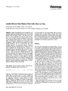
Fig. 1. Immunocytochemistry for CD 44 and variants. Immunocytochemical staining of the human pancreatic carcinoma cell line BxPC-3 (A,B) and the human SV40 immortalized duct cells E4 (C,D) using the antibodies SFF-2 against CD44st (A,C) and VFF-18 against CD44v6 (B,D). Note the intense staining of CD44st and CD44v6 in the BxPC-3 cells (A,B) and the weak expression in the ductal cell line E4 (C,D).
CD44 in Pancreatic Cancer Cell Lines
101
Fig. 2. FACS analysis histograms for CD44st and CD44 variants. The cell line Panc-1 showed a staining for CD44st, v3, v4, v5, v6, and v7. The closed line shows the CD44 antigen and the black diagram is the IgG isotype matched control mAb.
Our analyses revealed that all investigated cell lines, independent of the differentiation grade and the mutations in the p53 and Ki-ras genes, were reactive for CD44st, CD44v3, CD44v4, CD44v5, and CD44v6. The quantitative level of CD44st and v3, v4, v5, and v6 variants varied little between the individual cell lines. The well-differ-
102 Ringel et al.
TABLE I. Expression of CD44st and Variants in the Different Pancreatic Cancer Cell Lines Analyzed by FACS-Analyses*
BxPC-3 Panc-1 PaCa44 SW850 SW979
CD44st
CD44v3
CD44v4
CD44v5
CD44v6
CD44v7
CD44v7/8
CD44v10
ICAM-1
335 158 165 225 236
225 171 156 129 143
230 165 167 99 132
200 173 175 155 204
232 158 225 183 212
46 155 67 65 60
93 0 85 0 0
110 0 99 0 0
410 324 305 320 314
*The fluorescence mean channels represent the fluorescence intensity of the CD44 expression. The relative fluorescence mean channels for CD44 are subtracted from the fluorescence mean channels of the control antibody (IgG). The well differentiated cell line BxPC-3 showed an augmented CD44st staining in comparison to the CD44 variants. ICAM-1 was analyzed as a control. In all cell lines the ICAM-1 expression was higher then the CD44 staining levels. These are the representative results of one of three independent FACS analyses for each cell line.
CD44 in Pancreatic Cancer Cell Lines
103
entiated cell line BxPC-3 showed an augmented CD44st expression in comparison to the CD44 variant exons. Only two pancreatic cancer cell lines, the cell line BxPC3 and the poorly differentiated cell line PaCa44, expressed variant exon v10 weakly. Furthermore, the brightness of staining with the CD44v7 VFF-7 mAb was generally low. The differences in the observed pattern between the CD44v7 VFF-7 mAb and the CD44v7/8 VFF-17 mAb may be determined by the low expression grade, the different binding epitopes, and the binding affinities. The cell surface molecule ICAM-1 has been described to be overexpressed in pancreatic cancer cells [29]. Therefore, we used this molecule as a control for the flow cytometric analyses. However, in all cell lines the fluorescence intensity for CD44st and the variant exons revealed by flow cytometry was significantly lower than the expression level of ICAM-1 (Table I). Regulation of Expression of CD44st and CD44 Variant Exons
Growth factors like bFGF and TGF-β1 and cytokines like TNFα may directly and indirectly affect pancreatic carcinogenesis. Therefore, we investigated the possible regulation effects of bFGF, TGF-β1, EGF, TNFα, and IFNγ on the surface expression of CD44st and CD44v5, and v6 in the human pancreatic cell line Panc-1. IFNγ treatment results in a slight reduction of the CD44v6 surface expression, whereas CD44st and CD44v5 were unchanged (Fig. 3). These results were independent of the IFNγ concentration. Panc-1 cells treated with bFGF, EGF, and Tα showed no changes in the surface expression of CD44st, v5, and v6. Furthermore, incubation with TGF-β1 did not affect the expression (data not shown). In addition, we measured the CD44 protein expression in TGF-β1 transfected Panc-1 cells in comparison to MOCK transfected control cells to investigate the possible effects of a TGF-β1 overexpression. No significant difference was found in TGF-β1 transfected Panc-1 cells compared with the control cells (data not shown).
Fig. 3. CD44 expression in response to IFNγ treatment. Expression of CD44st, CD44v5, and CD44v6 on Panc-1 cells using flow cytometry after incubation with IFNγ (250 U/ml; 500 U/ml) in DMEM without FCS for 48 h. The fluorescence mean channels for CD44v6 as value for the fluorescence intensity were decreased after incubation with IFNγ independent of the concentration. This figure shows the fluorescence mean channels without the reduction of the fluorescence mean channels of the control antibody. The data represents the mean ± SD of three experiments. w/o = without IFN.
104
Ringel et al.
DISCUSSION
Altered expression of adhesion molecules on the surface of cancer cells has been shown to play an important role in cancer growth behavior and in cell migration, invasion, and metastasis [30,31]. CD44 isoforms are known to be involved in various biological processes affecting tumor behavior. These include invasion, metastasis, angiogenesis, cell proliferation, and apoptosis [4,5,32–34]. Furthermore, expression of CD44 molecules, especially CD44 variant forms, was found to be correlated with the clinical outcome of various cancer types [7,12]. Recently, a significant correlation was reported between CD44v6 and a decreased overall survival in pancreatic cancer [22]. Some investigators have found that CD44v5 and CD44v6 are expressed in human pancreatic adenocarcinoma cells but not in normal pancreatic tissue [20,23]. In one study involving a SCID mice metastasizing model, CD44st was found to be downregulated in metastatic cells while a large fraction of pancreatic cancer cells expressed on membrane the splice variants v5/v6 [18]. However, another report showed that CD44v6 is detectable in normal pancreatic tissue as well as in human pancreatic cancer samples [20]. The reasons for the discrepancies observed could be related to the different techniques used for the expression analyses of CD44. In most studies, the CD44 expression has been explored by RTPCR and/or immunohistochemistry [20–23]. Because different studies are performed with different techniques, antibodies, primers, and probes, it is very difficult to compare their results. Moreover, there is some evidence that not all CD44 isoforms detectable on RNA level are translated to the protein level [20]. Furthermore, the different results by using immunocyto- and immunohisto-chemistry may be caused by using different CD44 antibodies. Besides the different splicing forms of CD44 molecules, their expression levels may be important for the pathogenic behavior of human pancreatic cancer cells. We performed these studies to determine the protein expression of CD44st and CD44 variants, especially v5 and v6, in pancreatic cancer cell lines using immunocytochemistry and, for quantification, flow cytometry. We observed a staining for CD44st, CD44v5, and v6 as well as for CD44v3 and v4 in all investigated cancer cell lines. These results are supported by data published by other groups [19,20]. The surface level of CD44 forms was generally lower compared to the adhesion molecule ICAM1, which is proposed to be highly expressed in pancreatic cancer cells [30]. In addition, we investigated the expression of CD44st, v5, and v6 in two SV40 large T antigen transfected non-tumorigenic human ductal cell lines. Both cell lines showed only a weak staining for CD44st, CD44v5, and v6 compared to the stronger expression in the pancreatic cancer cell lines. These findings are interesting in the light of a published study that describes the expression of CD44v5 and v6 in almost all duct cell carcinomas and invasive cancers [19]. We also investigated possible regulation mechanism, which might influence the surface expression of CD44st, CD44v5, and v6. To date, little information is available about the regulation of CD44 in pancreatic cancer. In CAPAN-1 cells, a reduction of CD44-positive cells was detected after incubation with interleukin 1β and interleukin 1β [36]. In this study, we investigated the effects of bFGF, TGF-β1, EGF, TNFα, and IFNγ, which are believed to be involved in carcinogenesis of human pancreatic adenocarcinoma [1,36] also to affect the CD44 cell surface levels in other cell types [1,2,15]. Only IFN γ affected the staining intensity by downregulation of CD44v6 in the Panc-1 cell line. Interestingly, the CD44variant form v5 was not altered. In contrast to the findings on
CD44 in Pancreatic Cancer Cell Lines
105
the CAPAN-1 cells, we could not detect an increase of CD44 either after incubation with external TGF-b1 or after transfection of full length TGF-β1 in Panc-1 cells. The conflicting data about the TGF-β1 effects may be the results of the use of different pancreatic cancer cell lines, which are known to respond variably to TGF-β1. In conclusion, we demonstrated the protein expression of malignancy- related CD44 variant forms v5 and v6 in all cancer cell lines and only a weak expression in SV 40 large T transfected human ductal cells. According to our FACS analysis, the staining level for CD44st and variants in the cancer cells was lower than the ICAM1 expression. Furthermore, the regulation of CD44 expression in pancreatic cancer cells seems to differ from other cell types. REFERENCES 1. Friess H, Berberat P, Schilling M, Kunz J, Korc M, Büchler M. Pancreatic cancer: the potential clinical relevance of alterations in growth factors and their receptors. 1996;J Mol Med 74:35–42. 2. Kalthoff H, Roeder C, Gieseking J, Humburg I, Schmiegel W. Inverse regulation of human ERBB2 and epidermal growth factor receptors by tumor necrosis factor alpha. Proc Natl Adad Sci USA 1993;90:8972–8976. 3. Satoh K, Shimosegawa, T, Hirota M, Koizumi M, Toyota T. Expression of transforming growth factor 1 (TGF1) and its receptors in pancreatic duct cell carcinoma and in chronic pancreatitis. Pancreas 1998;16:468–474. 4. Lesley J, Hyman R, Kincade P. CD44 and its interaction with extracellular matrix. Adv Immunol 1993;54:271–335. 5. Lesley J, Hyman R. CD44 structure and function. Frontiers Biosci 1998;3:e616–e630. 6. Rudy W, Hofmann M, Schwartz-Albiez R, Zöller M, Heider K. H, Ponta H, Herrlich P. The two major CD44 proteins expressed on a metastatic rat tumor cell line are derived from different splice variants: each one individually suffices to confer metastatic behavior. Cancer Res 1993;53:1262– 1268. 7. Gotoda T, Matsumura Y, Kondo H, Ono H, Kanamoto A, Kato H, Watanabe H, Tachimori Y, Nakanishi Y, Kakizoe T. Expression of CD44 variants and prognosis in oesophageal squamous cell carcinoma. Gut 2000;46:14–19. 8. Combaret V, Coll J, Favrot M. Expression of integrin and CD44 adhesion molecules on neuroblastoma: the relation to tumor aggressiveness and embryonic neural-crest differentiation. In: Thiery J, Sordat B, editors. Invasion and metastasis: news and current views. Basel: Karger Verlag. 1995. p 56–163. 9. Dall P, Heider K. H, Hekele A, Kaufmann M, Ponta H, Herrlich P. Surface protein expression and messenger RNA-splicing analysis of CD44 in uterine cervical cancer and normal cervical epithelium. Cancer Res 1994;54:3337–3341. 10. Ermak G, Gerasimov G, Troshina K, Jennings T, Ross J. S, Figge J. Deregulated alternative splicing of CD44 messenger RNA transcripts in neoplastic lesions of human thyroid. Cancer Res 1995;55:4594–4598. 11. Heider KH, Dämmrich J, Skorch-Angel P, Müller-Hermelink K, Vollmers H. P, Herrlich P, Ponta H. Differential expression of CD44 splice variants in intestinal- and diffuse-type human gastric carcinomas and normal gastric mucosa. Cancer Res 1993;53:4197–4203. 12. Yamaguchi A, Zhang M, Goi T, Fujita T, Niimoto S, Katayama K, Hirose K. Expression of variant CD44 containing variant exon v8-10 in gallbladder cancer. Oncology 2000;7:541–544. 13. Naot D, Sionov R. V, Ish-Shalom D. CD44: structure, function, and association with the malignant process. Adv Cancer Res 1997;71:241–319. 14. Salmi M, Grön-Virta K, Sointu P, Kalimo H, Jalkanen S. Regulated expression of exon v6 containing isoforms of CD44 in man: Downregulation during malignant transformation of tumors of squamocellular origin. J Cell Biol 1993;122:431–442. 15. Hamada J, Sawamura Y, van Meir E. CD44 expression and growth factors. Frontiers Biosci 1998;3:e657–e664. 16. Takada M, Yamamoto M, Saitoh Y. The significance of CD44 in human pancreatic cancer. I. High expression of CD44 in human pancreatic adenocarcionoma. Pancreas 1994;9:748–752.
106
Ringel et al.
17. Takada M, Yamamoto M, Saitoh Y. The significance of CD44 in human pancreatic cancer. II. The role of CD44 in human pancreatic adenocarcinoma invasion. Pancreas 1994;9:753–757. 18. Piselli P, Vendetti S, Vismara D, Cicconi R, Poccia F, Colizzi V, Delplno A. Different expression of CD44, ICAM-1, and HSP60 on primary tumor and metastases of a human pancreatic carcinoma growing inscid mice. Anticancer Res 2000;20:825–831. 19. Satoh K, Shimosegawa T, Koizumi M, Toyota T. Expression of CD44 in duct cell carcinomas and in intraductal neoplasms of the pancreas. Anticancer Res 1997;17:215–219. 20. Gansauge F, Gansauge S, Zobywalski A, Scharnweber C, Link HK, Nussler A. K, Beger H. G. Differential expression of CD44 splice variants in human pancreatic adenocorcinoma and in normal pancreas. Cancer Res 1995;55:5499–5503. 21. Castella EM, Ariza A, Ojanguren I, Mate J. L, Roca X, Fernandez-Vasalo A, Navas-Palacios JJ. Differential expression of CD44v6 in adenocarcinoma of the pancreas: an immunohistochemical study. Virchows Arch 1996;429:191–195. 22. Gotoda T, Matsumura Y, Kondo H, Saitoh Y. Expression of CD44 variants and its association with survival in pancreatic cancer. Jpn J Cancer Res 1998;89:1033–1040. 23. Rall C. J, Rustgi A. K. CD44 isoform expression in primary and metastatic pancreatic adenocarcionoma. Cancer Res 1995;55:1831–1835. 24. Balague C, Audiz JP, Porchet N, Real FX. In situ hybridization shows distinct patterns of mucin gene expression in normal, benign, and malignant pancreas tissues. Gastroenterology 1995; 109:953–964. 25. Jesnowski R, Liebe S, Löhr M. Increasing the transfection efficacy and subsequent long-term culture of resting human pancreatic duct epithelial cells. Pancreas 1998;17:262–265. 26. Jesnowski R, Müller P, Schareck W, Liebe S, Löhr M. Immortalized pancreatic duct cells in vitro and in vivo. Ann NY Acad Sci 1999;880:50–65. 27. Mai, M, Brune, K, Jacoby, B, Kern, H.F, Mollenhauer, J. Laminin interactions with ductal pancreatic adenocarcinoma cells: identification of laminin- and collagen-binding proteins. J Cell Sci 1990;95:65–74. 28. Kalthoff H, Schmiegel W, Roeder C, Kasche D, Schmidt A, Lauer G, Thiele H. G, Honold G, Pantel K, Riethmüller G. p53 and K-RAS alterations in pancreatic epithelial cell lesions. Oncogene 1993;8:289–298. 29. Nebe B, Bohn W, Pommerenke H, Rychly J. Flow cytometric detection of the association between cell surface receptors and the cytoskeleton. Cytometry 1997;28:66–73. 30. Shimoyama S, Gansauge F, Gansauge S, Kaminishi M, Beger HG. Basal expression and cytokine induction of intercellular adhesion molecule-1 in human pancreatic cancer cell lines. J Exp Clin Cancer Res 1999;18:107–110. 31. Löhr M, Trautmann B, Peters S, Zauner I, Meier A, Klöppel G, Liebe S, Kreuser ED. Expression and function of extracellular matrix receptors in human ductal adenocarcinoma of the pancreas. Pancreas 1996;12:248–259. 32. Ellenrieder V, Adler G, Gress TM. Invasion and metastasis in pancreatic cancer. Ann Oncol 1999;10:46–50. 33. Günthert U. CD44: a multitude of isoforms with diverse function. Curr Topics Microbiol Immunol 1993;184:47–63. 34. Griffioen AW, Coenen MJ, Damen CA, Hellwig SM, van Weering DH, Vooys W, Blijham GH, Groenewegen G. CD44 is involved in tumor angiogenesis; an activation antigen on human endothelial cells. Blood 1998;90:1150–1159. 35. Stefani AL, Basso D, Panozzo MP, Greco E, Mazza S, Zancanaro F, De Franchis G, Plebani M. Cytokines modulate MIA PaCa 2 and CAPAN-1 adhesion to extracellular matrix proteins. Pancreas 1999;19:362–369. 36. Schmilau J, Kalthoff H, Roeder C, Schmiegel W. The role of cytokines in pancreatic cancer. Int J Pancreat 1996;19:157–163.