IEICE TRANS. INF. & SYST., VOL.Exx–??, NO.xx XXXX 200x
1
LETTER
Robust Watermarking Scheme Applied to Radiological Medical Images Raul RODRIGUEZ COLIN†a) , Member, Claudia FEREGRINO URIBE†b) , Nonmember, and Jose-Alberto MARTINEZ VILLANUEVA†c) , Nonmember
SUMMARY We present a watermarking scheme that combines data compression and encryption in application to radiological medical images. In this approach we combine the image moment theory and image homogeneity in order to recover the watermark after a geometrical distortion. Image quality is measured with metrics used in image processing, such as PSNR and MSE. key words: Watermarking, Data Hiding, Medical Images
1.
Introduction
Digital image watermarking has been proposed in the literature as a method to enhance medical data security, confidentiality and integrity, especially for data regarding patient information (i.e., personal data, studies and diagnosis) [1]. Medical image watermarking requires extreme care when embedding additional data in the medical images because the additional information must not affect the image quality to such an extent that this information could change the diagnosis. The aim of watermarking in the medical field is to invisibly embed a message in an image [2]. Recent modalities of Computed Radiology, Magnetic Resonance and Computed Tomography obtain digital images in DICOM format (standard for Digital Imaging and Communications in Medicine), store patient data, and study information and images. Given the increasing use of Internet in medicine, the need for security and the importance of the DICOM standard, we propose the use of DICOM metadata as a watermark to embed in medical images extracted from the DICOM file and converted to JPEG lossless format. By encrypting the message to be hidden, the transmission of medical images will be more secure. We have used the stream cipher RC4 [3]. 2.
Proposed Scheme
In this work we propose a blind watermarking scheme divided into two stages. The first stage consists in constructing the watermark as follows: 1) Generate the Manuscript revised August 10, 2007. Final manuscript received November 07, 2007. † The author is with the National Institute of Astrophysics, Optics and Electronics, Luis Enrique Erro No. 1 Sta. Maria Tonantzintla, Puebla, Mexico C.P. 72840 a) E-mail:
[email protected] b) E-mail:
[email protected] c) E-mail:
[email protected]
Fig. 1
Process to select pixels.
watermark using the DICOM file, 2) Compress the DICOM data in order to reduce the amount of data to embed (we apply a Huffman compression), and 3) Cipher the compressed data to enhance the security of the original message using RC4 scheme. The second stage consists in embedding the watermark into the image in JPEG lossless format. This format is useful to fairly compare the results with other watermarking methods that use 8 bits JPEG gray scale images. However, the method can be adapted to work with any bit sizes and formats. Fig. 1 shows the process of selecting the pixels where the data will be embedded. The complete process is as follows: 1. The image is scanned by applying a polar mapping and by using the centroid of the image as the origin of this scan. To obtain the centroid, we used the image moment theory [4], [5] in order to correct the geometric distortions before extracting the watermark. 2. For each pixel in polar form, the homogeneity is calculated using the variance (σ 2 ) of a block of k x k pixels, where k is the size of the window. If σ 2 ≥ T h then the pixel is selected to embed data in this position according to formulas (1) and (2). T h is a threshold for the homogeneity, and both T h and k are given by the user. k−1 k−1 1 XX σ = (f (x, y) − µ)2 kk x=0 y=0
(1)
k−1 k−1 1 XX µ= f (x, y) kk x=0 y=0
(2)
2
IEICE TRANS. INF. & SYST., VOL.Exx–??, NO.xx XXXX 200x
2
Fig. 2
Images used in the experiments.
The embedding process is described below: 1. A block of size (k x k) is obtained with its center in the position of each of the selected pixels 2. a) If the bit to embed is ”1”, then change the luminance value of the central pixel to make sure that: Lreal ≥ Lmean + δ1
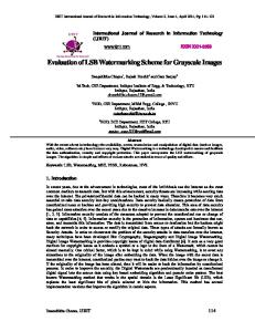
Fig. 3
M SE =
(3)
b) If the bit to embed is ”0”, then change the luminance value of the pixel to make sure that: Lreal < Lmean − δ2
1. Locate the changed pixel by using the polar mapping and by starting from the centroid of the image. 2. The decision threshold to extract the watermark is:
M −1 N −1 1 X X (f (x, y) − f 0 (x, y))2 M N x=0 y=0
P SN R = 10 ∗ Log10
2552 dB M SE
(5)
(6)
PR−1 PC−1
(4)
where δ1 and δ2 are calculated based on the homogeneity and luminance of the block (k x k), Lreal is the gray-scale level of the pixel, and Lmean is the gray-scale level mean of the block [6]. The extraction process is the following:
N CC =
0 y=0 (W (x, y) ∗ W (x, y)) PR−1 PC−1 2 x=0 y=0 |W (x, y)|
x=0
(7)
In Table 1, the degradation and distortion in the watermarked images are presented. In Fig. 3 one of the images after some attacks such as brightness, contrast, compression and rotation is shown. The percentage of recovered watermark after these attacks is shown in Table 2. From the results it can be seen our proposed scheme is robust to several attacks while allowing to recover the watermark message.
• If Lreal ≥ Lmean then the extracted bit is ”1”.
4.
• If Lreal < Lmean then the extracted bit is ”0”.
We have presented a blind watermarking scheme applied to medical images that has shown to be robust to several attacks. Additionally, the watermark has been compressed with the Huffman algorithm and ciphered with RC4 in order to diminish the message size and to add security to the whole system. The algorithm is robust by both embedding and extracting the watermark
The values of δ1 and δ2 are not required to extract the watermark. 3.
Different attacks in the watermarked image.
Experimental Results
The experiments were carried out in three DICOM files shown in Fig. 2.where a) and b) are 256x256x16 bits, and c) 1024x1024x12 bits gray scale medical images. The three images were resized to 512x512x8 bits when they are converted to JPEG lossless format. The homogeneity is calculated using a window of 3 x 3 pixels. The watermark consists of the DICOM metadata plus the medical diagnostic of the study, 4800 bits in total. In order to determine the degradation of the watermarked image with respect to the original image, we apply the PSNR and MSE metrics to measure the distortion produced after the embedding process [7], and we apply the NCC to evaluate the similarity between the original watermark and the extracted watermark.
Conclusions
Table 1
Imperceptibility using PSNR (dB) and MSE metrics. Image Brain Knee Shoulder
PSNR 37.1 42.3 41.1
MSE 12.5 4.2 5.4
Table 2 Recovered data after brightness, contrast, JPEG compression with and 35 degrees rotation. Image
Brightness
Contrast
Brain Knee Shoulder
98.5% 99.0% 97.0%
97.0% 96.0% 95.5%
JPEG Rotation Compression 94.5% 81.5% 90.5% 83.4% 91.5% 80.3%
LETTER
3
message. Additionally, using the homogeneity allows us to obtain a better accuracy in the extraction process than by using a simple LSB method. References [1] G. Coatrieux, et al., Relevance of watermarking in medical imaging, In IEEE-embs Information Technology Applications in Biomedicine, 250-255, 2000. [2] W. Puech, J. M. Rodrigues, A new crypto-watermarking method for medical images safe transfer, In Proceedings of the 12th European Signal Processing Conference, 1481-1484, 2004. [3] Bruce Schneier, Applied Cryptography, John Wiley and Sons, 2nd edition, 1996. [4] P. Dong et al., Digital Watermarking Robust to Geometric Distortions, In IEEE Transactions on Image Processing, 4, 12, 2005. [5] L. Tung-Lam, N. Thi-Hoang-Lan, Digital Image Watermarking with Geometric Distortion Corrections Using the Moment Image Theory, International Conference on Research Innovation and Vision for the Future (RIVF), 2004. [6] Y. Wang, A. Pearmain Blind image data hiding based on self reference, In Pattern Recognition Letters, 2, 15 1681-1689, 2004. [7] B. Plaintz, A. Maeder, Medical Image Watermarking: A Study on Image Degradation, In Proceedings of the Australian Pattern Recognition Society, 2005.