Neuroscience Research 31 (1998) 69 – 75
Effects of long-term physical exercise on age-related changes of spinal motoneurons and peripheral nerves in rats Kenro Kanda a,*, Ken Hashizume 1,b a
Department of Central Ner6ous System, Tokyo Metropolitan Institute of Gerontology, 35 -2 Sakaecho, Itabashi-ku, Tokyo 173, Japan b Department of Kinesiology, Tokyo Metropolitan Insitute of Gerontology, 35 -2 Sakaecho, Itabashi-ku, Tokyo 173, Japan Received 7 January 1998; accepted 6 March 1998
Abstract We examined the number and size of motoneurons and myelinated nerve fibers innervating the medial gastrocnemius (MG) muscle in the hindlimb of rats that were subjected to swimming for 10 months (30 min, 3 days/week) from 17 to 27 months of age. The number of MG motoneurons in exercised aged rats was slightly, but not significantly, greater than that in sedentary aged rats, and the number for both aged groups was significantly less compared to that in middle-aged (17-month-old) rats. The size of motoneuronal somata in the exercised aged rats was significantly larger than that in sedentary aged rats. Morphological parameters (fascicular area, fiber density, and axonal diameter) for the MG nerve in the exercised rats showed intermediate values between those for middle-aged and sedentary aged rats, although all of the differences in the means between groups were not significant. Thus, the overall changes seen in the exercised rats seem less compared to the changes in the sedentary rats, suggesting that long-term moderate exercise retards the progressive changes in motoneurons and peripheral nerves that develop in old age. © 1998 Elsevier Science Ireland Ltd. All rights reserved. Keywords: Motoneuron; Peripheral nerve; Myelinated fiber; Exercise; Aging; Rat
1. Introduction Age-related changes in spinal motoneurons and peripheral nerves appear to play an important role in the decline of motor function with age (Gutmann and Hanzlı´kova´, 1972; Wallace et al., 1980; Larsson, 1982a,b; Brooks and Faulkner, 1994; Larsson and Ansved, 1995). By studying the morphological properties of the spinal motor nucleus innervating the medial gastrocnemius (MG) muscle of rats of various ages, we have demonstrated a decrease in the number
* Corresponding author. Tel.: +81 3 39643241 (ext. 3083); fax: +81 3 35794776; e-mail:
[email protected] 1 Present address. Faculty of Health and Sport Sciences, Osaka University. 1-17, Machikaneyama, Toyonaka, Osaka 560, Japan. K. Hashizume was at the Department of Kinesiology, TMIG when the experiments were performed.
of motoneurons and an atrophy of cell soma in aged rats (Hashizume et al., 1988; Hashizume and Kanda, 1990). A decrease in the number of myelinated fibers, increases in fascicular area and axon diameter were also found in aging MG nerves (Hashizume and Kanda, 1995). Similar findings have been reported for other motoneuron groups and muscle nerves in rats (Ishihara and Araki, 1988; Ansved and Larsson, 1990). Spontaneous locomotion activity gradually decreases as rats age (Samorajski and Rolsten, 1975; Holloszy et al., 1985; Mondon et al., 1985; Brown et al., 1992). One of the possible factors facilitating the age-related changes in neuromuscular systems has been assumed to be reduced motor activity. It has been reported that physical exercise actually improves functional, morphological and histochemical changes in aging muscles (Larsson, 1982a,b; Brown et al., 1992; Coggan et al., 1992; Ishihara and Taguchi, 1993).
0168-0102/98/$19.00 © 1998 Elsevier Science Ireland Ltd. All rights reserved. PII S0168-0102(98)00026-1
70
K. Kanda, K. Hashizume / Neuroscience Research 31 (1998) 69–75
The effects of physical exercise on morphological and physiological properties of spinal motoneurons in young and middle-aged animals have been reported (Edstro¨m, 1957; Geinisman et al., 1971; Gerchman et al., 1975; Gilliam et al., 1977; Jasmin et al., 1988; Nakano et al., 1997). There are, however, few studies on the properties of aging spinal motoneurons. Retzlaff and Fontaine (1965) have merely shown that exercise delays the change in staining reaction of motoneurons to protargol in aging rats. Samorajski and Rolsten (1975) have demonstrated that the axon diameter in the posterior tibial nerve of exercised, aged mice is significantly greater compared to that in control, non-exercised, aged mice. It is, however, difficult to interpret their results in terms of the effects of exercise on aging of the peripheral nerves because they showed data obtained only from aged animals. In the present experiment, we investigated the effects of long-term, physical exercise on the number and the size of motoneurons and on some morphological parameters of the peripheral nerve innervating the MG muscle in aging rats. Preliminary reports have appeared elsewhere (Hashizume and Kanda, 1991, 1993).
2. Materials and methods
2.1. Experimental animals and exercise protocol Experiments were performed on male Fischer 344 rats, which were raised in a specific pathogen-free colony at our institute. The 75, 50, and 25% survival times of these rats were about 25.5, 28.0, and 31.5 months, respectively. They were kept in conventional housing, less than four per cage, without restrictions on feeding. They were randomly divided into three groups at the age of 17 months. One group was sacrificed immediately and the others were assigned to either the exercised or the non-exercised group. Rats in the exercised group (n=15) were subjected to swimming (30 min, 3 days/week) for 10 months until they were sacrificed at the age of 27 months. They were gradually adapted to swimming in a pool (40× 40 cm, water depth 60 cm). Within 2 weeks the daily exercise time was increased from 10 to 30 min. After this period, air was supplied from the bottom of the pool to make a current and a weight equal to 1 – 2% of body weights of the rats was attached to the tail until about 24 months of age. These procedures were necessary to force the rat to swim continuously. Usually, two rats swam in a pool together. The water temperature was maintained near 30°C. Non-exercised, control rats were raised under usual laboratory conditions without special exercise requirements to the age of 27 months. At 24 months of age, oxygen uptake (V: O2) during swimming was measured for all exercised rats by an
open-circuit system. A pool was covered with an acrylic resin board which had two small holes. Without air supply and a sinker on the tail, atmospheric air was let through a hole into the space surrounded by side walls and a ceiling board. Outgoing air (mixed gas) from the other hole passed through an absorption pump and a flow meter. Flow rate was adjusted to 1 l/min (ATPS), and 100 ml/min of this flow was passed through an oxygen analyzer (Toray, model LC-700E). V: O2 was calculated by determining the relationship between the difference in the percentage of oxygen (inlet O2%–outlet O2%) and airflow. Delay in oxygen measurements were taken into account. Values of V: O2 during exercise in the present study showed a mean value to 20 min from 10 min after start of exercise.
2.2. Surgical and analytic procedures The surgical and analytic procedures and techniques used in the present study have been described elsewhere (Hashizume et al., 1988; Hashizume and Kanda, 1995). In brief, at the completion of the exercise periods, small volumes (0.1–0.5 ml) of 40% horseradish peroxidase (HRP, Toyobo I-C) solution were injected bilaterally into the cut-and-tied MG nerve by using glass micropipettes and a pressure injection system under pentobarbital anesthesia (35–45 mg/kg; i.p.) and aseptic conditions. Prior to the surgical procedures, the range of motion at the knee joint in the maximum extension was also measured using a plastic protractor. After a survival period of 2 days, the rat was reanesthetized and perfused transcardially. Serial sections in the horizontal plane were cut at an 80 mm thickness from the lumber (L4–L6) spinal cord blocks, and were processed for demonstration of HRP using the tetramethyl benzidine (TMB) method (Mesulam, 1978). Individual HRP-labeled cells were identified and counted under microscopic observations with the aid of photomontage maps. The cross-sectional area (CSA) of the cell body was measured in individual motoneurons by using an image-processing system at ×400 magnification. A small piece of the MG nerve in either side was excised from the distal cut end and fixed in 2.5% glutaraldehyde. This was later osmicated in 1% OsO4. Semithin cross sections were cut at about 1 mm thickness from the specimen embedded in the Epon and stained with toluidine blue. The fascicular area of each nerve was measured by using an image-processing system at × 250 magnification. The total numbers of myelinated fibers were counted, and the average diameters of individual axons (one-half of the sum of the measured maximum and minimum diameters) were measured from these photographic prints (× 2070). Then, the density of myelinated fibers was calculated from the fascicular area and the total number of myelinated fibers. No correction for shrinkage during fixation was
K. Kanda, K. Hashizume / Neuroscience Research 31 (1998) 69–75
71
Table 1 Numbers and sizes of retrogradely labled motoneurons innervating the medial gastrocnemius muscle Group (n)
17-month-old (7) 27-month-old sedantary (8) 27-month-old exercised (10)
Number
133.8 9 5.5 121.29 3.3* 124.99 4.5*
Soma cross-sectional area a-motoneurons (m2)
g-motoneurons (m2)
939.7 979.9 861.6 9 152.1 1020.7 991.7**
353.8 9 37.3 311.3 948.7 383.6 9 34.7**
Values are mean 9S.D. * Significantly different from 17-month-old rats, PB0.05. ** Significantly different from 27-month-old, sedantary rats, PB0.05.
attempted in this study. A portion of the data has also been used for our previous paper (Hashizume and Kanda, 1995).
2.3. Statistical analysis For the motoneurons, the mean values obtained from both sides of the limb were averaged for the individual rats. We considered that the nerve data from one side of the leg (either left or right) represented the value for the individual rats. We used the multiple range test (Scheffe´ procedure) to examine for differences between mean values.
3. Results The mean number of strokes of one foot during swimming was about 2.1 s − 1 for all rats. The mean oxygen uptake during swimming in the trained rats was 42.0 (range 38.1 – 45.2) ml/min per kg at the age of 24 months. Three rats died during the exercise periods and two rats died after surgery. The mean body weight of the exercised rats was 4889 19 g (mean9 S.D., n = 15) at the beginning of the exercise period (at the age of 17 months). This decreased by about 35 g for the first 3 months and reached 4259 45 g (n=12) at the age of 27 months, which was not significantly different from that in the sedentary aged rats (398 9 48 g, n=8, P \0.2). The mean wet weight of the MG muscles was 596 974 mg for exercised rats and 560 937 mg for the sedentary aged rats. The difference between the exercised and sedentary rats was not significant (P \ 0.2). The weight of the soleus muscles, which were synergist of the MG muscles, in the exercised rats (1509 4 mg) was significantly heavier than that in the sedentary aged rats (1329 14 mg, PB0.01). The angle of the knee joint in the maximum extension was about 155° in most of the exercised rats. This value was comparable to that in the middle-aged rats. The limitation in the range of motion, which has been frequently seen in the sedentary
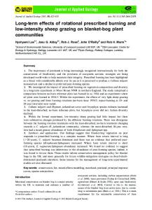
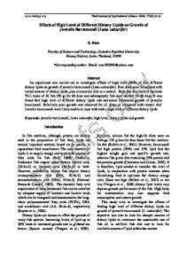
aged rats (about 135°, Hashizume and Kanda, 1995), was not observed. The summary of motoneuron data for the three groups (i.e., middle-aged, sedentary aged and exercised aged rats) is presented in Table 1. The mean number of motoneurons in the MG motor nucleus in both aged groups was significantly smaller than that of middle-aged rats. The value for the exercised rats was slightly, but not significantly (PB 0.2), larger than that for the sedentary rats. Fig. 1 shows the distribution of the CSA of cell soma of MG motoneurons. Motoneurons in individual rats were subdivided into two groups, presumed a-motoneurons and presumed g-motoneurons, by size using the distribution pattern of the frequency histograms and the cumulative curves (Hashizume et al., 1988). The mean CSA of both a- and g-motoneurons in the exercised aged rats was significantly larger than that in the sedentary aged rats. There was no significant difference in size between the middle-aged and sedentary aged rats, and between the sedentary and exercised groups. Fig. 2 shows the photomicrographs of representative semithin-sections of the MG nerves obtained from middle-aged, sedentary aged and exercised aged rats. The quantitative data on the morphological parameters of the MG nerves are summarized in Table 2. The mean fascicular area of exercised rats was significantly smaller than that of sedentary aged rats, and significantly larger than that of middle-aged rats. As to the number of myelinated fibers, no difference was found between the middle-aged and exercised aged rats. The value for the sedentary aged rats was significantly smaller than that for the middle-aged rats. The mean axon diameters in the sedentary aged rats was larger than that in the middle-aged and exercised rats. No difference was found between the middle-aged and exercised aged rats. The density of myelinated fibers in the exercised aged rats was intermediate between the values for the middle-aged and sedentary rats, and was significantly different from both of them.
72
K. Kanda, K. Hashizume / Neuroscience Research 31 (1998) 69–75
4. Discussion
4.1. Effects on motoneurons An atrophy of spinal motoneurons in aged rats has been reported previously (Hashizume et al., 1988; Hashizume and Kanda, 1995; Machado-Salas et al. (1977) for mouse spinal neurons). Motoneurons of the sedentary aged rats in the present experiment were also smaller than those of the middle-aged rats although the difference between them was not statistically significant (P\ 0.2 for presumed a-motoneurons and P\ 0.05 for presumed g-motoneurons). On the other hand, the size of motoneurons can be changed
Fig. 1. Histograms showing the frequency distribution of soma sizes of motoneurons innervating the medial gastrocnemius muscle. These histograms include all motoneurons measured: a total of 1845 neurons in 14 motor nuclei of seven 17-month-old rats, 2052 neurons in 16 motor nuclei of eight 27-month-old sedentary rats, and 2479 neurons in 20 motor nuclei of ten 27-month-old exercised rats. Note the slight left-ward shift of the distribution for the sedentary aged rats.
as a result of increased activity with physiological overload, although some inconsistency are noted in the literature. Acute bouts of swimming or running have been reported to increase, decrease, or not change the size of the soma (Gilliam et al. (1977)). The motoneuronal morphological alterations appear to regress to control levels as adaptation occurs during long term exercise (Edstro¨m, 1957; Burke and Edgerton (1975) for review). However, using x 2 analyses of contingency tables, Gilliam and his co-workers (1977) have revealed that there are significant distribution differences in the size of motoneurons between sedentary and exercised rats, but no difference in the means between the two groups after 12 weeks of chronic exercise. More recently, Nakano et al. (1997) have demonstrated that the soleus motoneurons of trained rats have a significantly larger soma size than those of control rats, whereas motoneurons innervating the extensor digitorum longus muscle do not show any alteration in soma size after running training. Hypokinesia by hindlimb suspension induces a soma atrophy in adult and aged rats (Kanda and Hashizume, unpublished data). In the present experiment, the rat were perfused three days after the last exercise day. Thus it is unlikely that the larger soma size of motoneurons in exercised rats is due to acute effects of the exercise bout (Gilliam et al., 1977). Hence, the present findings on the mean motoneuronal size seem to indicate that long-term exercise retards or prevents atrophy of motoneurons which commonly takes place in sedentary laboratory rats with advancing age. The number of MG motoneurons in the aged rats was significantly smaller than that in the middle-aged rats. The mean for the exercised rats was slightly, but not significantly greater than that for the sedentary rats (P\ 0.1). Recently, we have looked at the effects of overloading a muscle, which is induced by the removal of synergistic muscles, on age-related changes in the motor nucleus which innervates the muscle (Kanda et al., 1996). The number of motoneurons innervating overloaded muscle did not differ from that of motoneurons innervating the control muscle in an unoperated leg. However, it is possible that, under the conditions in these experiments, the exercise strength was not proper to prevent cell death. Cartee and Farrar (1987) reported V: O2max in trained aged rats (Fischer 344, 24 months of age, male) which had undergone a 6 month program of treadmill running. The mean value of V: O2max was 76.0 ml/min per kg, and that for age-matched, untrained rats was 65.6 ml/min per kg. Thus, exercise strength employed in the present experiment was judged to be moderate. We did not intend to test the effects of different strengths of exercise in this study, however this variable deserves further study.
K. Kanda, K. Hashizume / Neuroscience Research 31 (1998) 69–75
73
Fig. 2. Photomicrographs of tranverse semithin sections obtained from three representative MG nerves in 17-month-old (A), 27-month-old sedentary (B), and 27-month-old exercised rats (C), respectively. Sections were stained with toluodine blue. See Table 2 for detailed morphometric data. Calibration bar = 50 mm.
4.2. Effects on peripheral ner6es There have been few reports on the effects of longterm exercise on age-related changes of the peripheral nerve. The density of myelinated fibers was significantly higher in the exercised rats as has been reported previously (Samorajski and Rolsten, 1975). The number of myelinated fibers for the sedentary aged rats was significantly smaller than that for the middle-aged rats. The value for the exercised aged rats was intermediate between those for the middle-aged and sedentary aged rats, but the difference was not significant. Samorajski and Rolsten (1975) have also reported that the number of myelinated fibers in the posterior tibial nerve from exercised mice did not differ from those of the sedentary controls. Unfortunately, they only looked at aged mice, although the same author had reported in a different paper (Samorajski, 1974) that axon diameter did not alter from 20 to 30 months of age. Thus it seems to be difficult to directly compare our present findings with their results. The effect of aging on caliber size in the peripheral nerve is controversial in the literature. Enlargement (Grover-Johnson and Spencer, 1981; Inestrosa and Alvarez, 1988; Saitua and Alvarez, 1988; Hashizume and Kanda, 1995; and see also Ansved and Larsson (1990)), atrophy (Sharma et al., 1980; Krinke, 1983) and no changes in axon diameter (Samorajski, 1974; Knox et al., 1989) have been reported. The average diameter of individual axons of the MG nerves in the aged rats of the present experiment were larger than those in the middle-aged rats. The rate of transport of neurofilaments appears to be an important determinant of axon diameter (Hoffman et al., 1984). If the number of neurofilaments exported from the nerve cell body is relatively constant during aging, then the progressive decrease in the rate of neurofilament transport during aging (Komiya, 1980; McQuarrie et al., 1989) should tend to increase the diameter of the axons. An in-
creased axonal diameter in aged rats seems to support this hypothesis. The effects of exercise on nerves are also controversial. Both enlargement and shrinkage of nerve fiber diameter were found in different experiments (Okajima and Maloney, 1989). A significantly smaller axonal diameter in the exercised rats observed in the present experiment suggests the retardation of age-related decreases in axonal transport and an eventual increase in axonal diameter. Little is known, however, about the effects of exercise on the slow axonal transport. Another thing which should be noted is that an increased axonal diameter in the aged rats does not necessarily mean a faster conduction velocity of action potentials. Local shrinkage of axoplasma (Gilmore, 1972; Krinke, 1983) and/or demyelination (Thomas et al., 1980; Grover-Johnson and Spencer, 1981; Knox et al., 1989) may decrease longitudinal current greatly and thereby reduce conduction velocity. In fact, a decreased conduction velocity has been observed in the same peripheral nerve as that examined in this study, i.e. the MG nerve of aged rats (Kanda and Hashizume, 1989; Miwa et al., 1995).
4.3. Mechanisms underlying effects of exercise Most of the mean values of morphometric data of motoneurons as well as peripheral nerves in the exercised aged rats were between those in the sedentary aged and the middle-aged rats, or they were not different from those of middle-aged rats. Long-term moderate exercise, therefore, appears to retard degenerative changes in motoneurons and peripheral nerves with advancing age. How does exercise exert its beneficial effects on motoneurons and peripheral nerves? It has been reported that chronic as well as acute activity also induce changes in motoneuron metabolism (Burke and Edgerton, 1975). Thus, motoneurons seem to adapt biochemically to increased or decreased activity. Such
74
K. Kanda, K. Hashizume / Neuroscience Research 31 (1998) 69–75
Table 2 Morphological properties of the peripheral nerve innervating the medial gastrocnemius muscle Group (n)
Fascicular area (mm2)
Number of fibers
Axon diameter (mm)
Fiber density (n/mm2)
17-month-old (5) 27-month-old sedantary (8) 27-month-old exercised (7)
0.040 9 0.003 0.07090.009* 0.05890.003*,**
287.4 9 19.3 258.4 9 18.5* 273.3 925.9
5.6 9 0.5 6.6 9 0.4* 5.8 90.6**
7126 9 836 3732 9328* 4758 9680*,**
Values are mean 9 S.D. * Significantly different from 17-month-old rats, PB0.05. ** Significantly different from 27-month-old, sedantary rats, PB0.05.
biochemical alterations might help motoneurons to maintain life. It is known that neurotrophic factors play an important role in cell survival in developing and matured animals. Our recent studies suggest that a deficit in trophic factors causes motoneuronal death with advancing age (Kanda et al., unpublished data). A decrease in the rate of axonal transport with age (McMartin and O’Conner, 1979; Frolkis et al., 1985) may cause a deficit of trophic factors in the soma. Increased muscular activity may increase uptake of trophic substances because it has been reported that chronic endurance exercise increases the velocity and the amount of the fast axonal transport of proteins (Jasmin and Gardiner, 1987; Jasmin et al., 1988). Furthermore, it has been suggested that electrical activity itself is key to the effect of neurotrophic factors (Meyer-Franke et al., 1995). The direct effect of motoneuronal activity, as well as effects of exercise-induced changes in other regulatory systems such as various hormone releases and enzyme activities on neuronal cell death with advancing age, is still unclear and deserves further study.
Acknowledgements The authors thank to E. Nomoto for her technical assistance.
References Ansved, T., Larsson, L., 1990. Quantitative and qualitative morphological properties of the soleus motor nerve and the L5 ventral root in young and old rats. J. Neurol. Sci. 96, 269–282. Brooks, S.V., Faulkner, J.A., 1994. Skeletal muscle weakness in old age: underlying mechanisms. Med. Sci. Sports Exerc. 26, 432 – 439. Brown, M., Ross, T.P., Holloszy, J.O., 1992. Effects of ageing and exercise on soleus and extensor digitorum longus muscles of female rats. Mech. Ageing Dev. 63, 69–77. Burke, R.E., Edgerton, V.R., 1975. Motor unit properties and selective involvement in movement. Exerc. Sport Sci. Rev. 3, 31 – 81. Cartee, G.D., Farrar, R.P., 1987. Muscle respiratory capacity and V: O2max in identically trained young and old rats. J. Appl. Physiol. 63, 257 – 261.
Coggan, A.R., Spina, R.J., King, D.S., Rogers, M.A., Brown, M., Nemeth, P.M., Holloszy, J.O., 1992. Skeletal muscle adaptations to endurance training in 60 – 70-year-old men and women. J. Appl. Physiol. 72, 1780 – 1786. Edstro¨m, J.E., 1957. Effects of increased motor activity on the dimensions and the staining properties of the neuron soma. J. Comp. Neurol. 107, 295 – 304. Frolkis, V.V., Tanin, S.A., Marcinko, V.J., Kulchitsky, O.K., Yasechko, A.V., 1985. Axoplasmic transport of substances in motoneuronal axons of the spinal cord in old age. Mech. Ageing Dev. 29, 19 – 28. Geinisman, Y.Y., Larina, V.N., Mats, V.N., 1971. Changes of neurons dimensions as a possible morphological correlate of their increased functional activity. Brain Res. 26, 247 – 257. Gerchman, L.R., Edgerton, V.R., Carrow, R.E., 1975. Effects of physical training on the histochemistry and morphology of ventral motor neurons. Exp. Neurol. 49, 790 – 801. Gilliam, T.B., Roy, R.R., Taylor, J.F., Heusner, W.W., Van Huss, W.D., 1977. Ventral motor neuron alterations in rat spinal cord after chronic exercise. Experientia 33, 665 – 667. Gilmore, S.A., 1972. spinal nerve root degeneration in aging laboratory rats, a light microscopic study. Anat. Rec. 174, 251–258. Grover-Johnson, N., Spencer, P.S., 1981. Peripheral nerve abnormalitites in aging rats. J. Neuropathol. Exp. Neurol. 40, 155–165. Gutmann, E., Hanzlı´kova´, V., 1972. Age Changes in the Neuromuscular System. Bristol, Scientechnica. Hashizume, K., Kanda, K., 1990. Neuronal dropout is greater in hindlimb motor nuclei than in forelimb motor nuclei in aged rats. Neurosci. Lett. 113, 267 – 269. Hashizume, K., Kanda, K., 1991. Effects of swimming exercise on aging rat motoneuron. Neurosci. Res. Suppl. 14, S11. Hashizume, K., Kanda, K., 1993. Effects of swimming exercise on age-related changes in motoneurons and peripheral nerves in the rat. Soc. Neurosci. Abstr. 19, 1742. Hashizume, K., Kanda, K., 1995. Differential effects of aging on motoneurons and peripheral nerves innervating the hindlimb and forelimb muscles of rats. Neurosci. Res. 22, 189 – 196. Hashizume, K., Kanda, K., Burke, R.E., 1988. Medial gastrocnemius motor nucleus in the rat: age-related changes in the number and size of motoneurons. J. Comp. Neurol. 269, 425 – 430. Hoffman, P.N., Griffin, J.W., Price, D.L., 1984. Control of axonal caliber by neurofilament transport. J. Cell Biol. 99, 705–714. Holloszy, J.O., Smith, E.K., Vining, M., Adams, S., 1985. Effect of voluntary exercise on longevity of rats. J. Appl. Physiol. 59, 826 – 831. Inestrosa, N.C., Alvarez, J., 1988. Axons grow in the aging rat but fast transport and acetylcholinesterase content remain unchanged. Brain Res. 441, 331 – 338. Ishihara, A., Araki, H., 1988. Effects of age on the number and histochemical properties of muscle fibers and motoneurons in the rat extensor digitorum longus muscle. Mech. Ageing Dev. 45, 213 – 221. Ishihara, A., Taguchi, S., 1993. Effect of exercise on age-related muscle atrophy. Neurobiol. Aging 14, 331 – 335.
K. Kanda, K. Hashizume / Neuroscience Research 31 (1998) 69–75 Jasmin, B.J., Gardiner, P.F., 1987. Patterns of EMG activity of rat plantaris muscle during swimming and other locomotor activities. J. Appl. Physiol. 63, 713–718. Jasmin, B.J., Lavoie, P.-A., Gardiner, P.F., 1988. Fast axonal transport of labeled proteins in motoneurons of exercise-trained rats. Am. J. Physiol. 255, Cell Physiology C731–C736. Kanda, K., Hashizume, K., 1989. Changes in properties of the medial gastrocnemius motor units in aging rat. J. Neurophysiol. 61, 737 – 746. Kanda, K., Hashizume, K., Miwa, T., Miwa, Y., 1996. Overloading a muscle does not alter the rate of motoneuronal loss in aged rats. Neurobiol. Aging 17, 613–617. Knox, C.A., Kokmen, E., Dyck, P.J., 1989. Morphometric alteration of rat myelinated fibers with aging. J. Neuropath. Exp. Neurol. 48, 119 – 139. Komiya, Y., 1980. Slowing with age of the rate of slow axon flow in bifurcating axons of rat dorsal root ganglion cells. Brain Res. 183, 477 – 480. Krinke, G., 1983. Spinal radiculoneurophathy in aging rats, demyelination secondary to neuronal dwindling? Acta Neuropath. 59, 63– 69. Larsson, L., Ansved, T., 1995. Effects of ageing on the motor unit. Prog. Neurobiol. 45, 397–458. Larsson, L., 1982a. Physical training effects on muscle morphology in sedentary males at different ages. Med. Sci. Sports Exerc. 14, 203 – 206. Larsson, L., 1982b. Aging in mammalian skeletal muscle. Ch. 4, In: Motimer, J.A., Pirozzolo, F.J., Maletta, G.J., (Eds.). Advances in Neurogerontology. Vol. 3. The Aging Motor System. New York, Praeger, pp. 60 – 97. Machado-Salas, J., Scheibel, M.E., Scheibel, A.B., 1977. Neuronal changes in the aging mouse: Spinal cord and lower brain stem. Exp. Neurol. 54, 504 –512. McMartin, D.N., O’Conner, J.A., 1979. Effect of age on axoplasmic transport of cholinesterase in rat sciatic nerves. Mech. Ageing Dev. 10, 241 – 248. McQuarrie, I.G., Brady, S.T., Lasek, R.J., 1989. Retardation in the slow axonal transport of cytoskeletal elements during maturation and aging. Neurobiol. Aging 10, 359–365.
.
75
Mesulam, M.-M., 1978. Tetramethyl benzidine for horseradish peroxidase neurohistochemistry: a non-carcinogenic blue reactionproduct with superior sensitivity for visualizing neural afferents and efferents. J. Histochem. Cytochem. 26, 106 – 117. Meyer-Franke, A., Kaplan, M.R., Pfrieger, F.W., Barres, B.A., 1995. Characterization of the signaling interactions that promote the survial and growth of developing retinal ganglion cells in culture. Neuron 15, 805 – 819. Miwa, T., Miwa, Y., Kanda, K., 1995. Dynamic and static sensitivities of muscle spindle primary endings in aged rats to ramp stretch. Neurosci. Lett. 201, 179 – 182. Mondon, C.E., Dolkas, C.B., Sims, C., Reaven, G.M., 1985. Spontaneous running activity in male rats: effect of age. J. Appl. Physiol. 58, 1553 – 1557. Nakano, H., Masuda, K., Sasaki, S., Katsuta, S., 1997. Oxidative enzyme activity and soma size in motoneurons innervating the rat slow-twitch and fast-twitch muscles after chronic activity. Brain Res. Bull. 43, 149 – 154. Okajima, Y., Maloney, F.P., 1989. Overwork weakness in rats with acrylamide neuropathy. Am. J. Phys. Med. Rehabil. 68, 66–69. Retzlaff, E., Fontaine, J., 1965. Functional and structural changes in motor neurons with age. In: Welford, A.T., Birren, J.E., (Eds.). Behavior, Aging, and the Nervous System. Springfield, IL, pp. 340 – 352. Saitua, F., Alvarez, J., 1988. Do axons grow during adulthood? A study of caliber and microtubules of sural nerve axons in young, mature, and aging rats. J. Comp. Neurol. 269, 203 – 209. Samorajski, T., Rolsten, C., 1975. Nerve fiber hypertrophy in posterior tibial nerves of mice in response to voluntary running activity during aging. J. Comp. Neurol. 159, 553 – 558. Samorajski, T., 1974. Age differences in the morphology of posterior tibial nerves of mice. J. Comp. Neurol. 157, 439 – 452. Sharma, A.K., Bajada, S., Thomas, P.K., 1980. Age changes in the tibial and plantar nerves of the rat. J. Anat. 130, 417 – 428. Thomas, P.K., King, P.H.M., Sharma, A.K., 1980. Changes with age in the peripheral nerves of the rat. Acta Neuropathol. 52, 1–6. Wallace, J.F., Krauter, E.E., Canpbell, B.A., 1980. Motor and reflexive behavior in the aging rat. J. Gerontol. 35, 364 – 370.