Effects of West Nile Virus Dose on Temporal Infection of the Culex pipiens quinquefasciatus Say (Diptera: Culicidae) Midgut Sheri L. Anderson, Stephanie L. Richards, Chelsea T. Smartt, and Walter J. Tabachnick Florida Medical Entomology Laboratory, Department of Entomology and Nematology, University of Florida - IFAS, Vero Beach, FL
Introduction
Results
West Nile virus (WNV), family Flaviviridae, genus Flavivirus, has been detected in all 48 continental states in the U.S. since its introduction in New York in 1999. West Nile virus is maintained in nature in birds and mosquitoes in the genus Culex. Culex pipiens quinquefasciatus has been implicated as a primary vector of WNV in the southern U.S.1
•At 6 dpi, significantly more mosquitoes had infected midguts at the high dose (p=0.048) compared to the low dose but there was no significant difference in dissemination between doses on any dpi (Table 1, Fig. 2).
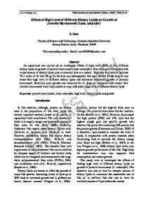
Fig. 1 IFA of Cx. p. quinquefasciatus midgut 6 dpi with a high dose of WNV.
•By 10 dpi, 100% of the mosquitoes, regardless of dose, had infected midguts (p=1.0).
The extrinsic incubation period (EIP) encompasses the time from ingestion of an infected blood meal until the mosquito can transmit the virus. The EIP for WNV is approximately 12 days at 30˚C2 and is influenced by temperature2,3, age3, and virus dose.3
Discussion Table 1. Percentage of midgut and disseminated WNV infections in Cx. p. quinquefasciatus by day post-infection and WNV dose.
Culex pipiens quinquefasciatus Say
West Nile virus
Day Post- Dose infection 4 High
% Midgut infection % Disseminated infection 4/4 (100%)
3/4 (75%)
4
Low
1/5 (20%)
0/1 (0%)
6
High
5/5 (100%)
4/5 (80%)
6
Low
1/5 (20%)
0/1 (0%)
Objective
8
High
5/5 (100%)
4/5 (80%)
The objective of this study was to determine the temporal pattern of midgut infection and dissemination of WNV in Cx. p. quinquefasciatus given two different doses of virus.
8
Low
3/5 (60%)
2/3 (67%)
10
High
5/5 (100%)
4/5 (80%)
•At 6 dpi, mosquitoes given the high virus dose blood meal show significantly greater midgut infection rates than mosquitoes given the low virus dose blood meal. This was also likely at 4 dpi, but the sample size of only four mosquitoes was too small for significance. •Culex p. quinquefasciatus fed the high virus dose in the blood meal become maximally infected and had complete disseminated infections by 4 dpi, sooner than the expected EIP of 12 days for WNV at 30˚C.2 These results should be considered in studies seeking to understand the role of mosquitoes in WNV transmission cycles. •These results suggest that the midgut escape barrier in Cx. p. quinquefasciatus infected with WNV is present on all dpi and is not influenced by virus dose under these environmental conditions.
Future directions
12
High
5/5 (100%)
4/5 (80%)
•Determine temporal patterns for other populations of Cx. p. quinquefasciatus and Culex species infected with WNV with increased sample sizes.
•Mosquitoes were given blood meals containing either a low dose (4.8 logs plaque forming units (pfu)/mL) or a high dose (6.7 pfu/mL) of freshly prepared WNV.
12
Low
5/5 (100%)
4/5 (80%)
•Determine the molecular mechanisms involved with midgut-virus interactions that are influenced by temperature and viral dose.
•Mosquitoes were held at 28˚C and five mosquitoes per group were dissected 4, 6, 8, 10, and 12 days post-infection (dpi).
Fig. 2 Percentage of midgut and disseminated WNV infection in Cx. p. quinquefasciatus by day post-infection and WNV dose.
1.Blackmore, CGM et al. 2003. Amer. J. Trop. Med. Hyg. 69: 141-150. 90
2.Dohm, DJ et al. 2002. J. Med. Entomol. 39: 221-225.
80
3.Richards, SL et al. 2007. Vector-Borne and Zoonotic Dis. 7: 629-636.
70
4.Girard, YA et al. 2004 Vector-Borne and Zoonotic Dis. 4: 109-122.
60
Midgut infection Disseminated infection
50 40 30
This work was supported by a grant from the National Institutes of Health (Grant no. 49326) to Cynthia C. Lord, WJT, and Jonathan F. Day.
10
8
8 10 10 12 12
Day post-infection and WNV dose
Low
Low
6
High
6
High
4
Low
0
4
5.SAS Version 8.2. 1999-2001. SAS Institute Inc., Cary, NC.
Acknowledgements
*
20
Low
•Comparisons were made using Fisher’s Exact Test.5
References
*
100
High
•Disseminated infection rate = # with disseminated infections / # with infected midguts (100).
4/5 (80%)
Low
•Midgut infection rate = # with infected midguts / # of mosquitoes engorged on an infectious blood meal (100).
5/5 (100%)
High
•Quantitative reverse transcription polymerase chain reaction (qRT-PCR) was used to test virus dissemination out of the midgut: Legs from the mosquitoes were removed and placed into BA-1 diluent for analysis. qRTPCR was performed according to methods described elsewhere.3
Low
High
•Immunofluorescent assay (IFA) was used to test midgut infection: IFA was performed following methods described elsewhere.4 Slides containing midguts were stained and read using an Olympus BH-2 microscope adapted for fluorescence to observed infected midguts (Fig. 1).
10
Percent infected
Methods