Expression of Vascular Endothelial Growth Factor Receptor-2 or Tie-2 on Peripheral Blood Cells Defines Functionally Competent Cell Populations Capable of Reendothelialization Grzegorz Nowak, Azza Karrar, Carolina Holmén, Silvia Nava, Mehmet Uzunel, Kjell Hultenby and Suchitra Sumitran-Holgersson Circulation 2004;110;3699-3707; originally published online Sep 20, 2004; DOI: 10.1161/01.CIR.0000143626.16576.51 Circulation is published by the American Heart Association. 7272 Greenville Avenue, Dallas, TX 72514 Copyright © 2004 American Heart Association. All rights reserved. Print ISSN: 0009-7322. Online ISSN: 1524-4539
The online version of this article, along with updated information and services, is located on the World Wide Web at: http://circ.ahajournals.org/cgi/content/full/110/24/3699
Subscriptions: Information about subscribing to Circulation is online at http://circ.ahajournals.org/subsriptions/ Permissions: Permissions & Rights Desk, Lippincott Williams & Wilkins, 351 West Camden Street, Baltimore, MD 21202-2436. Phone 410-5280-4050. Fax: 410-528-8550. Email:
[email protected] Reprints: Information about reprints can be found online at http://www.lww.com/static/html/reprints.html
Downloaded from circ.ahajournals.org by on December 23, 2006
Expression of Vascular Endothelial Growth Factor Receptor-2 or Tie-2 on Peripheral Blood Cells Defines Functionally Competent Cell Populations Capable of Reendothelialization Grzegorz Nowak, MD, PhD; Azza Karrar, MSc; Carolina Holmén, BSc; Silvia Nava, BSc; Mehmet Uzunel, PhD; Kjell Hultenby, PhD; Suchitra Sumitran-Holgersson, PhD Background—Receptor tyrosine kinases that include vascular endothelial growth factor (VEGFR)-1, VEGFR-2, and Tie-2 regulate cardiovascular development and physiological and pathological angiogenesis. We were interested in the phenotypic and functional characterization of peripheral blood cells expressing these receptors and their therapeutic potential in vascular injury. Methods and Results—VEGFR-1⫹, VEGFR-2⫹, and Tie-2⫹ cells constituted ⬇3.0⫾0.2%, 0.8⫾0.5%, and 2.0⫾0.3%, respectively, of the total population of mononuclear cells in blood. Phenotypic analysis demonstrated that all 3 cell populations mainly expressed markers of monocytic/macrophage lineage. Only VEGFR-2⫹ and Tie-2⫹ cells phenotypically, morphologically, and functionally differentiated to endothelial cells after culture, whereas VEGFR-1⫹ cells did not. None of the cell types proliferated in vitro. Only freshly isolated VEGFR-2⫹ or Tie-2⫹ cells but not VEGFR-2⫺ or Tie-2⫺ cell populations significantly contributed to efficient endothelialization of balloon-injured femoral arteries of nude mice. Furthermore, these cells also differentiated into ␣-actin–positive smooth muscle cells. Administration of bromodeoxyuridine to animals transplanted with human endothelial progenitor cells showed that VEGFR-2⫹ and Tie-2⫹ cells proliferated in vivo. Conclusions—These data demonstrate that expression of VEGFR-2 and/or Tie-2 on peripheral blood cells defines functionally competent cell populations that proliferate in vivo and that contribute to reendothelialization. These findings may have implications for a cell-based approach in vascular diseases. (Circulation. 2004;110:3699-3707.) Key Words: endothelium-derived factors 䡲 cells 䡲 revascularization
R
evascularization procedures such as percutaneous balloon angioplasty and bypass grafting are widely used in the treatment of coronary artery disease but are often prone to failure because of restenosis, thrombosis, and vasospasm.1 Endothelial cell loss is a major contributing factor to postangioplasty restenosis and graft failure.2 One therapeutic option would be endothelial progenitor cell (EPC) transplantation for reendothelialization of injured vessels or grafts. Several reports have documented the potential of EPCs as therapeutic tools for rescue from ischemic damage.3,4 One of the most important growth and survival factors for endothelium is vascular endothelial growth factor (VEGF).5 This factor induces angiogenesis and endothelial cell proliferation, and it plays an important role in regulating vasculogenesis.6,7 VEGF acts by interacting with a family of specific receptor tyrosine kinases that includes VEGF receptor (R)-1 (flt-1), VEGFR-2 (flk-1/kinase insert domain receptor [KDR]), and VEGFR-3/flt-4.8 Disruption of VEGFRs interferes with differentiation of endothelial cells, and it is lethal for the
embryo. Endothelial cells express all 3 different VEGFRs. Their expression is believed to be exclusively restricted to endothelial cells, but VEGFR-1 can also be found on monocytes.9 Another family of receptor tyrosine kinases, Tie-1 and Tie-2, or Tek, has also been identified in vascular endothelium and hematopoietic cells.10,11 Mice lacking Tie-1 or Tie-2 die in utero.11,12 In conjunction with Tie receptors, VEGFRs relay signals for processes essential in stimulation of vessel growth, vasorelaxation, induction of vascular permeability, endothelial cell migration, proliferation, and survival.8 So far, EPCs have been defined and isolated by expression of the hematopoietic stem cell markers CD34 and AC133.3,13,14 Because VEGFRs and Tie receptors are endothelium-specific receptors that control many aspects of vascular growth and angiogenic responses,6 –12 here we decided to isolate, from peripheral blood, cells that expressed endothelium-specific tyrosine kinase receptors and studied their phenotypic and functional capacity, including their ability to contribute to reendothelialization.
Received June 24, 2004; accepted July 22, 2004. From the Divisions of Transplantation Surgery (G.N., A.K., S.N., S.S.-H.), Clinical Immunology (C.H., M.U.), and Pathology (K.H.), Karolinska University Hospital-Huddinge, Karolinska Institute, Stockholm. Sweden. Correspondence to Dr Suchitra Sumitran-Holgersson, Department of Transplantation Surgery B56, Huddinge University Hospital, S-141 86 Stockholm, Sweden. E-mail
[email protected]. © 2004 American Heart Association, Inc. Circulation is available at http://www.circulationaha.org
DOI: 10.1161/01.CIR.0000143626.16576.51
3699 by on December 23, 2006 Downloaded from circ.ahajournals.org
3700
Circulation
December 14, 2004
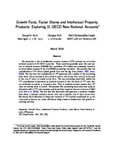
Figure 1. A, Percentage of flow cytometry–sorted VEGFR-1⫹, VEGFR-2⫹, and Tie-2⫹ in PBMCs. B, Western blotting of PBMC lysates with antibodies to VEGFR-1, VEGFR-2, and Tie-2 gave expected bands of ⬇200, 200, and 140 kDa, respectively. C, Electron microscopy illustrates that freshly sorted VEGFR-1⫹, VEGFR-2⫹, and Tie-2⫹ cell populations contained mixture of cells comprising ⬎95% mononuclear monocytes (M) and macrophages (M), 2% lymphocytes (L), and ⬍1% polymorphonuclear cells (PMN). D, Transmission electron photomicrographs illustrate that VEGFR-2⫹ and Tie-2⫹ cells differentiated into endothelial cells demonstrating presence of Weibel-Palade bodies (rounded arrow) and tight junctions typical for endothelial cells (arrow) and fenestrae (arrow). All other abbreviations are as defined in text.
Methods Protocol The local ethics committee approved the present study.
In Vitro Studies Isolation of VEGFR-1ⴙ, VEGFR-2ⴙ, and Tie-2ⴙ Cells From Peripheral Blood Peripheral blood mononuclear cells (PBMCs) expressing VEGFR-1, VEGFR-2, or Tie-2 were obtained first by isolating mononuclear cells by Ficoll density-gradient centrifugation of human blood buffy coats. PBMCs were distributed into several tubes, each containing 4⫻107 cells. Fifteen microliters of anti–VEGFR-1 (4 g/mL, R&D Systems), anti–VEGFR-2 (20 g/mL, Reliatech), or anti–Tie-2 (5 g/mL, Becton Dickinson) antibody were added to each tube and incubated for 30 minutes at room temperature. After being washed once with phosphate-buffered saline, the cells were labeled with 10 L of a 1:10-diluted secondary goat anti-mouse monoclonal antibody. Cells were sorted by fluorescence cytometry by using a stringent gate to obtain highly purified populations of these cells (Figure 1A–1C). To obtain VEGFR-2⫺ and Tie-2⫺ populations, cells were incubated with magnetic particles (Miltenyi Biotec) coated with anti–VEGFR-2 and anti–Tie-2 antibodies. The procedure was followed as described by the manufacturer. The negative fractions were used as VEGFR-2⫺ and Tie-2⫺ populations after confirming by flow cytometry to make sure that no contaminating VEGFR-2⫹ or Tie-2⫹ cells were present.
Western Blotting Lysates of PBMCs (106) were immunoblotted with anti–VEGFR-1, anti–VEGFR-2, and anti–Tie-2 antibodies (all 1:200) by standard sodium dodecyl sulfate–polyacrylamide gel electrophoresis and Western blots analysis. Horseradish peroxidase– conjugated F(ab⬘)2 fragments of goat anti-mouse secondary antibodies (1:2000, Jackson
Immunoresearch) were used. Antibody-binding components were detected by enhanced chemiluminescence (ECL kit, Amersham).
Flow Cytometry and Cell Cultivation Phenotyping of precursor endothelial cells was performed as described earlier15 with an array of antibodies to specific markers expressed on hematopoietic and endothelial cells (Table 1). Antibodies were purchased as follows: anti–von Willebrand factor, anti-CD142, and anti– human leukocyte antigen class I and II from DAKO; anti–acetylated LDL from Molecular Probes; anti–␣-actin and anti-fibroblast antibody from Serotec; anti-CD144, anti-CD141, and anti-human nuclei antibody from Chemicon; and Ulex europaeus from Sigma. All other antibodies were purchased from Becton Dickinson. Corresponding control isotypes were used for evaluation of nonspecific binding of monoclonal antibodies. The cells were analyzed on a Becton Dickinson fluorescence-activated flow cytometer (FACSorter). Sorted VEGFR-1⫹, VEGFR-2⫹, and Tie-2⫹ cells were cultivated on fibronectin-coated, tissue-culture plates in endothelium-selective medium (Clonetics). Human umbilical vein endothelial cells (HUVECs) were purchased from Clonetics and cultivated in the recommended medium. HUVECs were used as control cells.
Electron Microscopy
Sorted VEGFR-1⫹, VEGFR-2⫹, and Tie-2⫹ cells were grown on membrane filters on 24-well plates. The procedure for preparation of cells for scanning and transmission electron microscopy has been described elsewhere.16
Migration Assay
The migration assay with freshly isolated VEGFR1⫹, VEGFR-2⫹, and Tie-2⫹ cells was performed as reported elsewhere.17 Varying concentrations of VEGF (R&D Systems; 0, 0.5, 2, and 10 ng/mL) in medium containing 5% fetal calf serum and angiopoietin-1 (the ligand for Tie-2; R&D Systems) at concentrations of 0, 100, 200, and 300 ng/mL were used. Each experiment was performed in triplicate wells and repeated 4 times.
Nowak et al
Characterization of Circulating Endothelial Progenitors
3701
TABLE 1. Phenotypic Characteristics of Freshly Isolated VEGFR-1ⴙ, VEGFR-2ⴙ, and Tie-2ⴙ Cells From Peripheral Blood Antibodies to
VEGFR-1⫹ Cells (n⫽7), %
VEGFR-2⫹ Cells (n⫽7), %
Tie-2⫹ Cells (n⫽7), %
HUVECs, %
Monocyte/macrophage markers CD15
5.5⫾0.4
5⫾0.3
13⫾0.2
0
CD64
60⫾0.03
15⫾0.01
80⫾0.05
0
CD14
93⫾0.02
80⫾0.04
75⫾0.25
0
CD11b
97⫾0.02
81⫾0.1
90⫾0.01
0
CD11c
72⫾0.2
54⫾0.4
65⫾0.1
0
CD68
0
0.4⫾0.05
CD83
0.49⫾0.05
3.7⫾0.05
0.07⫾0.01
0
1.0⫾0.02
0.19⫾0.1
5
2⫾0.5
0
Stem/progenitor/mature endothelial cell markers CD34
0.05⫾0.1
CD123
4⫾0.03
2⫾0.02
5⫾0.03
0
AC133
0.21⫾0.02
0.2⫾0.01
0.8⫾0.01
0
CD90
0.7⫾0.3
1.3⫾0.1
2⫾0.3
100
100
Ac-LDL CD31 CD105
75⫾0.04 100
70⫾0.2 100
100 77⫾0.03 100
0 100 100 100
VEGFR-2
52⫾0.4
92⫾0.03
6⫾0.4
12
VEGFR-1
95⫾0.03
5⫾0.03
2.7⫾0.01
7
Tie-2
4.3⫾0.03
CD144
0
55⫾0.2 6⫾0.03
94⫾0.03
12
2⫾0.25
100
CD141
1⫾0.05
6⫾0.2
5⫾0.13
100
CD142
4⫾0.06
3⫾0.02
4⫾0.02
100*
HLA class I
90⫾0.03
83⫾0.03
80⫾0.05
100
HLA-DR
20⫾0.03
23⫾0.03
29⫾0.01
100*
Ulex europaeus
100
100
100
100
Control markers
␣-Actin Fibroblasts
1⫾0.03
1.0⫾0.01
1.0⫾0.07
0
6.6⫾0.01
38.45⫾0.01
0.1⫾0.05
0
94⫾0.03
63⫾0.03
CD45
100
CD3
0
0
0
0
0
CD56⫹16
0
0
0
0
Ac indicates acetylated; HLA, human leukocyte antigen. *All cells expressed HLA-DR on activation with interferon-␥ and tumor necrosis factor-␣.
Capillary-Like Tubule Formation Assay The formation of capillary-like tubular structures was assessed in Matrigel-coated multiwell plates as described previously.18
Cell Invasion Assay Cell invasion through reconstituted basement membrane Matrigel was assayed by a method reported previously.19 Migrated cells were counted in 5 different fields under a light microscope at ⫻100 magnification. Each experiment was performed in triplicate wells and repeated 4 times.
In Vivo Studies Because our in vitro studies had indicated that the VEGFR-1⫹ cell population had the fewest endothelium-like cell characteristics, the in vivo studies were performed with only VEGFR-2⫹ and Tie-2⫹ cells.
Mice Balloon injury of the right femoral artery was performed as follows. Nude C57 black mice (Taconic M&B, Ejby, Denmark) with a body
weight of 20 to 25 g were anesthetized with isoflurane, and the right femoral artery was exposed to the level of the bifurcation through a transabdominal incision. Microclamps (S&T) were placed on the lower aorta, left iliac artery, and the distal part of the right femoral artery. A 2F Fogarty balloon catheter (Baxter) was introduced into the right femoral artery, inflated, and withdrawn 3 times with rotation. Inflation was performed through the cannula and 1 mL of Ringer’s solution with free outflow through the microincision. Cells (5⫻105 cells per animal) in 100 L of MCDB medium (VEGFR-2⫹, n⫽6; Tie-2⫹, n⫽6; VEGFR-2⫺, n⫽6; and Tie-2⫺, n⫽6) were instilled through the same cannula and incubated in the freshly injured arterial bed for 15 to 20 minutes, whereas controltransplanted mice received only culture medium (n⫽4 mice). After incubation, unbound cells were aspirated, the catheter was removed, and the microincision was sutured with 11-0 nylon (S&T) interrupted sutures. Blood flow was restored by removing the microclamps. All animal procedures were performed in accordance with institutional guidelines and conformed to the Guide for the Care and Use of Laboratory Animals at Huddinge University Hospital in Sweden. Animals were humanely killed at 2 and 4 weeks after human cell
3702
Circulation
December 14, 2004
transplantation, and the right femoral artery was harvested for histopathological examinations. Vessels were embedded in OCT compound and frozen in LN2. To demonstrate activation of DNA synthesis in the human VEGFR-2⫹ and Tie-2⫹ cells in vivo, the thymidine analogue bromodeoxyuridine (BrdU, Sigma) was given as single intraperitoneal injections, 100 mg/kg body weight, 12 and 24 hours before termination.
TABLE 2. Phenotypic Characteristics of VEGFR-2ⴙ and Tie-2ⴙ Cells After 2 Weeks in Culture Antibodies to
VEGFR-2⫹ Cells (n⫽7), %
Tie-2⫹ Cells (n⫽7), %
CD141
95⫾4.5
90⫾3.1
CD142*
98⫾1.9
96⫾4.2
Immunohistochemistry
CD144
95⫾3.8
91⫾3.8
To localize human cells in the mouse artery, 5-m cryosections were stained with a mouse anti-human nuclei monoclonal antibody (Chemicon),20 developed in diaminobenzidine with the Vectastain Elite ABC kit (Sigma), and counterstained with hematoxylin. BrdU incorporation was detected with use of a commercial kit (Sigma). In some instances, detection of BrdU was performed using 3-m cryosections. The phenotype of transplanted human cells was detected by immunofluorescence studies. Sections were doubly stained with antibodies to human nuclei (1:50) and CD31 (1:100), von Willebrand factor (1:100), or ␣-actin (1:200), followed by staining with secondary anti-mouse subclass-specific fluorescein isothiocyanate/Texas red– conjugated antibodies (1:500). Endothelialization was calculated as the ratio of the surface covered by human diaminobenzidine-Nickel–positive cells and the total luminal surface. For histomorphology (3 animals per group, 12 sections/femoral artery), cross sections of the artery were stained with hematoxylin/eosin and examined for vessel diameter, media area, and intima-media area ratio.
CD14
Reverse Transcriptase–Polymerase Chain Reaction Total RNA was extracted from 3 human-mouse chimeric murine femoral arteries and 3 sham-transplanted mice at 4 weeks after transplantation with use of the Micro-FastTrack RNA isolation kit (Invitrogen). We used human-specific primers to detect human CD31 in the EPC-transplanted mouse arteries. We selected primers for CD31 and glucose-6-phosphate dehydrogenase (G6PD, the housekeeping gene) by using Primer Express software, version 2.0 (Applied Biosystems). Primer sets were commercially synthesized by CyberGene. Primer sequences were as follows: CD31 forward, 5⬘-CCA CTG CAG AGT ACC AGG TGT TGG-3⬘; CD31 reverse, 5⬘-ATC GAG AAG GAG CGT TTC T-3⬘, with an expected product size of 230 bp; G6PD forward, 5⬘-TGC CCC CGA CCG TCT AC-3⬘; and G6PD reverse, 5⬘-ATG CGG TTC CAG CCT ATC TG-3⬘, with an expected product size of 76 bp. The PCR was carried out as described earlier.16
Statistical Analysis Where applicable, results are presented as mean⫾SEM. An unpaired t test was used for comparisons between cell-transplanted and sham (medium only)-treated groups. P⬍0.05 was considered significant.
Results In Vitro Studies VEGFR-1ⴙ, VEGFR-2ⴙ, and Tie-2ⴙ Cells From Peripheral Blood Belong to the Monocyte/Macrophage Lineage We found that VEGFR-1⫹, VEGFR-2⫹, and Tie-2⫹ cells constitute ⬇3⫾0.2%, 0.8⫾0.5%, and 2⫾0.3%, respectively, of the total population of mononuclear cells in blood (Figure 1A). Expression of these receptors on PBMCs was further supported by the fact that PBMC lysates, when immunoblotted with anti–VEGFR-1, anti–VEGFR-2, and anti–Tie-2 antibodies, gave the expected bands of ⬇200, 200, and 140 kDa, respectively (Figure 1B). Phenotypic analysis of the 3 populations demonstrated a mixture of cell types belonging mainly to the monocyte/ macrophage cell lineage (Table 1). In addition, all 3 populations expressed the CD45 marker, indicating the hematopoietic origin of these cells. Electron microscopic studies of freshly isolated VEGFR-1⫹, VEGFR-2⫹, and Tie-2⫹ cells further supported the
CD31 CD106* CD62E*
0 92⫾7.2 100 20⫾4.3
0 90⫾6.8 100 20⫾6.2
CD11b
0
0
CD15
0
0
CD68
0
0
0.07⫾3.2
0.08⫾1.5
0.5⫾0.8
0.4⫾0.2
Fibroblasts
␣-Actin
*Expressed only after activation of cells with cytokines interferon-␥ and tumor necrosis factor-␣.
findings of the phenotypic analysis that all 3 populations contained a mixture of cells, including mainly monocytes/macrophages (⬎95%), lymphocytes, and some polymorphonuclear cells (⬍1%) (Figure 1C). Cultivated VEGFR-2ⴙ and Tie-2ⴙ Cells Exhibit Exclusively Endothelial Cell Markers Cultivation of these cells on fibronectin-coated culture wells showed 2 populations of adherent (80%) and nonadherent (20%) cells. The nonadherent cells were discarded after 1 week and could not be further cultivated under the conditions described for endothelial cells. After 2 weeks in culture, the majority of adherent cells in the VEGFR-2⫹ and Tie-2⫹ cultures expressed the endothelial cell markers CD144 and CD141 and, on activation, expressed CD106 and CD142 (Table 2). Further analysis by electron microscopy confirmed the endothelial morphology of these cells, demonstrating the presence of fenestrae, Weibel-Palade bodies, and tight junctions typical of endothelial cells (Figure 1D). On the other hand, only a small fraction of VEGFR-1⫹ cells remained adherent after 2 weeks, and sufficient cells were not obtained for further analysis. Neither VEGFR-2⫹ nor Tie-2⫹ cells proliferated in culture. However, these populations could be kept viable in culture without proliferation for ⬎4 weeks. VEGFR-2ⴙ and Tie-2ⴙ Cells Have Migratory and Invasive Capacity and Form Tubule-Like Structures Freshly isolated VEGFR-2⫹ and Tie-2⫹ but not VEGFR-1⫹ cells migrated toward VEGF and angiopoietin-1 (Figure 2Aand 2B). However, 60% of VEGFR-1⫹, 70% of VEGFR2⫹, and 65% of Tie-2⫹ cells added to Matrigel migrated to the lower chamber in the invasion assay, indicating that all 3 cell types had invasive capacity (Figure 2C). In Matrigel, these freshly isolated cells showed that only VEGFR-2⫹ and Tie-2⫹ cells could form tubule-like structures, but VEGFR-1⫹ cells did not have the ability to do so (Figure 2D–2G). Thus, these data indicate that freshly isolated VEGFR-2⫹ and Tie-2⫹ cells phenotypically belonged to the monocyte/ macrophage lineage. However, cultured VEGFR-2⫹ and
Nowak et al
Characterization of Circulating Endothelial Progenitors
3703
Figure 2. A and B, In migration assay, VEGFR-2⫹ and Tie-2⫹ but not VEGFR-1⫹ cells migrated toward VEGF and angiopoietin-1. C, However, all 3 populations of cells showed invasive capacity in Matrigel cell invasive assay. Results are presented as mean⫾SEM. D–G, VEGFR-2⫹ and Tie-2⫹ but not VEGFR-1⫹ cells formed capillary-like tubules in Matrigel. HUVECs served as control cells. Abbreviations are as defined in text.
Tie-2⫹ cells phenotypically and morphologically resembled endothelial cells, whereas VEGFR-1⫹ cells did not exhibit endothelial cell–like characteristics. We therefore concluded that the EPCs were derived from monocytes.
In Vivo Studies VEGFR-2ⴙ/CD14ⴙ and Tie-2ⴙ/CD14ⴙ Cells Significantly Contribute to Efficient Repopulation of Denuded Mouse Femoral Arteries and Proliferate In Vivo EPC-transplanted vessels showed no sign of thrombus. The specificity of the human nuclei antibody was first determined by using a normal human and mouse artery. Human cells stained black (Figure 3A), whereas the control mouse artery did not stain with the human nuclei antibody (Figure 3B). At 2 weeks after transplantation, human cells were detected on the luminal surface of VEGFR-2⫹– or Tie-2⫹– seeded, balloon-injured femoral artery (diaminobenzidineNi–positive; Figure 3C and 3D). At 4 weeks after transplantation, a high degree of repopulation was seen, and ⬇77⫾6% or 68⫾4% of the lesion was covered with VEGFR-2⫹or Tie-2⫹ human cells, respectively. However, this was not observed in animals transplanted with
VEGFR-2⫺ or Tie-2⫺ cells, even 4 weeks after transplantation (Figure 3E and 3F). The presence of human cells in the mouse artery was once again confirmed in the immunofluorescence studies (Figure 3G–3I). The endothelial cell phenotype in vivo was confirmed by staining with antibodies to von Willebrand factor and VE-cadherin (CD144, Figure 4A and 4B). Each section was doubly stained with the anti-human nuclear antibody to differentiate human cells from host cells (yellow staining indicating doubly positive cells). Interestingly, transplanted human cells were found not only on the luminal surface of the seeded arteries but also in the interstitial media. When labeled with an antibody to ␣-actin, cells in the media stained positive, indicating the presence of smooth muscle cells (Figure 4C). Thus, both VEGFR-2⫹ and Tie-2⫹ cells differentiated in vivo not only into endothelial cells but also into smooth muscle cells. Transcription of Human Endothelium-Specific Gene in Mice Transplanted With Human Endothelial Progenitor Cells We further confirmed engraftment of the transplanted cells by determining the expression of human genes in the transplanted mice. We analyzed femoral arteries of the mice killed 1 month
3704
Circulation
December 14, 2004
Figure 3. A and B, Human nuclei antibody–stained cells (black) in human but not control mouse artery. Balloon-injured mouse femoral artery showed localization of transplanted human cells on luminal surface of seeded artery. C, VEGFR-2⫹ cells (arrows) at 4 weeks and Tie-2⫹ cells (D) (arrows) at 2 weeks. E–F, However, no localization of transplanted VEGFR-2⫺ and Tie-2⫺ cells was observed at 4 weeks. Original magnification, ⫻60. Immunofluorescence staining of (G) normal human artery, (H) injured mouse artery transplanted with human VEGFR-2⫹ cells and (I), VEGFR-2⫺–transplanted mouse artery with the anti– human nuclei antibody (arrows). Abbreviations are as defined in text. Tissue sections in A through F are 5 m thick.
after transplantation with human EPCs by RT-PCR and primers specific for the human endothelium-specific gene CD31. RNA from the arteries of 3 sham-transplanted nude mice resulted in amplification for G6PD, which was used as a control for the integrity of the RNA. The CD31 primers were species-specific for humans, because they did not amplify the respective mouse gene (Figure 4D). These results show that the transplanted human EPCs engrafted the damaged mouse artery. VEGFR-2ⴙ and Tie-2ⴙ Cells Proliferate In Vivo Neither VEGFR-2⫹ nor Tie-2⫹ cells proliferated in vitro (data not shown), but to obtain evidence for proliferation in the
transplanted VEGFR-2⫹ and Tie-2⫹ cells, BrdU incorporation studies were performed in 8 animals (VEGFR-2⫹, n⫽4; Tie-2⫹, n⫽4). Analysis of tissue at 7 days showed extensive BrdU incorporation in transplanted human cells. Because both the anti-BrdU and the human nuclei antibodies stain DNA, double staining of the same tissue section was not possible. Therefore, 3-m consecutive sections were stained with anti-BrdU (brown) and the anti-human nuclei antibody (black) to show proliferation in the transplanted cells (Figure 5A–5F). These findings show that VEGFR-2⫹ and Tie-2⫹ cells not only repopulated the injured arteries but also proliferated in vivo.
Nowak et al
Characterization of Circulating Endothelial Progenitors
3705
Figure 4. Endothelial cell phenotype of transplanted (Tx) human cells was confirmed by double staining with human nuclei antibody and antibodies to (A) von Willebrand factor for VEGFR-2⫹ cells (doubly positive cells are stained yellow, arrows) and (B) VE-cadherin for Tie-2⫹ cells. C, When labeled with antibody to ␣-actin, cells (green, filamentous staining pattern) in media stained positive, indicating presence of smooth muscle cells. Original magnification, ⫻60. Tissue sections in A through C are 5 m thick. D, Human CD31 was detected in balloon-injured femoral arteries of mice that received human EPCs but not in sham-transplanted mice. G6PD was used as housekeeping gene. All other abbreviations are as defined in text.
Reendothelialization With EPCs Did Not Result in Neointima Proliferation After Balloon Injury of Mouse Femoral Arteries Cross sections of injured arterial segments were examined at 2 and 4 weeks after transplantation for histomorphological changes. The balloon injury procedure in the mouse femoral artery resulted in dilation of the lesioned arterial segment, so that the vessel diameter was significantly increased from 0.362⫾0.060 mm in uninjured control arteries to 0.598⫾0.026, 0.559⫾0.057, and 0.601⫾0.036 mm in injured vessels that were incubated with VEGFR-2⫺ or Tie-2⫺ cells or medium alone, respectively (P⬍0.0.001). This widening was not found in femoral arteries of mice that had received transplantation of human cells (Figure 5G). A reduction in cross-sectional media area from 0.020⫾0.004 to 0.010⫾0.009 mm2 was observed in injured arteries. Thinning of media area was not seen in VEGFR-2⫹ and Tie-2⫹ cell–transplanted arteries (Figure 5H). Reendothelialization mediated by EPC seeding did not result in neointima formation in femoral arteries at 2, 4, or 6 weeks after injury. In fact, intima-media area ratio was not significantly higher in VEGFR-2⫹– and Tie-2⫹–transplanted vessel segments (normal uninjured artery, 0.01⫾0.01; medium control,
0.02⫾0.01; VEGFR-2⫹, 0.02⫾0.01; Tie-2⫹, 0.02⫾0.01; P⫽NS).
Discussion Much debate currently exists as to how an endothelial progenitor is truly defined. Several cell populations defined by phenotypic markers have been suggested as candidates for EPCs. Currently, EPCs are defined by the expression of antigens shared by embryonic endothelial progenitors and hematopoietic stem cells (CD34/VEGFR-2, CD133/VEGFR-2, and CD34).3,13,14 However, EPC populations defined by expression of other markers have also been isolated.21 Thus, the lineage and exact phenotype of EPCs are not yet known. In the present study, our aim was to identify and isolate a population of human cells expressing endothelium-specific markers regardless of the expression of hematopoietic stem cell markers. We hoped to find a population(s) of cells that (1) is present at a higher frequency in blood compared with the previously described EPCs and (2) would perform the functions of cells involved in reendothelialization of denuded vessels. For this purpose, we isolated 3 populations of cells from peripheral blood expressing endothelium-specific tyrosine
3706
Circulation
December 14, 2004
Figure 5. Representative 3-m consecutive sections of (A and B) sham-transplanted mouse artery and (C–F) transplanted mouse artery stained with anti-BrdU (brown cells) and human nuclei antibody (black), showing proliferation in transplanted VEGFR-2⫹ and Tie-2⫹ cells, respectively. G and H, Balloon-injury procedure in mouse femoral artery resulted in dilation of lesioned arterial segment and reduction in cross-sectional media area in injured vessels incubated with control cells and medium alone but not in femoral arteries of mice that had received VEGFR-2⫹ and Tie-2⫹ human cells. Abbreviations are as defined in text.
kinase receptors. Our results demonstrated that PBMCs expressing the tyrosine kinase receptors VEGFR-1, VEGFR-2, or Tie-2 phenotypically represent a mixed population of cells mainly belonging to the monocyte/macrophage lineage. VEGFR-2⫹ and Tie-2⫹ cell populations demonstrated features required by cells involved in revascularization. Both cell populations demonstrated the ability to migrate toward angiogenic growth factors VEGF or angiopoietin-1, had invasive capacity, formed capillary-like tubules, and responded to vascular injury by restoring the endothelial lining of damaged arteries. In addition, these cells also differentiated into smooth muscle cells. These observations indicate that VEGFR-2⫹ and/or Tie-2⫹ cells can be recruited to sites of vascular damage and may take part in reendothelialization during vascular injury. Importantly, neither VEGFR-2⫺ nor Tie-2⫺ populations demonstrated any of the aforementioned functional properties. Compared with previous reports, the percentages of VEGFR-1, VEGF-R2, and Tie-2
expression among PBMCs are slightly higher in our study. This discrepancy may be explained by the fact that expression of a specific hematopoietic marker was not a prerequisite for our definition of EPCs. It has been reported that peripheral blood EPCs are derived from a monocyte/macrophage cell lineage.22 Recently, Urbach et al23 demonstrated that EPCs can be generated from both monocytic (CD14⫹) and nonmonocytic (CD14⫺) peripheral blood cells. This finding demonstrates that expression of the monocytic lineage marker per se does not define a functionally active cell capable of improving neovascularization. Our results, on the other hand, indicate that expression of endothelium-specific tyrosine kinase receptors on peripheral blood cells, irrespective of cell lineage, defines a functionally active cell population capable of reendothelialization. We believe that in the study reported by Urbich et al both the monocytic and nonmonocytic populations expressed the tyrosine kinase receptor VEGFR-2
Nowak et al
Characterization of Circulating Endothelial Progenitors
and therefore, were equally capable of neovascularization. The same study also reported no functional improvement of neovascularization when freshly isolated monocytes (CD14⫹) were used. However, in the present study, when freshly isolated VEGFR-2⫹ and Tie-2⫹ cells were injected into denuded grafts, these cells repopulated the injured vessels and proliferated in vivo. It would be interesting to study the functional capacity of CD14⫹/VEGFR-2⫹ or CD14⫹/Tie-2⫹ and CD14⫹/VEGFR-2⫺ or CD14⫹/Tie-2⫺ subpopulations in reendothelialization. Such studies are under way at our center. In our study, VEGFR-1⫹ cells did not exhibit characteristics of endothelial cells. The reason for this is unclear. It is possible that the anti–VEGFR-1 antibodies used for isolation of the VEGFR-1⫹ population in the present study were blocking antibodies that prevented in vitro migration, tubule formation, and in vivo functional capacity of reendothelialization. Another possible explanation could be that the weak signaling abilities of VEGFR-1 might act as a negative regulator of angiogenesis.8 Interestingly, a very low percentage of VEGFR-1⫹ cells expressed VEGFR-2. Thus, lack of VEGFR-2 on these cells may also contribute to the “nonendothelial” character of VEGFR-1⫹ cells. Interestingly, both VEGFR-2⫹ and Tie-2⫹ cells differentiated not only into endothelial cells but also into smooth muscle cells. Studies have demonstrated the presence of functional VEGFRs (VEGFR-2 and VEGFR-4) on vascular smooth muscle cells.24 In fact, a common progenitor to endothelial and smooth muscle cells has been proposed.25 In the presence of platelet-derived growth factor, murine embryonic stem cells expressing VEGFR-2 also gave rise to smooth muscle, actin⫹ mural cells.26 Our results further support this concept and indicate that transplantation of VEGFR-2⫹ or Tie-2⫹ cells may augment healing in vessels with both intima and media destruction. Our data indicate a time-dependent increase in the area covered by the transplanted cells, indicating proliferation of these cells in vivo. Balloon injury may have provided a microenvironment that was conducive to the proliferation of EPCs by producing several factors, including growth factors, cytokines, and other proteins, and may help explain why the cells proliferated in vivo but not in vitro. This exciting finding suggests that probably smaller numbers of cells without the need for extensive in vitro expansion may be used for transplantation. Reendothelialization with VEGFR-2⫹ and Tie-2⫹ cells restored vessel diameter and media thickness, and importantly, the proliferative capacity of the cells did not result in neointima formation in balloon-injured mouse femoral arteries. In summary, our data show 2 novel findings: (1) when freshly isolated circulating VEGFR-2⫹ or Tie-2⫹ cells belonging to the monocytic/macrophage lineage were transplanted, they exhibited unique functional competence to improve reendothelialization in balloon-injured femoral arteries and (2) these cells proliferated in vivo. Thus, by restoring an intact endothelium, these cells may participate in the maintenance of vascular homeostasis and may be used as cell-based therapy for vascular diseases.
3707
Acknowledgments The present study was financed by grants from the Lars Erik Gelins Foundation and the Swedish Research Council No. K2002-06X14004-02B to Dr Holgersson.
References 1. Bennett MR, O’Sullivan MO. Mechanisms of angioplasty and stent restenosis: implications for design of rational therapy. Pharmacol Ther. 2001; 91:149 –166. 2. Schwartz RS. Pathophysiology of restenosis: interaction of thrombosis, hyperplasia, and/or remodeling. Am J Cardiol. 1998;81:14E–17E. 3. Asahara T, Murohara T, Sullivan A. Isolation of putative progenitor endothelial cells for angiogenesis. Science. 1997;275:964 –967. 4. Shintani S, Murohara T, Ikeda I, et al. Therapeutic potential of ex vivo expanded endothelial progenitor cells for myocardial ischemia. Circulation. 2001;103:897–903. 5. Yancopoulos GD, Davis S, Gale NW, et al. Vascular-specific growth factors and blood vessel formation. Nature. 2000;407:242–248. 6. Veikkola T, Alitalo K. VEGFs, receptors and angiogenesis. Semin Cancer Biol. 1999;9:211–220. 7. Neufeld G, Cohen T, Gengrinovitch S, et al. Vascular endothelial growth factor (VEGF) and its receptors. FASEB J. 1999;13:9 –22. 8. Karkkainen MJ, Petrova T. Vascular endothelial growth factor receptors in the regulation of angiogenesis and lymphangiogenesis. Oncogene. 2000;19:5598 –5605. 9. Sawano A, Iwai S, Sakurai Y, et al. Flt-1, vascular endothelial growth factor receptor 1, is a novel cell surface marker for the lineage of monocyte-macrophages in humans. Blood. 2001;97:785–791. 10. Partanen J, Dumont DJ. Functions of Tie1 and Tie2 receptor tyrosine kinases in vascular development. Cur. Top Microbiol Immunol. 1999; 237:159 –172. 11. Merenmies J, Parada LF, Henkemeyer M. Receptor tyrosine kinase signaling in vascular development. Cell Growth Diff. 1997;8:3–10. 12. Jones N, Dumont DJ. Tek/Tie2 signaling: new and old partners. Cancer Met Rev. 2000;19:13–17. 13. Choi K, Kennedy M, Kazarov A, et al. A common precursor for hematopoietic and endothelial cells. Development. 1998;125:725–732. 14. Yin AH, Miraglia S, Zanjani ED, et al. AC133, a novel marker for human hematopoietic stem and progenitor cells. Blood. 1997;90:5002–5012. 15. Vermehren D, Sumitran-Holgersson S. Isolation of precursor endothelial cells from peripheral blood for donor-specific crossmatching before organ transplantation. Transplantation. 2002;14:1479 –1486. 16. Bo X, Broomé U, Uzunel M, et al. Capillarization of hepatic sinusoid by liver endothelial cell-reactive autoantibodies in patients with cirrhosis and chronic hepatitis. Am J Pathol. 2003;163:1275–1289. 17. Ming WJ, Bersani L, Mantovani A. Tumor necrosis factor is chemotactic for monocytes and polymorphonuclear leukocytes. J Immunol. 1987;138: 1469–1474. 18. Farina AR, Tacconelli A, Cappabianca L, et al. Thioredoxin inhibits microvascular endothelial capillary tubule formation. Exp Cell Res. 2003; 291:474 – 483. 19. Lakka SS, Jasti SL, Gondi C, et al. Downregulation of MMP-9 in ERK-mutated stable transfectants inhibits glioma invasion in vitro. Oncogene. 2002;21:5601–5608. 20. Kohama I, Lankford KL, Preiningerova J, et al. Transplantation of cryopreserved adult human Schwann cells enhances axonal conduction in demyelinated spinal cord. J Neurosci. 2001;21:944 –950. 21. Burger PE, Coetzee S, McKeehan WL, et al. Fibroblast growth factor receptor-1 is expressed by endothelial progenitor cells. Blood. 2002;100:3527–3535. 22. Rehman J, Li J, Orschell CM, et al. Peripheral blood ‘endothelial progenitor cells’ are derived from monocytes/macrophages and secrete angiogenic growth factors. Circulation. 2003;107:1164 –1169. 23. Urbich C, Heeschen C, Aicher A, et al. Relevance of monocytic features for neovascularization capacity of circulating endothelial progenitor cells. Circulation. 2003;108:2511–2516. 24. Ishida A, Murray J, Saito Y, et al. Expression of vascular endothelial growth factor receptors in smooth muscle cells. J Cell Physiol. 2000;188:359–368. 25. Simper D, Stalboerger PG, Panetta CJ, et al. Smooth muscle progenitor cells in human blood. Circulation. 2002;106:1199 –1204. 26. Yamashita J, Itoh H, Hirashima M, et al. Flk1-positive cells derived from embryonic stem cells serve as vascular progenitors. Nature. 2000;408:92–96.
Correction The article “Expression of Vascular Endothelial Growth Factor Receptor-2 or Tie-2 on Peripheral Blood Cells Defines Functionally Competent Cell Populations Capable of Reendothelialization” by Nowak et al, which originally appeared online on September 20, 2004, and which published in the present issue of Circulation, has been modified. The editors of the journal asked the authors of this article to revise Figure 5 and all text pertaining to Figure 5 because of a problem detected by a reader of the original online version. Those revisions are reflected in this issue.
(Circulation. 2004;110:3741.) © 2004 American Heart Association, Inc. Circulation is available at http://www.circulationaha.org
DOI: 10.1161/01.CIR.0000152260.41340.00
3741