H-NS family members function coordinately in an opportunistic pathogen Sandra Castang1, Heather R. McManus1, Keith H. Turner, and Simon L. Dove2 Division of Infectious Diseases, Children’s Hospital, Harvard Medical School, Boston, MA 02115 Edited by John J. Mekalanos, Harvard Medical School, Boston, MA, and approved October 9, 2008 (received for review August 19, 2008)
MvaT 兩 MvaU 兩 Pseudomonas aeruginosa
T
he histone-like nucleoid structuring protein H-NS is a prominent global regulator of gene expression found in a number of different Gram-negative bacteria. Although H-NS silences the expression of hundreds of genes, it also plays a role in DNA compaction (1). Furthermore, several genome-wide association studies in enteric bacteria have revealed that H-NS displays a marked preference for binding AT-rich regions of DNA (2–5). In so doing H-NS provides a mechanism for selectively silencing so called xenogeneic DNA (DNA acquired from a foreign source), such as that acquired through horizontal transfer, and whose AT-content is typically higher than that of the cognate genome (3, 5–7). Although traditionally thought of as a nonsequence-specific DNA-binding protein, recent evidence suggests that H-NS binds specific sites on the DNA with high affinity (8). Once bound to these sites, which are themselves AT-rich, H-NS is thought to oligomerize across adjacent AT-rich regions of DNA, forming an extensive nucleoprotein complex that facilitates gene silencing (8). Many bacteria contain more than one H-NS family member (9). Of the two in Escherichia coli K-12 (H-NS and its paralog StpA), only H-NS appears to play a significant role in controlling gene expression. StpA is less abundant than H-NS, and although it can associate with H-NS, and can functionally substitute for H-NS, it is not thought to influence the expression of any H-NS-regulated gene (1). However, in uropathogenic E. coli, H-NS and StpA are thought to have both overlapping and distinct functions, with one regulon controlled exclusively by H-NS, and a second smaller regulon under the control of both H-NS and StpA (10). Nevertheless, in most bacteria it is not known whether H-NS family members have overlapping or distinct functions. The Gram-negative bacterium Pseudomonas aeruginosa is an opportunistic pathogen that is best known through being the principal cause of morbidity and mortality in cystic fibrosis (CF) patients (11). The organism uses an impressive battery of virulence factors to intoxicate the human host, and persists in the chronically infected CF lung as a biofilm (a community of cells encased in a polymeric matrix), a lifestyle that engenders inwww.pnas.org兾cgi兾doi兾10.1073兾pnas.0808215105
creased resistance to antibiotics, and exacerbates the problem of eradicating the organism from the lung. Foremost among the regulators of virulence gene expression and biofilm formation in P. aeruginosa is MvaT, one of the two H-NS family members present in this organism (12–14). That MvaT belongs to the H-NS family, despite sharing limited sequence similarity with H-NS, is suggested by multiple functional similarities and several structural predictions (15–19). MvaT was initially identified in P. aeruginosa as a global regulator of virulence gene expression (12). Subsequent microarray analyses revealed that MvaT controls the expression of at least 150 genes in P. aeruginosa (13, 14), including genes encoding virulence factors, house-keeping genes, and a preponderance of genes encoding proteins associated with the cell surface, some of which play important roles in biofilm formation (13, 14). However, it is not known which of these genes are regulated directly by MvaT. In addition, very little is known about which genes are regulated by the MvaT paralog MvaU. Although MvaU can interact with MvaT (18), and plays a role in regulating the expression of at least one MvaT-regulated gene, deletion of mvaU had no effect on the expression of several others (13, 18). Thus, the extent to which the MvaT and MvaU regulons overlap was unknown. Here we present evidence that the H-NS family members MvaT and MvaU regulate expression of the same set of target genes in P. aeruginosa. In particular, using chromatin immunoprecipitation (ChIP) coupled with fully tiled high density DNA microarrays (ChIP-on-chip) we show that MvaT and MvaU occupy the same regions of the chromosome, suggesting that MvaT and MvaU function coordinately. In addition to identifying those genes that are regulated directly by MvaT and MvaU, and extending the MvaT (/MvaU) regulon, these analyses reveal that, like H-NS from enteric bacteria, MvaT and MvaU bind preferentially to AT-rich regions of the DNA. Furthermore, using a ClpXP protease-based protein degradation system we show that the loss of both MvaT and MvaU from the cell cannot be tolerated. Depletion of MvaT in cells of an mvaU mutant strain results in overexpression of target genes, suggesting that the synthetic lethality that ensues is due to misregulated expression of certain target genes. These findings have implications for xenogeneic silencing in P. aeruginosa, and the function of H-NS and its paralogs in other bacteria. Author contributions: S.C., H.R.M., K.H.T., and S.L.D. designed research; S.C., H.R.M., and K.H.T. performed research; S.C., H.R.M., and K.H.T. contributed new reagents/analytic tools; S.C., H.R.M., K.H.T., and S.L.D. analyzed data; and S.C., H.R.M., K.H.T., and S.L.D. wrote the paper. The authors declare no conflict of interest. This article is a PNAS Direct Submission. 1S.C.
and H.R.M. contributed equally to this work.
2To
whom correspondence should be addressed at: Division of Infectious Diseases, Children’s Hospital, Enders Building, Room 754, 300 Longwood Avenue, Boston, MA 02115. E-mail:
[email protected].
This article contains supporting information online at www.pnas.org/cgi/content/full/ 0808215105/DCSupplemental. © 2008 by The National Academy of Sciences of the USA
PNAS 兩 December 2, 2008 兩 vol. 105 兩 no. 48 兩 18947–18952
MICROBIOLOGY
The histone-like nucleoid structuring protein, H-NS, is a prominent global regulator of gene expression. Many Gram-negative bacteria contain multiple members of the H-NS family of proteins. Thus, a key question is whether H-NS family members have overlapping or distinct functions. To address this question we performed genomewide location analyses with MvaT and MvaU, the two H-NS family members present in Pseudomonas aeruginosa. We show that MvaT and MvaU bind the same chromosomal regions, coregulating the expression of ⬇350 target genes. We show further that like H-NS in enteric bacteria, which functions as a transcriptional silencer of foreign DNA by binding to AT-rich elements, MvaT and MvaU bind preferentially to AT-rich regions of the chromosome. Our findings establish that H-NS paralogs can function coordinately to regulate expression of the same set of target genes, and suggest that MvaT and MvaU are involved in silencing foreign DNA elements in P. aeruginosa.
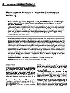
Fig. 1. MvaT and MvaU occupy the same AT-rich regions of the chromosome. (A) The percent GC content for 1 Mb of the PAO1 chromosome (in windows of 1000 bp across the genome with a window step size of 500 bp) is plotted (in red) against the locations of MvaT-V (plotted as log2 ratios in blue) and MvaU-V (plotted as log2 ratios in green) as determined by ChIP-on-chip. Association of MvaT-V and MvaU-V with the cgrA-cupA1 intergenic region (B), and with the lecA promoter and coding region (C). Log2 ratio values were normalized and averaged across three replicate arrays.
Results ChIP-on-Chip Reveals Extensive Overlap Between the MvaT and MvaU Regulons. To identify those genes that are controlled directly by
MvaT and MvaU, and determine the extent of the overlap between the MvaT and MvaU regulons we used ChIP-on-chip. To facilitate the immunoprecipitation of MvaT and MvaU we constructed two strains of P. aeruginosa PAO1 that synthesized epitope-tagged forms of MvaT and MvaU at native levels. In one of these strains (PAO1 MvaT-V) the native copy of the mvaT gene specified MvaT with a vesicular stomatitis virus glycoprotein (VSV-G) epitope tag fused to its C terminus (MvaT-V). The function of MvaT was not impaired by the presence of the VSV-G tag as MvaT-V fully repressed expression of the MvaTregulated cupA gene (data not shown). In the other strain (PAO1 MvaU-V) the native copy of the mvaU gene specified MvaU with a VSV-G epitope tag fused to its C terminus (MvaU-V). ChIP-on-chip with cells of the PAO1 MvaT-V strain revealed a total of 111 distinct genomic regions that were significantly enriched for MvaT (Fig. 1, see supporting information (SI) Fig. S1 and Table S1). Peaks corresponding to genomic regions associated with MvaT were located throughout the chromosome and encompassed both intergenic and coding regions (Fig. 1). Peaks ranged in size from ⬇0.2 to 22 kilobases, with an average width of ⬇2.5 kb (Table S1), consistent with the idea that MvaT is a member of the H-NS family of proteins that typically oligomerize across long stretches of target DNA (1, 6). To validate the MvaT ChIP-on-chip results, independent ChIP 18948 兩 www.pnas.org兾cgi兾doi兾10.1073兾pnas.0808215105
experiments were conducted and analyzed by using quantitative real-time PCR (qPCR). Of the 21 MvaT peaks tested, no false positives were detected and none of the 9 negative control regions showed significant enrichment (Fig. S2 A and data not shown). Furthermore, trends in the magnitude of foldenrichment of a specific target region were similar between ChIP-on-chip and ChIP datasets (Fig. S2 A). ChIP-on-chip and ChIP control experiments conducted using a strain that synthesized MvaT with a C-terminal tandem affinity purification (TAP) tag (PAO1 MvaT-TAP) (18), yielded results similar to those obtained with the PAO1 MvaT-V strain (Fig. S2B and data not shown), illustrating the robust nature of these results. An additional control performed with a strain synthesizing a TAPtagged form of the putative factor PA2896 confirmed the specificity of the association of MvaT with target regions (Fig. S2B). Finally, qPCR analyses of mock ChIP samples revealed no appreciable enrichment of any of the DNA regions that were tested (Fig. S2B and data not shown). ChIP-on-chip analyses of MvaU-V revealed a total of 102 distinct genomic regions that were significantly enriched for MvaU binding (Fig. 1, Fig. S1, and Table S2). Similar to our findings with MvaT, peaks corresponding to those regions of the chromosome associated with MvaU were found throughout the genome with each region spanning anywhere from approximately 0.1 to 21 kb, with an average width of approximately 2 kb. Independent ChIP experiments followed by qPCR were conducted to validate the MvaU-V results, with no false positives evident among the 14 peaks tested, and no appreciable enrichment of MvaU-V at any of the 5 negative control regions that were analyzed (Fig. S3). Comparison of the MvaT and MvaU-associated regions revealed a striking correspondence between the two (Fig. 1 and Fig. S1). In particular, 99 of the 102 regions associated with MvaU were also associated with MvaT. Furthermore, minimally relaxing the stringency of the criteria we used to identify peaks resulted in MvaU being enriched in all 111 of the regions associated with MvaT (data not shown). The extent of the overlap in the genomic regions associated with MvaT and MvaU strongly suggests that these two proteins control the expression of essentially the same set of genes. MvaT and MvaU Preferentially Associate with AT-Rich Regions of the Chromosome. In Salmonella enterica and E. coli (2–5), H-NS has
been shown to preferentially associate with AT-rich regions of the genome. To determine whether MvaT and MvaU show a similar binding preference, the percentage GC content within 1,000 bp windows across the PAO1 genome was calculated and compared with the average fold enrichment of MvaT-V and MvaU-V (expressed as a log2 ratio) within each window. A strong correlation between MvaT or MvaU enrichment and genomic AT content was found (Fig. 1, Fig. S1, and Fig. S4), suggesting that like H-NS, MvaT and MvaU display a distinct preference for binding AT-rich regions of the DNA and may be involved in xenogeneic silencing in P. aeruginosa. Consistent with this idea we found that MvaT and MvaU were associated with approximately two thirds of the so-called regions of genomic plasticity (PGRs) in P. aeruginosa PAO1 that represent strainspecific segments of DNA that may contribute to the ability of P. aeruginosa to adapt to particular environments (20). Many PGRs are thought to have been acquired by horizontal gene transfer and contain regions where the AT content is higher than that of the rest of the genome. Direct Targets of MvaT and MvaU. Our ChIP-on-chip analyses
revealed a total of 394 and 311 genes associated directly with MvaT-V and MvaU-V peaks, respectively. Although these numbers are in good agreement with one another, we suggest that the difference between them likely reflects differences in the threshCastang et al.
Extension of the MvaT Regulon. Our analyses identified an addi-
tional 311 MvaT target genes that were not known to belong to the MvaT regulon (Table S1) (13, 14). Among these are several of the phz genes that are required for synthesis of the toxic secondary metabolite pyocyanin (21), and genes encoding critical regulators of quorum sensing and virulence such as mvfR, lasR, rsaL, lasI, pprB, rpoS, and rsmZ (22). These findings likely explain several of the phenotypes observed for an mvaT mutant, including overproduction of pyocyanin and increased synthesis of the C4-HSL and 3O-C12-HSL quorum-sensing signals (12). Other newly identified MvaT target genes include many known and putative virulence genes such as pldA encoding phospholipase D (23), the algA and algD genes that are involved in the synthesis of alginate (11), the exsE and exsD genes encoding regulators of the type III secretion system (24), the mvaT gene itself, and the PA1656, clpV3, PA2372, and vgrG3 genes present on the recently identified HSI-II and HSI-III type VI secretion islands, together with the type VI secretion related genes hcp2 (hcpA) and vgrG2 (25). Therefore, our findings both confirm identified members of the MvaT regulon and identify previously unknown members of the regulon. Mechanistic Implications. Defining the specific locations of MvaT
and MvaU along the P. aeruginosa chromosome by ChIP-on-chip suggests specific mechanistic hypotheses for how MvaT and MvaU control the expression of individual target genes. For example, the results depicted in Fig. 1B suggest that MvaT mediates its effects on expression of the cupA fimbrial genes by (i) directly controlling the expression of the cgr regulatory genes that positively regulate cupA gene expression (26), and by (ii) directly binding the cupA promoter and cupA1 coding region. In regard to the latter it is interesting to note that H-NS can inhibit transcription initiation by occupying regions both upstream and downstream of a promoter (27). As a second example, the results depicted in Fig. 1C show that MvaT and MvaU bind both the promoter and entire coding region of the lecA gene, raising the possibility that MvaT and MvaU might exert a portion of their repressive effects on lecA expression by interfering with transcription elongation (28). Because many regulators mediate their effects on gene expression by relieving the silencing effects of H-NS proteins (1, 7), our findings concerning the locations of MvaT and MvaU provide a resource for those interested in understanding whether particular regulators in P. aeruginosa exert their effects by relieving transcriptional silencing mediated by MvaT and MvaU. MvaT Has a Marked Effect on the Ability of MvaU to Associate with a Subset of Genomic Regions. We have shown that MvaT and MvaU
associate with one another (18), raising the possibility that they bind the same genomic regions because they function exclusively Castang et al.
Fig. 2. MvaT can influence the association of MvaU with target sites. Association of MvaU-V with the cgrA-cupA1 intergenic region (A) and with the lecA promoter and coding region (B), in the presence (green) or absence (orange) of MvaT. Log2 ratio values were normalized and averaged across three replicate arrays.
as MvaT-MvaU heteromers. To determine whether MvaT is required in order for MvaU to associate with any genomic region we performed ChIP-on-chip with MvaU-V in a ⌬mvaT mutant strain (PAO1 ⌬mvaT MvaU-V) and compared it with our MvaU ChIP-on-chip data obtained in the presence of MvaT (i.e., from PAO1 MvaU-V). We found that all genomic regions that showed MvaU-V enrichment in the presence of MvaT were still enriched for MvaU-V when MvaT was no longer present (Table S2), suggesting that MvaU-V does not require MvaT to interact with genomic DNA. However, in the absence of MvaT, the log2 enrichment ratios for all MvaU-V peaks were slightly lower, with the majority of peaks (51%) showing a ⬍3-fold reduction in enrichment in the absence of MvaT (Table S2). Interestingly, the log2 ratio of some peaks decreased substantially in the ⌬mvaT MvaU-V dataset, with ⬇16% of the peaks showing between a 5and 10-fold reduction (Fig. 2 and Table S2). For example, in Fig. 2 A, the degree of enrichment of MvaU within the cgrA-cupA1 region is substantially less in the absence of MvaT than in the presence of MvaT (10-fold versus 64-fold enrichment, respectively, at the maximum peak height) whereas in Fig. 2B, the degree of association of MvaU with the lecA region is similar in the absence or presence of MvaT (74-fold enrichment versus 135-fold, respectively). Taken together, our findings suggest that MvaT and MvaU need not necessarily function as heteromers, and that MvaT can influence the association of MvaU with specific genomic regions. MvaT and MvaU Are Essential in the Absence of the Partner Regulator.
Because MvaT and MvaU regulate a common set of genes, we reasoned that removal of both MvaT and MvaU might result in particularly pronounced effects on the expression of MvaT/ MvaU target genes. Repeated attempts at constructing a ⌬mvaT ⌬mvaU mutant failed, suggesting that loss of both MvaT and MvaU resulted in lethality. Therefore, to test our idea we made derivatives of our ⌬mvaT and ⌬mvaU mutant strains (18) in which the respective partner regulator could be depleted. Our strategy for depleting either MvaU or MvaT is indicated in Fig. 3A, and involved adapting a ClpXP protease-based controllable protein degradation system (29) for use in P. aeruginosa. This technique utilizes a small epitope tag that can be fused to the C terminus of a particular protein to mark it for degradation in a controllable fashion. The degradation tag (referred to here as DAS4) contains a low-affinity binding site for the ClpXP protease complex and a high-affinity binding site for SspB—an adaptor protein that feeds the tagged protein to the ClpXP machinery. Degradation of a tagged protein depends on ClpXP and SspB, with the rate of degradation being dependent on the intracellular concentration of SspB (29). To begin to address the question of whether the combined activities of MvaT and MvaU are essential, we made a version of PNAS 兩 December 2, 2008 兩 vol. 105 兩 no. 48 兩 18949
MICROBIOLOGY
old values used to define peaks. Note that these tallies likely underestimate the total number of genes regulated directly by MvaT and MvaU as they do not take into account genes that are not bound directly by MvaT/MvaU but lie within operons whose regulatory regions are. To determine whether our results are in accordance with known regulatory roles of MvaT, we compared genes associated with MvaT-bound regions with known MvaT-regulated genes. Of the 104 genes whose expression was shown to be repressed by MvaT (13), 84 (81%) were found to have peaks associated with them or with their corresponding regulatory regions. Conversely, only 7 (14%) of the 49 genes whose expression was activated by MvaT were located in the vicinity of MvaT-associated regions, and 6 of these genes were in the same operon. Taken together, these findings suggest that MvaT functions directly as a repressor, and likely mediates many of its positive effects on gene expression indirectly.
Fig. 3. Combined loss of MvaT and MvaU results in lethality. (A) ClpXP-based controllable protein degradation system. Representation of the VSVG-DAS4 tag integration vector and its use in making strains synthesizing MvaT-V-DAS4 and MvaU-V-DAS4. The VSV-G epitope tag (blue) and the DAS4 tag (green) are shown together with the gentamicin resistance determinant (GentR), the mobilization region (mob) and FRT sites. (B) Depletion of MvaT-V-DAS4 in a ⌬mvaU mutant strain and depletion of MvaU-V-DAS4 in a ⌬mvaT mutant strain results in loss of viability. Plasmids pV-SspB and pPSPK-SspB express V-sspB and sspB respectively in an IPTG-inducible fashion. Plasmids pPSV35 and pPSPK are empty vector controls. Colonies of plasmid-containing cells were resuspended in LB to the same OD600. Cells were serially diluted to the indicated OD600 and spotted onto LB plates that either did (⫹) or did not (⫺) contain 2 mM IPTG. Row 1, PAO1 ⌬sspB cupA lacZ MvaT-V-DAS4 carrying pV-SspB. Row 2, PAO1 ⌬sspB ⌬mvaU cupA lacZ MvaT-V carrying pV-SspB. Row 3, PAO1 ⌬sspB ⌬mvaU cupA lacZ MvaT-V-DAS4 carrying pPSV35. Row 4, PAO1 ⌬sspB ⌬mvaU cupA lacZ MvaT-V-DAS4 carrying pV-SspB. Row 5, PAO1 ⌬sspB MvaU-V-DAS4 carrying pPSPK-SspB. Row 6, PAO1 ⌬sspB ⌬mvaT MvaU-V carrying pPSPK-SspB. Row 7, PAO1 ⌬sspB ⌬mvaT MvaU-V-DAS4 carrying pPSPK. Row 8, PAO1 ⌬sspB ⌬mvaT MvaU-V-DAS4 carrying pPSPK-SspB.
our PAO1 cupA lacZ reporter strain that carried an in-frame deletion of the sspB gene (PAO1 ⌬sspB cupA lacZ). We then tagged the native copy of the mvaT gene in this strain such that it specified MvaT with a VSV-G and a DAS4 tag fused to its C terminus (MvaT-V-DAS4) (see Fig. 3A). The resulting MvaT depletion strain (PAO1 ⌬sspB cupA lacZ MvaT-V-DAS4) therefore made MvaT-V-DAS4 at native levels, and allowed us to determine that the MvaT-V-DAS4 fusion was functional; the MvaT-V-DAS4 fusion fully repressed expression of the cupA lacZ reporter (data not shown). To determine whether MvaT was essential in the absence of MvaU, we next made a version of the MvaT depletion strain in which the mvaU gene was deleted (PAO1 ⌬sspB ⌬mvaU cupA lacZ MvaT-V-DAS4). A vector directing the synthesis of SspB in an IPTG-inducible fashion was then introduced into each of the MvaT depletion strains, and cells from single colonies of each transformed strain were serially diluted and spotted onto LB agar plates, or LB agar plates containing IPTG to induce expression of sspB and deplete the DAS4-tagged MvaT. Depletion of MvaT resulted in at least a 104-fold decrease in colony forming units only in the absence of MvaU (Fig. 3B, rows 1 and 4). Additional controls revealed that the loss of viability that results upon depletion of MvaT in the ⌬mvaU mutant strain depended on the presence of SspB and the presence of the DAS4 tag on MvaT-V-DAS4 (Fig. 3B). These findings suggest that MvaT is essential when MvaU is absent but not when MvaU is present. Using an analogous strategy we tested the prediction that MvaU is essential in the absence of MvaT. Depletion of MvaUV-DAS4 resulted in at least a 104-fold decrease in colony forming units only in the ⌬mvaT mutant background (Fig. 3B, rows 5 and 8). Taken together, our findings suggest that MvaT and MvaU have shared functions that are essential in P. aeruginosa. They further suggest that a degree of functional redundancy exists between MvaT and MvaU, because cells can tolerate loss of either protein, but not the loss of both. Transcriptional Effects of the Combined Loss of MvaT and MvaU. We next asked whether we could use our depletion system to determine the transcriptional effects of removing both MvaT and MvaU. For these experiments we used the mvaU⫹ and ⌬mvaU pair of MvaT depletion strains described earlier (PAO1 ⌬sspB cupA lacZ MvaT-V-DAS4, and PAO1 ⌬sspB ⌬mvaU cupA lacZ MvaT-V-DAS4). Cells of each of these MvaT depletion strains were transformed with a vector directing the IPTG18950 兩 www.pnas.org兾cgi兾doi兾10.1073兾pnas.0808215105
dependent synthesis of a VSV-G tagged version of SspB (VSspB), and an empty vector control, and grown to mid-log. (Use of V-SspB and MvaT-V-DAS4 allowed the simultaneous analysis of both proteins by western blotting.) Cultures were then divided into two and grown for a further 30 min either in the absence or presence of IPTG. Cells were harvested for western blot analyses and for RNA isolation. As additional controls, cells of a wildtype strain (PAO1 cupA lacZ), cells of a ⌬mvaT mutant strain (PAO1 ⌬mvaT cupA lacZ), and cells of a ⌬mvaU mutant strain (PAO1 ⌬mvaU cupA lacZ) were grown in the same way as the cells used for the depletion studies and were harvested for RNA isolation. The results depicted in Fig. 4A show that the addition of IPTG to cells of each MvaT depletion strain containing the sspB expression vector resulted in both induction of V-SspB synthesis and concomitant depletion of MvaT-V-DAS4 to the extent that MvaT-V-DAS4 could no longer be detected. Thus, induction of V-SspB is rapid and causes depletion of MvaT-V-DAS4. An additional control revealed that the ability of V-SspB to promote depletion of MvaT-V-DAS4 depended on the presence of the DAS4 tag as MvaT-V accumulated in both the presence and absence of SspB (Fig. S5A). We next determined what effects the combined loss of MvaT and MvaU had on the expression of target genes, and whether the transcriptional effects of depleting MvaT were any different in the presence or absence of MvaU. To do this we quantified the transcripts of a subset of MvaT/MvaU target genes after MvaT depletion in the mvaU⫹ and ⌬mvaU mutant cells by quantitative real time RT-PCR (qRT-PCR). The same transcripts were also quantified in control cells containing the empty vector in which MvaT was not depleted. The results depicted in Fig. 4B show that the combined loss of MvaT and MvaU resulted in changes in target gene expression that ranged anywhere from an approximate 3-fold to 90-fold increase. These differences in magnitude presumably reflect, at least in part, differences in the affinity of MvaT/MvaU for a particular region, and/or differences in the mechanism by which the binding of MvaT/MvaU to a particular regulatory region or gene facilitates repression. Furthermore, in most cases, depletion of MvaT had a greater effect on the expression of MvaT/MvaU target genes in the absence of MvaU than in the presence of MvaU (Fig. 4B), confirming that the effects observed upon depletion of MvaT in the ⌬mvaU mutant strain are dependent on the absence of MvaU. Note that the effects on gene expression observed upon depletion of MvaT in the ⌬mvaU mutant cells were not simply because of an effect on Castang et al.
MvaU had a much greater effect on the expression of MvaT/ MvaU target genes than the loss of either MvaT or MvaU alone. In fact, deletion of mvaU had little effect on the expression of any of the MvaT/MvaU target genes tested (Fig. 4C). Furthermore, the results depicted in Fig. 4 B and C suggest that depletion of MvaT in the presence of MvaU tended to have less of an effect on the expression of target genes than did deletion of mvaT alone, suggesting that we may not be depleting all of the available MvaT-V-DAS4, or that we may not be depleting it quickly enough to fully recapitulate the effects of deleting mvaT.
Fig. 4. Combined loss of MvaT and MvaU has a larger effect on the expression of target genes than loss of MvaT or MvaU alone. Cells of the MvaT depletion strains containing plasmid pV-SspB (expressing V-SspB in an IPTG-inducible manner) or the empty control vector pPSV35, were first grown to mid-log (time 0), then grown for a further 30 min either in the absence (⫺) or presence of IPTG (⫹). Cells were harvested at the indicated time points and analyzed for protein and RNA. (A) Depletion of MvaT-V-DAS4 and induction of V-SspB analyzed by western blot. (Upper) Western blot analysis probed with antibody against VSV-G tag. (Lower) Western blot probed with antibody against the ␣ subunit of RNA polymerase serves as a control for sample loading. PAO1 ⌬sspB cupA lacZ MvaT-VDAS4 carrying either pPSV35 (lanes 1–3) or pV-SspB (lanes 4 – 6). PAO1 ⌬sspB ⌬mvaU cupA lacZ MvaT-V-DAS4 carrying either pPSV35 (lanes 7–9) or pV-SspB (lanes 10 –12). (B) Effect on target gene expression of depleting MvaT in the absence and presence of MvaU. Abundance of transcripts in cells PAO1 ⌬sspB ⌬mvaU cupA lacZ MvaT-V-DAS4 (⌬mvaU strain) carrying pV-SspB relative to those carrying pPSV35 (black bars). Abundance of transcripts in cells of PAO1 ⌬sspB cupA lacZ MvaT-V-DAS4 (mvaU⫹ strain) carrying pV-SspB relative to those carrying pPSV35 (gray bars). The indicated transcripts were quantified by qRT-PCR. The dotted line at the bottom of the graph represents transcript abundance in cells of the strains carrying the pPSV35 control vector. (C) Effect of ⌬mvaT and ⌬mvaU mutations on target gene expression. Abundance of transcripts in cells of PAO1 ⌬mvaT cupA lacZ (black bars) and cells of PAO1 ⌬mvaU cupA lacZ (gray bars) relative to those in cells of the WT control PAO1 cupA lacZ. The dotted line represents the transcript levels of PAO1 cupA lacZ. Error bars in B and C represent relative expression values calculated from ⫹/⫺1 SD from the mean ⌬⌬Ct.
growth rate as the mvaU⫹ cells and the ⌬mvaU mutant cells grew at similar rates over the course of the experiment. An additional control revealed that the induction of V-SspB alone had little effect on the expression of the MvaT/MvaU target genes (Fig. S5B). Comparison of the effects of removing both MvaT and MvaU (Fig. 4B) with the effects of deleting either mvaT or mvaU (Fig. 4C) revealed that in general the combined loss of MvaT and Castang et al.
Coordinate Activity of MvaT and MvaU in P. aeruginosa. We propose that the coordinate activity of MvaT and MvaU may allow for the integration of multiple environmental signals. If the various species of MvaT and MvaU (MvaT homomers, MvaT-MvaU heteromers, and MvaU homomers) (18) recognized certain sites with different affinities, and if the expression of mvaT and mvaU responded to different environmental signals, this would allow these signals to fine-tune the expression of the MvaT/MvaU regulon by changing the intracellular concentration and thus activity of each protein. Our findings with MvaT and MvaU suggest that H-NS and its paralogs might also function coordinately in other bacteria, a possibility that is consistent with several previous observations (10, 19, 30). Using a ClpXP protease-based protein depletion system (29) that we adapted for use in P. aeruginosa, we found that the loss of both MvaT and MvaU from the cell resulted in loss of viability. Because neither MvaT nor MvaU on its own is essential, these findings suggest that MvaT and MvaU are, at least by one measure, functionally redundant. We propose, however, that the roles of MvaT and MvaU may not simply be to provide a backup system in the event that the gene encoding one or the other regulator is lost or in the event that the activity of either regulator is inhibited (7). Although the functional redundancy between MvaT and MvaU would be of obvious benefit under these circumstances, both MvaT and MvaU are evidently involved in controlling the same set of genes in WT cells, which may be their primary role. Although we do not yet know why loss of both MvaT and MvaU results in cell death in P. aeruginosa, we speculate that the uncontrolled expression of a subset of MvaT/MvaU target genes is responsible. Indeed, in Salmonella H-NS appears to be essential; hns cannot be deleted in WT cells but can in cells carrying mutations in other global regulatory genes including rpoS and phoP, suggesting that the essential activity of H-NS involves repression of a gene or set of genes that are positively regulated by rpoS and phoP (3, 5). The reciprocity between MvaT and MvaU, i.e., the fact that deletion of the gene encoding one of these regulators leads to an increase in production of the other (18), likely contributes to the ability of cells to tolerate loss of MvaT or MvaU, and likely masks the individual contributions of MvaT and MvaU to the regulation of target genes. Our ChIP-on-chip analyses revealed that MvaT and MvaU were associated with both the mvaT and mvaU genes (Table S1 and Table S2), suggesting that the reciprocal effects of deleting mvaT and mvaU result from a direct repressive PNAS 兩 December 2, 2008 兩 vol. 105 兩 no. 48 兩 18951
MICROBIOLOGY
Discussion We have obtained evidence that the two H-NS family members in P. aeruginosa, MvaU and MvaT, regulate expression of the identical set of target genes. Specifically, using ChIP-on-chip, we have found that MvaT and MvaU associate with the same regions of the chromosome. These findings provide a demonstration that, on a genome-wide level, a pair of H-NS family members functions coordinately. In addition, our analysis indicates that MvaT and MvaU preferentially associate with AT rich regions suggesting that MvaT and MvaU are involved in silencing foreign DNA elements in P. aeruginosa.
effect of MvaT and MvaU on the expression of their respective genes. Xenogeneic Silencing in P. aeruginosa. In enteric bacteria H-NS displays a distinct preference for AT-rich regions of DNA, and in so doing provides a mechanism for silencing xenogeneic DNA (7); this presumably explains why H-NS plays such a prominent role in controlling the expression of virulence genes in enteric pathogens, many of which are located on pathogenicity islands or other AT-rich foreign DNA elements (1, 6, 7). Xenogeneic silencing is thought to minimize any potential fitness cost of inheriting DNA acquired from a foreign source, and at the same time facilitate integration of any inherited genes into the existing regulatory networks of the cell. Despite MvaT and MvaU being somewhat distant homologs of H-NS, we found that MvaT and MvaU display a distinct preference for binding AT-rich regions of the P. aeruginosa chromosome. Based on these findings, and the evidence that MvaT and MvaU function as repressors through their DNA-binding (and possible DNA-bridging activities) (17), we propose that MvaT and MvaU function coordinately as xenogeneic silencers in P. aeruginosa. H-NS can evidently recognize specific sites on the DNA with high affinity, and once bound to these sites oligomerize across adjacent AT-rich stretches of DNA (8). We suggest, therefore, that if MvaT homomers, MvaT-MvaU heteromers, and MvaU homomers recognized different specific sites with high affinity, but could still form higher order oligomers with one another, this would expand the number of potential sites that could nucleate the silencing of foreign DNA. Our findings with MvaT and MvaU not only imply that MvaT paralogs are likely involved in xenogeneic silencing in other pseudomonads, they also suggest that xenogeneic silencing is mediated by H-NS family members in bacteria other than those of the enterobacteriaceae. 1. Dorman CJ (2004) H-NS: A universal regulator for a dynamic genome. Nat Rev Microbiol 2:391– 400. 2. Grainger DC, Hurd D, Goldberg MD, Busby SJW (2006) Association of nucleoid proteins with coding and non-coding segments of the Escherichia coli genome. Nucleic Acids Res 34:4642– 4652. 3. Lucchini S, et al. (2006) H-NS mediates the silencing of laterally acquired genes in bacteria. PLoS Pathog 2:e81. 4. Oshima T, Ishikawa S, Kurokowa K, Aiba H, Ogasawara N (2006) Escherichia coli histone-like protein H-NS preferentially binds to horizontally acquired DNA in association with RNA polymerase. DNA Res 13:141–153. 5. Navarre WW, et al. (2006) Selective silencing of foreign DNA with low GC content by the H-NS protein in Salmonella. Science 313:236 –238. 6. Dorman CJ (2007) H-NS, the genome sentinel. Nat Rev Microbiol 5:157–161. 7. Navarre WW, McClelland M, Libby SJ, Fang FC (2007) Silencing of xenogeneic DNA by H-NS—facilitation of lateral gene transfer in bacteria by a defense system that recognizes foreign DNA. Genes Dev 21:1456 –1471. 8. Bouffartigues E, Buckle M, Badaut C, Travers A, Rimsky S (2007) H-NS cooperative binding to high-affinity sites in a regulatory element results in transcriptional silencing. Nat Struct Mol Biol 14:441– 448. 9. Tendeng C, Bertin PN (2003) H-NS in Gram-negative bacteria: A family of multifaceted proteins. Trends Microbiol 11:511–518. 10. Muller CM, et al. (2006) Role of histone-like proteins H-NS and StpA in expression of virulence determinants of uropathogenic Escherichia coli. J Bacteriol 188:5428 –5438. 11. Govan JRW, Deretic V (1996) Microbial pathogenesis in cystic fibrosis: Mucoid Pseudomonas aeruginosa and Burkholderia cepacia. Microbiol Rev 60:539 –574. 12. Diggle SP, Winzer K, Lazdunski A, Williams P, Camara M (2002) Advancing the quorum in Pseudomonas aeruginosa: MvaT and the regulation of N-acylhomoserine lactone production and virulence gene expression. J Bacteriol 184:2576 –2586. 13. Vallet I, et al. (2004) Biofilm formation in Pseudomonas aeruginosa: Fimbrial cup gene clusters are controlled by the transcriptional regulator MvaT. J Bacteriol 186:2880 –2890. 14. Westfall LW, et al. (2006) mvaT mutation modifies the expression of the Pseudomonas aeruginosa multidrug efflux operon mexEF-oprN. FEMS Microbiol Lett 255:247–254. 15. Tendeng C, Soutourina OA, Danchin A, Bertin PN (2003) MvaT proteins in Pseudomonas spp.: A novel class of H-NS-like proteins. Microbiology 149:3047–3050. 16. Rescalli E, et al. (2004) Novel physiological modulation of the Pu promoter of TOL plasmid. J Biol Chem 279:7777–7784. 17. Dame RT, et al. (2005) DNA bridging: A property shared among H-NS-like proteins. J Bacteriol 187:1845–1848.
18952 兩 www.pnas.org兾cgi兾doi兾10.1073兾pnas.0808215105
Materials and Methods Plasmids and Strains. Construction of the plasmids and bacterial strains used in this study is described in the SI Text. Chromatin Immunoprecipitation (ChIP). Cultures were inoculated at a starting OD600 of ⬇0.05 and grown with aeration to an OD600 of ⬇0.6 at 37 °C in LB. ChIP was then performed with 20 ml of culture by using anti-VSV-G agarose beads (BETHYL laboratories) for VSV-G-tagged proteins, and IgG Sepharose beads (GE Healthcare) for TAP-tagged proteins (see SI Text for detailed protocol). Quantitative PCR. qPCR was performed by using iTaq SYBR green with ROX (BioRad) and an Applied Biosystems StepOnePlus detection system. ChIP fold enrichment values were calculated as described (31) and represent the relative abundance of a sequence of interest compared with a negative control region. All ChIP fold enrichment values represent the average of three biological replicates, except for those reported in Fig. S2B which are the values obtained from a single experiment. ChIP-on-Chip. For ChIP-on-chip experiments, high-density oligonucleotide arrays corresponding to the P. aeruginosa PAO1 genome were designed and manufactured by Roche NimbleGen. Each array contains 385,382 probes (50 mers) tiled ⬇20 bp apart, and provides almost complete sequence coverage of the PAO1 genome. ChIP and input DNA labeling, hybridization, detection, data analyses, together with additional materials and methods are described in the SI Text. ACKNOWLEDGMENTS. We thank Isabelle Vallet-Gely for initial ChIP experiments and strain construction, Andrew Dutton (Children’s Hospital, Boston), Arne Rietsch (Case Western Reserve University), and David Rudner (Harvard Medical School, Boston) for plasmids, Renate Hellmiss for artwork, Joseph Wade for advice on ChIP, William Navarre for the Perl script on which the GC content analysis was based, and Ann Hochschild and Bryce Nickels for comments on the manuscript. We also thank Tae-Kyung Kim, Jesse Gray, and Michael Greenberg for help and advice with ChIP-on-chip data analysis. This work was supported by National Institutes of Health Grants AI069007 and AI057754 (to S.L.D.).
18. Vallet-Gely I, Donovan KE, Fang R, Joung JK, Dove SL (2005) Repression of phasevariable cup gene expression by H-NS-like proteins in Pseudomonas aeruginosa. Proc Natl Acad Sci USA 102:11082–11087. 19. Baehler E, et al. (2006) Two novel MvaT-like global regulators control exoproduct formation and biocontrol activity in root-associated Pseudomonas fluorescens CHAO. Mol Plant Microbe Interact 19:313–329. 20. Mathee K, et al. (2008) Dynamics of Pseudomonas aeruginosa genome evolution. Proc Natl Acad Sci USA 105:3100 –3105. 21. Mavrodi DV, et al. (2001) Functional analysis of genes for biosynthesis of pyocyanin and phenazine-1-carboxamide from Pseudomonas aeruginosa PAO1. J Bacteriol 183:6454 – 6465. 22. Schuster M, Greenberg EP (2006) A network of networks: Quorum-sensing gene regulation in Pseudomonas aeruginosa. Int J Med Microbiol 296:73– 81. 23. Wilderman PJ, Vasil AI, Johnson Z, Vasil ML (2001) Genetic and biochemical analyses of a eukaryotic-like phospholipase D of Pseudomonas aeruginosa suggest horizontal acquisition and a role for persistence in a chronic pulmonary infection model. Mol Microbiol 39:291–303. 24. Yahr T, Wolfgang MC (2006) Transcriptional regulation of the Pseudomonas aeruginosa type III secretion system. Mol Microbiol 62:631– 640. 25. Mougous JD, et al. (2006) A virulence locus of Pseudomonas aeruginosa encodes a protein secretion apparatus. Science 312:1526 –1530. 26. Vallet-Gely I, Sharp JS, Dove SL (2007) Local and global regulators linking anaerobiosis to cupA fimbrial gene expression in Pseudomonas aeruginosa. J Bacteriol 189:8667– 8676. 27. Nagarajavel V, Madhusudan S, Dole S, Rahmouni AR, Schnetz K (2007) Repression by binding of H-NS within the transcription unit. J Biol Chem 282:23622–23630. 28. Dole S, Nagarajaval V, Schnetz K (2004) The histone-like nucleoid structuring protein H-NS represses the Escherichia coli bgl operon downstream of the promoter. Mol Microbiol 52:589 – 600. 29. McGinness KE, Baker TA, Sauer RT (2006) Engineering controllable protein degradation. Mol Cell 22:701–707. 30. Beloin C, Deighan P, Doyle M, Dorman CJ (2003) Shigella flexneri 2a strain 2457T expresses three members of the H-NS-like protein family: Characterization of the Sfh protein. Mol Genet Genomics 270:66 –77. 31. Aparicio O, Geisberg JV, Struhl K (2004) in Current Protocols in Molecular Biology, eds Ausubel FA, et al. (Wiley, New York), pp 21.3.1–21.3.17.
Castang et al.