30th Annual International IEEE EMBS Conference Vancouver, British Columbia, Canada, August 20-24, 2008
Intravascular Irreversible Electroporation: Theoretical and Experimental Feasibility Study Elad Maor, Antoni Ivorra, Boris Rubinsky
Abstract— Irreversible electroporation (IRE) employs microsecond scale, mega-volt/m electric field pulses to impair the cell membrane. IRE is emerging as a valuable new minimally invasive technique. Of central importance in using IRE is the existence of electric fields, which while impairing the cell membrane, do not cause thermal Joule heating induced damage to the tissue. Our recent studies suggest that IRE could become an important technique to ablate vascular smooth muscle cells of the arterial wall and attenuate restenosis following angioplasty. This study was done to support the use of IRE in treatment of restenosis and is a fundamental investigation on the electric field parameters that can produce non-thermal IRE ablation of cells on the arterial wall. The study combines time-dependant finite-element models of the electric field equation and of the bio-heat equation with Henriques and Moritz thermal damage integral to evaluate the range of non-thermal IRE fields for use in blood vessels. The theoretical analysis is supported by temperature measurements during intravascular IRE of rodent carotid arteries, showing no significant temperature rise.
R
I. INTRODUCTION
ESTENOSIS following angioplasty is a major clinical cardiovascular problem.[3] Restenosis involves both arterial remodeling as well as vascular smooth muscle cells proliferation.[4] Numerous minimally-invasive intravascular devices have been introduced in recent years, all of which focus on preventing the proliferation of the vascular smooth muscle cell population.[5] Irreversible electroporation (IRE) is a modality whereby microsecond electric pulses are applied to generate destabilizing electric field across biological membranes and cause the formation of nano-scale defects in the lipid bilayer. These defects cause the lipid bilayer to loose its semi permeability, thus leading to cell death. IRE is unique among all the ablation techniques because it affects only the cell membrane while the cell molecules, tissue scaffold and connective tissue remain intact. This facilitates safe cell ablation near large blood vessels.[6] Our group has recently shown in a long term, in-vivo rodent model, that IRE can successfully and efficiently
Manuscript received April 16, 2008. E. Maor is with the Biophysics Graduate Group, University of California at Berkeley, Berkeley CA 94720 (phone/fax 510-643-1866; e-mail:
[email protected]). A. Ivorra and B. Rubinsky are with the Department of Mechanical Engineering and the Department of Bioengineering, University of California at Berkeley, Berkeley CA 94720 USA; B. Rubinsky is also with the School of Computer Science and Engineering Hebrew University of Jerusalem, Givat Ram Campus, Jerusalem, Israel .
978-1-4244-1815-2/08/$25.00 ©2008 IEEE.
ablate vascular smooth muscle cells.[7] We have also shown that this method might be able to attenuate restenosis following arterial angioplasty.[9] However, the electric field used during IRE is in the range of mega-volts per meter and is bound to induce Joule heating. Therefore, one of the main concerns in designing an IRE treatment protocol for blood vessels is to ensure that the electric field does not cause thermal damage to the tissue due to Joule heating effect.[2] In the special case of the arterial wall, the use of IRE might cause thermal damage to extra cellular matrix, including damage to important proteins such as collagen and elastin. This damage, along with persistent arterial pressure and pulsatile flow, could cause weakening of the arterial wall, culminating in aneurysm formation or even failure of wall integrity. The goal of this report is to examine whether it is feasible to find IRE electric fields that can cause cell ablation without inducing thermal damage. We chose to focus on the most severe possible cases, and used both mathematical modeling as well as in vivo temperature measurements. At the end of the study, we will present an histological example of the effects of intravascular IRE on restenosis progression. II. MATERIALS AND METHODS A. Mathematical Model In this part we examine how the Joule heating associated with electric pulse thermally affects the arterial wall. Our problem is modeled as two-dimensional one, with the arterial wall modeled as an infinitely long tube. We modeled an artery with an inner diameter of 4 mm, arterial wall thickness of 0.2 mm, and thin copper conductive intravascular sheet of 0.1 mm. The artery is located in the center of a larger 1 cm3 cube of tissue. Electrical, thermal and biological parameters that were used for this analysis are summarized in Table 1. Pennes’ bioheat equation was used to solve the heat transfer problem. This equation accounts for metabolism and blood flow:
∇ ⋅ (k∇T ) + ωbcb (Ta − T ) + q' ' ' = ρc p
∂T ∂t
(1)
where k is the thermal conductivity of the tissue, T is the temperature, ωb is the blood perfusion, cb is the heat capacity of the blood, Ta is the arterial temperature, q’’’ is the metabolic heat generation, ρ is the tissue density and cp is the heat capacity of the tissue. The temperature distribution associated with an
2051
Quantity
Symbol
Units
Value
Ref.
Tissue electrical conductivity
σ
S/m
0.1
[1]
Tissue thermal conductivity
K
W/m K
0.5
[2]
τ
Ω(t ) = ∫ Ae
⎡ E ⎤ −⎢ ⎥ dt ⎣ ℜT ⎦
0
(8) Tissue heat capacity Cp J/kg K 3750 [2] where Ω is a dimensionless indicator of damage, Tissue density ρ Kg/m3 1000 [2] A is a measurement of molecular collision Blood heat capacity Cb J/Kg K 3640 [2] frequency (s-1), E is an energy barrier that Blood density 1080 Kg/m3 1000 [8] molecules surmount in order to denature Blood perfusion rate ωb 1/s 0.05 [8] (J/mole), R is the gas constant (J/mole-K), T is Electroporation pulse duration msec 1 [7] the temperature (K), and t is the time (s). The Molecular collision frequency A 1/sec 5.6x1063 [10] values of A and E are unique and are based on Energy Barrier E J/mole 4.3x10⁵ [10] experiments in different tissue evaluating Electrode conductivity σe S/m 6x10⁻⁷ different kinds of damage. Our analysis is based on values of A and E that are appropriate for Electrode thermal conductivity Ke W/m K 100 human arterial tissue. Initial temperature T0 K 310.15 The objective of the simulation was to simulate Gas Constant R J/mole K 8.13 the thermal effects of 10 pulses of 100 microseconds at a frequency of 10 Hz. However, Table 1 - Biophysical properties used in the mathematical analysis we used a single equivalent 1 millisecond electroporation pulse, which can be interpreted electroporation pulse can be calculated by simultaneously as an upper limit estimate for the amount of heating of these solving the bioheat equation with the Laplace equation for 10 pulses. potential distribution:
∇ ⋅ (σ∇ϕ ) = 0
(2) where φ is the electric potential and σ is the electrical conductivity. The electrical boundary condition of the inner part of the arterial wall that is in contact with conductive copper sheath is defined as φ=Vo (3) where Vo is the applied voltage. The electrical boundary condition at the outer surface of the arterial wall is defined as φ=0 (4) The solution of the Laplace equation enables to calculate the associated Joule heating as heat generation rate per unit volume (p):
p = σ ∇ϕ
2
B. In-vivo experimental model Eight Sprague-Dawley female rats weighting 300-350 grams were used in this study. All animals received humane care from a properly trained professional in compliance with both the Principals of Laboratory Animal Care and the Guide for the Care and Use of Laboratory Animals, published by the National Institute of Health (NIH publication No. 85-23, revised 1985).
(5)
this term is added to equation (1) to represent the heat generated from the electroporation pulse:
∇ ⋅ (k∇T ) + ωbcb (Ta − T ) + q' ' '+ p = ρc p
∂T ∂t
(6)
Equation (6) is readily solvable with finite-element package (Comsol Multiphysics ver 3.3), and yields the temperature distribution in the analyzed domain. In order to simulate temperature change as a function of time, a time dependant solver was used, using a time dependant version of equation (6): ∂T (7) ∇ ⋅ (k∇T ) + ω c (T − T ) + q' ' '+ p = δ ρc b b
a
ts
p
∂t
where δts is a time-scaling constant with a value of 1. Using the result of equation (7), a kinetic model of thermal damage base on the Arrhenius formulation was used.[11] This model, first introduced by Henriques and Moritz uses the now-common damage integral:
2052
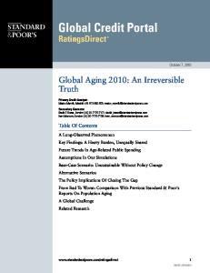
Figure 1 - Modeled temperature (ºK) as a function of time due to electric field of 5,000[V/cm]. Logarithmic scale was used due to the rapid increase in temperature during the first few.
analysis. Each slice was fixed with 10% buffered formalin, embedded in paraffin, and sectioned with a microtome (5µm-thick). III. RESULTS
Each animal was anaesthetized throughout the procedure. The left common carotid artery was exposed, intimal denudation was performed as previously described[9], and a 0.8 cm conductive cylinder was inserted into the interior of the artery. The outer surface of the artery was coated with highly conductive gel, connected to a second electrode that was used as ground. A sequence of 10 direct current pulses of 50 Volts (i.e. electric field of approximately 3300 V/cm assuming rodent carotid artery thickness of about 150μm), 100 µs each, at a frequency of 10 pulses per second, was applied between the inner electrode and the outer conductive gel using a high voltage pulse generator intended for electroporation procedures (ECM 830, Harvard Apparatus, Holliston, MA). Temperature was measured before and immediately following the electroporation pulses using fiber optic temperature sensor with a response time below 200 ms (Neoptix Inc., Quebec, Canada). C. Histological analysis Animals were euthanized with an overdose of Phenobarbital after 28 days of follow-up. The arterial tree was perfused with 10% buffered formalin for 40 minutes, and the left and right carotid arteries were exposed near the bifurcation of the internal and external carotid arteries. Three slices of approximately 2.6 mm were taken from each artery, at the core of the treated area, and were used for histological Animal # 1 2 3 4 5 6 7 8
Temperature before (ºC) 29.8 29.6 29 28.3 29.9 29.9 29.8 29,9
B. Experimental data Temperature measurements before and immediately after electroporation pulses are summarized in Table 2. The measured average temperature change due to the pulse was 0.6 ± 0.2 ºC. An analysis of temperature change during and immediately after electroporation pulse of a single case is shown in Figure 3. A single histological example is shown in Figure 4. This is the appearance of a single section of a treated artery 28 days after IRE (Figure 4b), compared with a control non-treated artery (Figure 4a). The histological examples show less neointimal formation in the treated artery, as well as scarcity of vascular smooth muscle cells, compared with the control
Temperature after (ºC) 30.5 30.5 29.8 29 30.1 30.5 30.5 30.4
32.8 32.7 Temperature (Celsius degrees)
Figure 2 - Logarithm of thermal damage as a function of the applied electric field .
A. Mathematical Analysis Our simulation focused on 50, 100, 200 300 and 400 volts of 1-milisecond pulses. These pulses are comparable to electric fields of 2,500, 5,000, 10,000 15,000 and 20,000 V/cm respectively. Time dependent solution for the temperature in the arterial wall yielded maximal temperatures of 38.8 ºC, 44.0 ºC, 65.0 ºC, 99.8 ºC and 149.1 ºC for the different fields (Figure 1). The complete 5-seconds interval solution for the temperature in the arterial wall due to 5,000 V/cm electric field is shown in a logarithmic scale in Figure 1. The results of the three time dependant solutions were integrated along time in order to calculate Ω for each electric field. The results were plotted (Figure 2), and using a linear regression technique (R2=0.998) the following second order polynomial correlation established : log Ω 5 · 10 8.122 (9) Where is Ω is a dimensionless evaluation of thermal damage and E is the electric field (V/cm).
32.6 32.5 32.4 32.3 32.2 32.1 32 31.9 31.8 0.4
0.9
1.4
1.9
2.4
Time (seconds)
Figure. 3 - Experimental results of the temperature of the arterial wall during two seconds time interval that includes the time before, during and immediately after the IRE pulses
Table 2 - Measurement of arterial wall temperature before and immediately after electroporation pulses (10 pulses of 100μs at 10Hz, with electrical field of approximately 3.500V/cm)
2053
artery. IV. DISCUSSION Our theoretical analysis, supported by experimental data, shows for the first time, that intravascular submicrosecond, mega-volt-per-meter electric field pulses can be applied to the arterial wall without significant thermal damage. The dimensionless indicator of thermal damage, Ω, is a function of two process parameters (molecular collision frequency and energy barrier) together with the transient analysis of temperature. In fact, Ω is the logarithm of the relative concentration of “reactants”:
⎛ C (0) ⎞ ⎟⎟ Ω(τ ) = ln⎜⎜ ⎝ C (τ ) ⎠
(10)
Therefore at Ω=1, a standard point of comparison 63.2% of the molecules in the arterial wall have already been changed into damaged state.[11] If we consider Ω=0.1 as the threshold for thermal damage, using equation (9) , our analysis shows that 1 millisecond electric field pulse of up to 12,000 V/cm is safe and will cause no significant damage. This result is approximately threefold higher than the electric field used successfully in our arterial wall ablation experiments.[7, 9] It should be stressed that our analysis used a single 1 ms pulse instead of 10 pulses of 100 μs at a frequency of 10 Hz. Using low frequency pulses naturally allows the use of even higher electric fields without significant damage. Our experiment data showed an average increase of 0.6 ºC, due to 10 100 μs pulses in a frequency of 10 Hz, with an estimated electric field of 3,300 Volt/cm. This result, which
Figure 4a. Non IRE-treated carotid artery 28 days following intimal damage. This example illustrates significant neointimal damage, as well as normal high density of vascular smooth muscle cells in the arterial wall
was measured during an in vivo experiment, is comparable to the theoretical model. The rise in temperature in the experiments is less significant compared with our theoretical model. The first reason is that our model assumed a single 1microsecond pulse, while in the experiment we used 10 shorter separate pulses. The second reason is that our experiment was done in an open surgery settings that allowed heat transfer with room-temperature air. Our single histological example is not a proof for the ability of intravascular IRE to attenuate restenosis. However, this example shows the feasibility of this technique, as well as its ability to cause a sustainable vascular cell ablation along the arterial wall that is apparent even after 28-days. We believe that intravascular IRE is a promising and safe technique. It is a promising tool that will enable physicians to resolve many vascular pathologies in which vascular smooth muscle cells play a pivotal role. We believe that the three most common and important pathologies are atherosclerosis, restenosis following angioplasty and diabetic peripheral vascular disease. REFERENCES [1] S. Gabriel, R. W. Lau, and C. Gabriel, "The dielectric properties of biological tissues: II. Measurements in the frequency range 10 Hz to 20 GHz " Physics in medicine & biology vol. 41, pp. 2251-2269, 1996. [2] R. V. Davalos, B. Rubinsky, and L. M. Mir, "Theoretical analysis of the thermal effects during in vivo tissue electroporation," Bioelectrochemistry, vol. 61, pp. 99-107, 2003. [3] J. Al Suwaidi, P. B. Berger, and D. R. Holmes, Jr., "Coronary Artery Stents," JAMA, vol. 284, pp. 1828-1836, 2000. [4] M. R. Ward, G. Pasterkamp, A. C. Yeung, and C. Borst, "Arterial Remodeling : Mechanisms and Clinical Implications," Circulation, vol. 102, pp. 1186-1191, 2000. [5] G. W. Stone, S. G. Ellis, D. A. Cox, J. Hermiller, C. O'Shaughnessy, J. T. Mann, M. Turco, R. Caputo, P. Bergin, J. Greenberg, J. J. Popma, M. E. Russell, and T.-I. V. I. the, "A Polymer-Based, Paclitaxel-Eluting Stent in Patients with Coronary Artery Disease," N Engl J Med, vol. 350, pp. 221231, 2004. [6] R. V. Davalos, L. M. Mir, and B. Rubinsky, "Tissue Ablation with Irreversible Electroporation," Annals of Biomedical Engineering, vol. 33, pp. 223-231, 2005. [7] E. Maor, A. Ivorra, J. Leor, and B. Rubinsky, "The effect of irreversible electroporation on blood vessels.," Technology in Cancer Research and Treatment, vol. 6, pp. 307-12, 2007. [8] L. Jing, L. Jing, and L. X. Xu, "Estimation of blood perfusion using phase shift in temperature response to sinusoidal heating at the skin surface Estimation of blood perfusion using phase shift in temperature response to sinusoidal heating at the skin surface," Biomedical Engineering, IEEE Transactions on, vol. 46, pp. 1037-1043, 1999. [9] E. Maor, A. Ivorra, J. Leor, and B. Rubinsky, "Irreversible Electroporation Attenuetes Neointimal Formation after Angioplasty," IEEE transactions on bio-medical engineering, vol. In Print, 2008. [10] N. T. Wright, "On a Relationship Between the Arrhenius Parameters from Thermal Damage Studies," Journal of Biomechanical Engineering, vol. 125, pp. 300-304, 1992. [11] K. R. Diller and J. A. Pearce, "Issues in Modeling Thermal Alterations in Tissues a," Annals of the New York Academy of Sciences, vol. 888, pp. 153-164, 1999.
Figure 4b. IRE-treated carotid artery 28 days following intimal damage and IRE ablation. This example illustrates minor neointimal formation, as well as scarcity of vascular smooth muscle cells in the arterial wall.
2054