Mutant huntingtin is present in neuronal grafts in Huntington's disease patients
Cicchetti F, PhD*1,2, Lacroix S, PhD1,3, Cisbani G, MSc1, Vallières N, MSc1,, Saint-Pierre M, DEC1, St-Amour I, PhD1, Tolouei R, PhD4, Skepper JN, PhD5, Hauser RA, MD6, Mantovani D, PhD4, Barker RA, MD, PhD7, Freeman TB, MD8,9
1Centre
de recherche du CHU de Québec (CHUQ), Québec, QC, Canada G1V 4G2; 2Département de psychiatrie et neurosciences, Université Laval, Québec, QC, Canada, G1K 7P4; 3Département de médecine moléculaire, Université Laval, Québec, QC, Canada, G1K 7P4; 4Laboratoire de biomatériaux et bioingénierie, Hôpital Saint-François d’Assise and Département de génie des mines, de la métallurgie et des matériaux, Université Laval, Québec, QC, Canada G1V 0A6, 5Cambridge Advanced Imaging Centre, Anatomy building, University of Cambridge, Cambridge, UK, CB2 3DY, 6Departments of Neurology; Pharmacology and Experimental Therapeutics, Parkinson’s Disease and Movement Disorders National Parkinson’s Foundation Center of Excellence, University of South Florida, Tampa, FL, USA, 33606, 7John van Geest Centre for Brain Repair, Department of Clinical Neurosciences, University of Cambridge, Cambridge, UK, CB2 0PY, 8Department of Neurosurgery and Brain Repair, 9Center of Excellence for Aging and Brain Repair, University of South Florida, Tampa, FL, USA, 33606
Correspondence Francesca Cicchetti, PhD Centre de recherche du CHU de Québec (CHUQ) Axe Neurosciences, T2-50 2705, Boulevard Laurier Québec, QC, G1V 4G2, Canada Tel #: (418) 656-4141 ext. 48853 Fax #: (418) 654-2753 E-mail:
[email protected] Running head: Transfer of mutant huntingtin to normal tissue Number of characters in the title: 80 Number of characters in the running head: 46 Number of words in the abstract: 213 Number of words in the body of the manuscript: 3501 Number of figures: 10 Number of colour figures: 9 Number of tables: 1 This article has been accepted for publication and undergone full peer review but has not been through the copyediting, typesetting, pagination and proofreading process which may lead to differences between this version and the Version of Record. Please cite this article as an ‘Accepted Article’, doi: 10.1002/ana.24174
Annals of Neurology
Cicchetti et al., 2014 Abstract Objective. Huntington’s disease (HD) is caused by a genetically encoded pathological protein (mutant huntingtin (mHtt)), which is thought to exert its effects in a cellautonomous manner. Here, we tested the hypothesis that mHtt is capable of spreading within cerebral tissue by examining genetically unrelated fetal neural allografts within the brains of patients with advancing HD. Methods. The presence of mHtt aggregates within the grafted tissue was confirmed using 3 different types of microscopy (brightfield, fluorescence and electron), 2 additional techniques consisting of western immunoblotting and infrared spectroscopy, and 4 distinct antibodies targeting different epitopes of mHtt aggregates. Results. We describe the presence of mHtt aggregates within intracerebral allografts of striatal tissue in three HD patients who received their transplants approximately a decade earlier and then died secondary to the progression of their disease. The mHtt+ aggregates were observed in the extracellular matrix of the transplanted tissue while in the host brain they were seen in neurons, neuropil, extracellular matrix and blood vessels. Interpretation. This is the first demonstration of the presence of mHtt in genetically normal and unrelated allografted neural tissue transplanted into the brain of affected HD patients. These observations raise questions on protein spread in monogenic neurodegenerative disorders of the central nervous system characterized by the formation of mutant protein oligomers/aggregates.
2
John Wiley & Sons
Page 2 of 47
Page 3 of 47
Annals of Neurology
Cicchetti et al., 2014 Introduction Huntington’s disease (HD) is an autosomal dominant genetic disorder characterized by a clinical triad of a movement disorder, cognitive dysfunction and psychiatric problems1 combined with a pathological CAG expansion in exon 1 of the huntingtin gene leading to the production of mutant huntingtin protein (mHtt).1,2 Wild type Htt is a soluble protein that is ubiquitously expressed but is present in higher concentrations especially in the brain. This cytoplasmic and nuclear protein is associated with several organelles, microtubules and vesicular membranes, pointing to a role in intracellular trafficking, exocytosis, endocytosis and therefore synaptic functions.3 In HD, like all genetic trinucleotide disorders of the central nervous system, it has been suggested that the abnormal mutant protein causes cellular dysfunction through a cell-autonomous process in which only genotypically mutant cells exhibit the mutant phenotype. This results in mHtt aggregation, inclusion body formation and cell death, although how these events relate to each other is still debated.4
HD is incurable, and different experimental therapeutic strategies have been tested including transplantation of fetal striatal tissue.5,6 This approach was predicated on the grounds that the primary pathology involves the striatum and that replacing it with unaffected allografts of fetal striatal tissue would be of benefit, as has been shown experimentally in non-transgenic animal models of HD.7-10 To date, these transplants have generally produced transient or no clinical benefits despite evidence that they survive in the short-term.11 This failure of clinical response to such targeted grafts may relate to the widespread pathology now recognised in HD from an early disease stage, but may also relate to the fact that these grafts survive poorly in HD patients and degenerate in a disease-
3
John Wiley & Sons
Annals of Neurology
Cicchetti et al., 2014 like manner, as we have described previously.12 In this respect, we now show for the first time that mHtt aggregates can be found within the genetically normal transplanted tissue in three HD patients in whom there was long-term graft survival (patients reported previously in 11-14).
Methods Patient information Three of the original 7 HD patients grafted as part of the transplant trial conducted at the University of South Florida5,11,12,14 were analysed. They all had a 42 CAG repeat and were transplanted at 58, 64 and 59 years of age, respectively. An additional 28-year-old patient with 53 CAG repeats was also studied. All patients had manifest disease (ranging between 6 and 17 years of symptom duration) at the time that they received bilateral fetal striatal transplants. They all showed mild clinical improvements lasting at most one year, except for patient five whose Unified Huntington’s disease rating scale (UHDRS) score worsened following grafting.11,12,14 They died between 9 and 12 years post-transplantation12,13 (see Fig. 1). All post-mortem analyses of human brain tissue were approved by the Comité d'éthique de la recherche du CHU de Québec (#A13-02-113).
Immunohistochemistry Brains were processed using methods previously published.11-13 Autolysis time was 5, 4, 5 and 2 hours for patients 1, 3, 5 and 7 respectively. Standard histology was undertaken to identify the grafts macroscopically. Additional post-mortem analyses included double immunohistochemical staining for neuronal elements using an antibody against neuronal
4
John Wiley & Sons
Page 4 of 47
Page 5 of 47
Annals of Neurology
Cicchetti et al., 2014 nuclei (NeuN, anti-mouse, MAB377, Millipore, 1:2500) as well as the anti- Htt antibodies EM48 (mouse anti-human huntingtin clone EM48, MAB5374, Millipore, 1:2000) and MW7 (mouse anti-human, obtained from the Developmental Studies Hybridoma Bank, 1:100) to identify mHtt+ aggregates. Photomicrographs were taken using Picture Frame software (Microbrightfield) linked to an E800 Nikon microscope (Nikon Instruments).
Stereology for EM48+ aggregate count The density of EM48+ mHtt deposits, reported as the number of aggregates/mm2 of tissue, was assessed in the cortex and putamen of the host brains as well as in p-zones and non pzones of the grafts using standard techniques (see13). For aggregate size, the perimeter of sampled aggregates was delineated and assessed in the above-mentioned structures.
Statistical analyses To calculate the density of aggregates, a negative binomial distribution was used. Stepdown Bonferonni correction was further employed to ensure that the overall significance level of the multiple comparison tests was 0.05. One-way ANOVA was used to compare aggregate size. All statistical analyses were performed using the MIXED and GENMOD procedures of SAS (version 9.2, SAS: Cary, North Carolina).
Immunofluorescence A series of double and triple immunofluorescence stainings was also performed to localize mHtt, using either EM48 (1:200) or MW7 (1:100) (see Table S1). The following antibodies were used for the detection of specific cell populations: astrocytes (rabbit anti-GFAP,
5
John Wiley & Sons
Annals of Neurology
Cicchetti et al., 2014 Z0334, Dako Cytomation, 1:500), microglia (rabbit anti-Iba1, 019-19741, Wako Chemicals, 1:800), neurons (rabbit anti-MAP2, 17490-1-AP, Protein Tech, 1:500 or anti-chicken Neurofilament H, AB5539, Millipore, 1:400), oligodendrocytes (rabbit anti-CAII, 1:2000), perivascular macrophages (rabbit anti-CD163, NBP1-30148, Novus biological), vascular endothelium (rabbit anti-Laminin, Z0097, Dako Cytomation, 1:500) and extracellular matrix (rat anti-phosphocan, MAB2688, R&D system, 1:100). Ubiquitinated aggregates were also identified using an anti-rabbit ubiquitin antibody (Z0458, DAKO, 1:100). Some of these antibodies required an additional post-fixation with paraformaldehyde (PFA) 4% pH 7.4 (Sigma-Aldrich) for 1 hr prior to staining (Table S1). Sections were stained using standard procedures (see13). All sections were observed at 60X and imaged on a Fluoview FV1000 confocal microscope system equipped with 559nm and 635nm laser diodes and an Ar 488nm laser (Olympus Canada Inc.).
All immunohistochemistry and immunofluorescence experiments included multiple controls such as normal and HD matched brain sections (both free floating and paraffin embedded sections; provided by the NIHR funded Cambridge Brain Bank, UK) and negative controls where the primary antibody was omitted from the incubation media. Control conditions further included a range of dilutions for the EM48 antibody (1:50 to 1:5000), all of which yielded positive immunoreactivity for mHtt aggregates in the grafted tissue as revealed with the nickel intensified DAB method and an optimized DAB protocol which was different from that used previously by us.11,12
6
John Wiley & Sons
Page 6 of 47
Page 7 of 47
Annals of Neurology
Cicchetti et al., 2014 Sequential method for chromogenic immunohistochemistry We undertook an additional series of experiments to investigate the co-localization of EM48 with MW7 and ubiquitin. In order to do this, we applied a sequential staining protocol using the chromogen 3-amino-9-ethylcarbazole (AEC).15-18 To confirm the complete removal of the primary antibody, the sections were also incubated solely in the secondary antibody. Photomicrographs were taken by Picture Frame software (Microbrightfield) linked to an E800 Nikon microscope (Nikon Instruments).
Electron microscopy For electron microscopy, specific areas of a 30µm nickel-intensified DAB-stained brain section were prepared by flat embedding in epoxy resin (Hayat, M.A.) The embedded samples were then serially sectioned in parallel at 60nm with a diamond knife (DDK, Delaware, USA) on an ultramicrotome (Ultracut E, Reichert-Jung, Austria). The sections were placed onto a single-hole copper/formvar grid and stained with 3% aqueous uranyl acetate for 5 min. After drying, observation was performed with an electron microscope, JEM-1230 (JEOL, Japan) set at 80 kV.12
Protein extraction from fixed tissue Brain sections were selected and dissected to isolate pieces of the cortex in both HD and control patients, as well as the grafted tissue. This was performed on 15 sections for each condition. The dissected tissue was collected into eppendorf tubes, weighed and frozen on dry ice and homogenized in 4 volumes of extraction buffer (50mM HEPES, 150mM NaCl, 10mM EDTA, 1% triton X-100 (Sigma-Aldrich), 0.5% deoxycholate (Sigma-Aldrich), 0.1%
7
John Wiley & Sons
Annals of Neurology
Cicchetti et al., 2014 BSA (Bishop), 200mM DTT, 2% SDS (Sigma-Aldrich), 20mM Tris HCl pH 8.8 (SigmaAldrich),19,20 containing a cocktail of protease (Roche) and phosphatase inhibitors (SigmaAldrich)) and sonicated. The homogenate was then incubated at 100°C for 20 min followed by an incubation of 1 h at 80°C on a shaker (80rpm). Finally, the tubes were spun for 20 min at 4°C at 13000rpm and the supernatant was collected and stored at -80°C until used. Total protein concentration was determined using the Pierce 660nm protein assay (Thermo Fisher).
Western blotting All reagents and chemicals used for immunoblotting were purchased from Sigma-Aldrich, unless otherwise specified. Fifteen µg of protein was prepared in the sample buffer (250 mM Tris-HCl, 2% w/v lithium dodecyl sulphate, 100 mM DTT, 0.4 mM EDTA, 10% v/v glycerol (Fisher Scientific), 0.2 mM bromophenol red, 0.2 mM Brilliant Blue G, pH 8.5) and heated for 10 min at 70°C. The proteins were then separated for 5 hrs at 150 V on a 3 - 8% gradient hand-cast polyacrylamide gel (37.5:1 acrylamide: bisacrylamide solution (J.T. Baker) in 0.2 M Tris-acetate buffer, pH 7.4 using a midi electrophoresis system (15 x 15 cm) in an electrophoresis buffer (50 mM Tricine, 50 mM Tris, 0.1% SDS, 1.3 mM sodium bisulfite, pH 8.2), as previously described.21 Proteins were electroblotted onto 0.45 μm Immobilon PVDF membranes (EMD Millipore, Billerica, MA, USA) overnight at 15V in the transfer buffer (20% methanol (Fisher Scientific) 25 mM Bicine, 25 mM Bis-Tris, 1 mM EDTA, 1.3 mM sodium bisulfite, pH 7.2). After being blocked in a solution of 5% skimmed milk, 0.5% bovine serum albumin (Bioshop Canada Inc, Burlington, ON, Canada) in PBS, 0.1% Tween-20 (Thermo Scientific) the membranes were immunoblotted with MW1 (anti-
8
John Wiley & Sons
Page 8 of 47
Page 9 of 47
Annals of Neurology
Cicchetti et al., 2014 mouse antibody, 1:1000) overnight at 4°C followed with horseradish peroxidase (HRP)conjugated goat anti-mouse antibody (Jackson Immunoresearch, 1:30,000) and detected by the addition of chemiluminescence reagents (Luminata forte, EMD Millipore). The membranes were then stripped and reprobed for total Htt (anti-mouse, clone: 1HU-4C8, MAB2166, EMD Millipore, 1:1000), glyceraldehyde 3-phosphate dehydrogenase (antimouse GAPDH, G041, Applied Biological Materials Inc, 1:7500), and neuronal nuclei (antimouse NeuN, MAB377, EMD Millipore, 1:1000). Images of the membranes were acquired using myEcl Imager (Thermo Fisher). Band intensities were quantified using Carestream Molecular Imaging Software (version 4.0.5f7, Eastman Kodak). The graphs were generated using the Prism software (version 4.0, GraphPad Software Inc.) and assembled in illustrator CS5.
Infrared spectroscopy Infrared spectroscopy was used to detect structural protein changes such as the β-sheets of mHtt within the HD host brain and grafted tissue of unstained sections.22,23 FTIR-FPA maps were acquired using a 32 × 32 detector with a pixel size of 5.5 µm giving 1024 spectra/map. The IR spectra were collected on a Nicolet Magna-IR 550 FT spectrometer (Thermo Nicolet, Madison, WI, USA) equipped with a deuterated triglycine sulfate (DTGS) detector and a germanium-coated KBr beam splitter. IR data were treated with Omnic E.S.P. 5.1 (Thermo Nicolet, Madison, WI, USA) and Grams 32 (Spectral Notebase version 4.11, Galactic Industries Corporation, Salem, NH, USA).
9
John Wiley & Sons
Annals of Neurology
Page 10 of 47
Cicchetti et al., 2014 Image preparation All the images were prepared using Adobe Photoshop CS5 and when necessary, brightness and contrast adjustments were made. All panels were assembled using Adobe Illustrator CS5.
Results Surviving neuronal transplants with a compartmentalized organization were observed in three of four transplant recipient brains that we have reported previously11-13 (Figs. 1 and 2). Within the surviving transplants of all three patients, we observed patches of striatallike neuronal tissue referred to as p-zones, as well as areas where there was no expression of striatal phenotypes (non p-zones).24 In the fourth patient (patient three (M.C.) from our original series), no surviving transplants were found.12
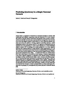
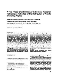
Aggregated mHtt protein was observed in the genetically unrelated transplanted tissue in all three HD patients with surviving grafts (Figs. 1 and 2). Aggregates identified with either EM48 or MW7 were frequently found within both the p-zones (Figs. 1A-C, F-I, 2A-C) and non p-zones of the grafts (Figs. 1A, C-D, F, H-I, 2A, C-D). The size of the aggregates was similar in both the grafts and surrounding non-transplanted putamen. Despite similar morphology, the number of aggregates was slightly less in the graft compartments than in the host putamen. In the host cortex, mHtt deposits were the largest and the most numerous (Fig. 1, bar graphs).
10
John Wiley & Sons
Page 11 of 47
Annals of Neurology
Cicchetti et al., 2014 Within the transplanted tissue, mHtt+ aggregates were localized to the extracellular matrix of both the p- and non p-zone throughout the grafts, as confirmed using two distinct antibodies (EM48 and MW7, Fig. 3A-B). EM48+ and MW7+ aggregates within transplants were not observed in neurons (Fig. 3A-B), microglia (Figs. 3C, 4A), astrocytes (Figs. 3D, 4B), perivascular macrophages (Figs. 3E, 4C), oligodendrocytes (data not shown), endothelial cells (data not shown) or blood vessels (Fig. 3F, data not shown for MW7).
In contrast in these same patients, mHtt protein aggregates found within the nontransplanted cortex were localized to the neurons and neuropil (Figs. 4D, 5A-D), as previously described.25,26 mHtt was also frequently observed within the extracellular matrix of the host cortex (Fig. 5E), a finding that has not been previously reported. Additionally, aggregates of mHtt protein were observed within the vascular space of the HD brain (Fig. 5F) including within cells of the vascular space (Fig. 5F inset – EM48/DAPI staining). Aggregates of mHtt protein (stained either with MW7 or EM48) were not observed in host microglia (Figs. 4E and 5G), astrocytes (Figs. 4F and 5H), perivascular macrophages (Figs. 4G and 5I), oligodendrocytes (Fig. 5J) nor within endothelial cells (data not shown).
Co-localization of EM48+ aggregates and the protein ubiquitin was confirmed within the grafted tissue (Fig. 6A) and the cortex (Fig. 6B). Using a technique of chromogen stripping, 15,16,18
we further demonstrated that EM48, ubiquitin and MW7 co-localize in the cortical
tissue of transplanted HD patients (Fig. 7). In order to confirm the presence of these aggregates, we further performed electron microscopy (Fig. 8), western immunoblotting
11
John Wiley & Sons
Annals of Neurology
Page 12 of 47
Cicchetti et al., 2014 (Fig. 9) and infrared spectroscopy (Fig. 10), all of which corroborated our original immunohistochemical findings of aggregates in the graft and host brain.
Discussion Here we describe for the first time that mHtt can be found within genetically unrelated tissue grafted into the brains of patients with advancing HD. The mHtt was localized to the extracellular matrix of the transplants. This differs from the localization of mHtt within the non-grafted regions of the brain of patients with HD, where the mHtt protein is primarily localized to neurons and the neuropil – as has been described before in HD.25,26
The fetal striatal allografts implanted in these patients with HD are derived from normal donors not carrying the mutant gene and thus mHtt. This raises two questions: 1) how did the mHtt protein from the patient become localized within the transplanted tissue and 2) did this localization of mHtt within the transplants contribute to their compromised survival?11-13 Several possible mechanisms can be put forward to explain these findings.
mHtt protein transmission, deposition or diffusion into the transplant. In another neurodegenerative disorder of the central nervous system, Parkinson’s disease (PD), it has been demonstrated that the pathologically associated protein α-synuclein (i.e. Lewy body pathology) could spread into the allografted ventral mesencephalic tissue.27,28 This was hypothesized to occur in a prion-like fashion.27,29-32 Both in vitro and in vivo studies have subsequently demonstrated that α-synuclein can be released and taken up by neurons33,34 and seed pathology. Indeed, intracerebral inoculation of brain homogenates derived from 12
John Wiley & Sons
Page 13 of 47
Annals of Neurology
Cicchetti et al., 2014 α-synuclein transgenic mice, or injection of synthetic α-synuclein preformed fibrils accelerates the formation of α-synuclein protein aggregates and precipitates neurological dysfunction in animals.35 A similar propagation of tau pathology has been described in a model of early Alzheimer’s disease (AD) and tauopathies.36,37 It is therefore possible that mHtt was transferred from the patients’ brains to the grafts via a similar mechanism. However, unlike the host brain, where mHtt aggregates were mainly seen within the neuropil, inclusions in the grafts were only observed within the extracellular matrix and not within any cellular elements. Therefore, within these case reports, we do not have direct evidence of cell-to-cell propagation of mHtt.
Findings from in vivo models of AD suggest that neurodegeneration in this disease result from tau pathology being spread via transynaptic connections.30,38 It is possible that mHtt could similarly be transmitted transynaptically to the transplants from the diseased cortex, and in this respect we have previously demonstrated synaptic connections between the diseased cortex and the grafted neurons in these HD patients.12,39 Although we did not observe mHtt within grafted neurons, the extracellular localization of aggregates within the p-zones could be derived from dying cortico-striatal synaptic terminals that innervated the transplant 12 and expressed mHtt, possibly leading to graft cell lysis and degeneration with release of the aggregates within transplant neuropil. However, extracellular localization of mHtt was also observed within non p-zones of the grafts, structures that do not receive direct projections from cortical neurons. This would suggest that, at most, this mechanism is partially involved in the mHtt spread and other mechanisms of protein transport may exist. 13
John Wiley & Sons
Annals of Neurology
Page 14 of 47
Cicchetti et al., 2014
An alternative possibility related to this is that aberrant cortical striatal neurons containing the mHtt protein may leave axonal debris within transplant extracellular spaces in the process of undergoing cell death. In support of this hypothesis, a large number of aggregates that did not colocalize with MAP2+ grafted neurones were nonetheless found in close proximity to these neuronal elements.
Regardless of the mechanism of cortical interactions with the putamen and graft (transynaptic transmission of mHtt vs. mHtt deposits via synaptic debris), our results suggest a time-dependent influence of the cortex on the putamen and transplant. The mHtt deposits are the most abundant and largest in the host cortex. They are smaller and less abundant in the putamen. The mHtt deposits in the grafts are the same size but less abundant than deposits in the host putamen. The transplants have been exposed to the disease process for less time than the putamen as the patients already had the disease at the time of transplantation. This suggests that the number of deposits correlates with the amount of time that the striatal tissue is exposed to the disease process if it originates in the cortex.
In vivo models of AD have also demonstrated that pathology may be associated with diffusion of the soluble form of Aβ in the extracellular space with uptake by cells in the vicinity.30,40 Ren et al.41 also described that fibrillar polyglutamine peptide aggregates can be internalized by mammalian cells in vitro. It is similarly possible that the localization of mHtt in our transplants results from diffusion of mHtt from the putamen to the transplants 14
John Wiley & Sons
Page 15 of 47
Annals of Neurology
Cicchetti et al., 2014 via the extracellular matrix. Uptake of this mutant protein may then compromise the viability of the cells in the graft that have endocytosed it. However, in our post-mortem samples we have no evidence of mHtt in any cell type within the graft, which would argue against such a mechanism compromising the transplants.
Oxidative stress, excitotoxicity, inflammation and poor trophic support. These mechanisms have been implicated in neuronal transplant pathology in both PD and HD.5,39,42,43 CAG repeat length gains may occur in non-dividing cells,44,45 independent of the DNA replication process. It is possible that oxidative stress, excitotoxicity, inflammation or a poor trophic milieu may induce pathological polyglutamine expression in the transplants. However, Shelbourne et al.44 and Kennedy et al.45 noted this CAG expansion within neurons and glia whereas we only saw the abnormal CAG in the extracellular milieu of the grafts. Furthermore, if such a mechanism was dominant, one would expect to see similar inclusions in other types of grafts such as ventral mesencephalon transplants in PD, which is not the case.
Hematogenous transport of mHtt. mHtt aggregates were occasionally observed within the HD cerebral blood vessels (Fig. 3F), within cells of these blood vessels (Fig. 3F, insert) and possibly within perivascular macrophages of the host brain (Fig. 8). These new observations raise the possibility that mHtt could be transported from the host to the transplant via blood-born cells such as those of the immune system. Interestingly, it was recently shown that certain types of immune cells can contribute to disease progression in animal models of HD.46,47 This mechanism is further supported by the diffuse localization of
15
John Wiley & Sons
Annals of Neurology
Page 16 of 47
Cicchetti et al., 2014 mHtt within both the p-zone and non p-zone aspects of the graft. This potential mechanism of HD pathology in man has not been previously reported. If true, then the morphological and quantitative differences in mHtt depositions between the cortex and the putamen and grafts would suggest that these different regions of the brain have different susceptibility to mHtt or differences in vascularity and/or blood-brain-barrier permeability.
Previously, we and others have reported the absence of mHtt aggregates within transplants in HD patients.11,12,48 Our new observation that mHtt inclusions are indeed present within grafts differs from these previous reports and can be explained in two ways. As in fetal ventral mesencephalon grafts in PD, α-synuclein depositions developed in a timedependent manner and were only observed in grafts exposed to the disease process for over a decade.27,28 Likewise, Freeman et al.11 and Keene et al.48 examined transplants in HD patients that were exposed to the disease process for comparatively short-times (18 months, 6 and 7 years respectively). In Cicchetti et al., 200912, one of the 3 patients did not show graft survival precluding histological evaluation. However, in the other 2 subjects in this paper, we did not observe mHtt aggregates using standard techniques of the time. We therefore sought to re-examine these cases in this report using several techniques that were not available at the time of the original publication, including some techniques developed for this study, and we now can show that these grafts do indeed contain mHtt.
This is the first demonstration in vivo of the transmission of a gene product to genetically unrelated cells. In genetic disorders such as HD, pathogenesis is thought to occur via cellautonomous mechanisms. The localization of mHtt expression within grafted tissue 16
John Wiley & Sons
Page 17 of 47
Annals of Neurology
Cicchetti et al., 2014 suggests that non cell-autonomous mechanisms may also play an important role in mHtt spread within the HD brain. Further research is needed to determine the scientific, clinical and therapeutic implications of these findings for patients with HD and possibly other genetic disorders.
17
John Wiley & Sons
Annals of Neurology
Page 18 of 47
Cicchetti et al., 2014 Acknowledgments This work was supported by a grant from the International Organization of Glutaric Acidemia (IOGA) awarded to Francesca Cicchetti. Salary support for Francesca Cicchetti and Steve Lacroix was provided by the Fonds de recherche du Québec en santé (FRQS). Giulia Cisbani was supported by the Bourse d’excellence du Centre thématique de recherche en neurosciences (Université Laval). The Cambridge Brain Bank is supported by an NIHR funded Biomedical Research Centre to the University of Cambridge/Addenbrooke’s Hospital. The authors would like to thank Mr. Richard Janvier for his very skilful electron microscopy sample preparation and analyses, Marie Leroy (MSc), Pascale Chevalier (PhD), Lucie Levesque (MSc) and Gaétan Laroche (PhD) for FTIR analysis as well as Greg Sutter and Pamela Pierce who participated in the procurement of the brain from patient seven. The MW7 and MW1 antibodies developed by Dr P.H. Patterson were obtained from the Developmental Studies Hybridoma Bank developed under the auspices of the NICHD and maintained by The University of Iowa, Department of Biology, Iowa City, IA 52242.
18
John Wiley & Sons
Page 19 of 47
Annals of Neurology
Cicchetti et al., 2014 Authorship FC made the observation of the presence of mHtt within the grafted tissue, was involved in experimental designs, image acquisition and data interpretation. She supervised the project and wrote the manuscript. SL was involved in data interpretation and manuscript writing. GC performed most of the immunohistochemical and immunofluorescent stainings, aggregate quantifications, some of the image acquisition and was responsible of assembling all figure panels. NV helped troubleshoot immunofluorescent protocols and performed all the confocal image acquisition. MSP performed some of the immunohistochemical and immunofluorescent stainings as well as some of the image acquisition. ISA performed the western immunoblotting. RT performed the infrared spectroscopy. JNS helped with the electron microscopy analyses/interpretation. RAH ensured the clinical follow-up of the patients. DM provided this expertise for the infrared spectroscopy. RAB was involved in manuscript writing. TBF was involved in data interpretation and manuscript writing.
19
John Wiley & Sons
Annals of Neurology
Page 20 of 47
Cicchetti et al., 2014 References 1.
Phillips W, Shannon KM, Barker RA. The current clinical management of Huntington's disease. Mov Disord 2008;23:1491–1504.
2.
Zuccato C, Valenza M, Cattaneo E. Molecular mechanisms and potential therapeutical targets in Huntington's disease. Physiol Rev 2010;90:905–981.
3.
Steffan JS, Kazantsev A, Spasic-Boskovic O, et al. The Huntington's disease protein interacts with p53 and CREB-binding protein and represses transcription. Proc Natl Acad Sci USA 2000;97:6763–6768.
4.
Ross CA, Tabrizi SJ. Huntington's disease: from molecular pathogenesis to clinical treatment. Lancet Neurol 2011;10:83–98.
5.
Freeman TB, Cicchetti F, Bachoud-Lévi AC, Dunnett SB. Technical factors that influence neural transplant safety in Huntington's disease. Exp Neurol 2011;227:1–9.
6.
Peschanski M, Cesaro P, Hantraye P. Rationale for intrastriatal grafting of striatal neuroblasts in patients with Huntington's disease. Neuroscience 1995;68:273–285.
7.
Isacson O, Brundin P, Kelly PA, et al. Functional neuronal replacement by grafted striatal neurones in the ibotenic acid-lesioned rat striatum. Nature 1984;311:458– 460.
8.
Wictorin K. Anatomy and connectivity of intrastriatal striatal transplants. Prog Neurobiol 1992;38:611–639.
9.
Pritzel M, Isacson O, Brundin P, et al. Afferent and efferent connections of striatal 20
John Wiley & Sons
Page 21 of 47
Annals of Neurology
Cicchetti et al., 2014 grafts implanted into the ibotenic acid lesioned neostriatum in adult rats. Exp Brain Res 1986;65:112–126. 10.
Deckel AW, Robinson RG, Coyle JT, Sanberg PR. Reversal of long-term locomotor abnormalities in the kainic acid model of Huntington's disease by day 18 fetal striatal implants. Eur J Pharmacol 1983;93:287–288.
11.
Freeman TB, Cicchetti F, Hauser RA, et al. Transplanted fetal striatum in Huntington's disease: phenotypic development and lack of pathology. Proc Natl Acad Sci USA 2000;97:13877–13882.
12.
Cicchetti F, Saporta S, Hauser RA, et al. Neural transplants in patients with Huntington's disease undergo disease-like neuronal degeneration. Proc Natl Acad Sci USA 2009;106:12483–12488.
13.
Cisbani G, Freeman TB, Soulet D, et al. Striatal allografts in patients with Huntington's disease: impact of diminished astrocytes and vascularization on graft viability. Brain 2013;136:433–443.
14.
Hauser RA, Furtado S, Cimino CR, et al. Bilateral human fetal striatal transplantation in Huntington's disease. Neurology 2002;58:687–695.
15.
Kim M, Soontornniyomkij V, Ji B, Zhou X. System-wide immunohistochemical analysis of protein co-localization. PLoS ONE 2012;7:e32043.
16.
Glass G, Papin JA, Mandell JW. SIMPLE: a sequential immunoperoxidase labeling and erasing method. J Histochem Cytochem 2009;57:899–905.
21
John Wiley & Sons
Annals of Neurology
Page 22 of 47
Cicchetti et al., 2014 17.
Becher MW, Kotzuk JA, Sharp AH, et al. Intranuclear neuronal inclusions in Huntington's disease and dentatorubral and pallidoluysian atrophy: correlation between the density of inclusions and IT15 CAG triplet repeat length. Neurobiol Dis 1998;4:387–397.
18.
Pirici D, Mogoanta L, Kumar-Singh S, et al. Antibody elution method for multiple immunohistochemistry on primary antibodies raised in the same species and of the same subtype. J Histochem Cytochem 2009;57:567–575.
19.
Addis MF, Tanca A, Pagnozzi D, et al. Generation of high-quality protein extracts from formalin-fixed, paraffin-embedded tissues. Proteomics 2009;9:3815–3823.
20.
Weiss A, Träger U, Wild EJ, et al. Mutant huntingtin fragmentation in immune cells tracks Huntington's disease progression. J Clin Invest 2012;122:3731–3736.
21.
Cubillos-Rojas M, Amair-Pinedo F, Tato I, et al. Simultaneous electrophoretic analysis of proteins of very high and low molecular mass using Tris-acetate polyacrylamide gels. Electrophoresis 2010;31:1318–1321.
22.
André W, Sandt C, Dumas P, et al. Structure of Inclusions of Huntington's Disease Brain Revealed by Synchrotron Infrared Microspectroscopy: Polymorphism and Relevance to Cytotoxicity. Anal Chem 2013;85:3765–3773.
23.
Khare SD, Ding F, Gwanmesia KN, Dokholyan NV. Molecular origin of polyglutamine aggregation in neurodegenerative diseases. PLoS Comput Biol 2005;1:230–235.
24.
Graybiel AM, Liu FC, Dunnett SB. Intrastriatal grafts derived from fetal striatal
22
John Wiley & Sons
Page 23 of 47
Annals of Neurology
Cicchetti et al., 2014 primordia. I. Phenotypy and modular organization. J Neurosci 1989;9:3250–3271. 25.
Gutekunst CA, Li SH, Yi H, et al. The cellular and subcellular localization of huntingtinassociated protein 1 (HAP1): comparison with huntingtin in rat and human. J Neurosci 1998;18:7674–7686.
26.
DiFiglia M, Sapp E, Chase KO, et al. Aggregation of huntingtin in neuronal intranuclear inclusions and dystrophic neurites in brain. Science 1997;277:1990–1993.
27.
Kordower JH, Chu Y, Hauser RA, et al. Lewy body-like pathology in long-term embryonic nigral transplants in Parkinson's disease. Nat Med 2008;14:504–506.
28.
Li JY, Englund E, Holton JL, et al. Lewy bodies in grafted neurons in subjects with Parkinson's
disease
suggest
host-to-graft
disease
propagation.
Nat
Med
2008;14:501–503. 29.
Brundin P, Melki R, Kopito R. Prion-like transmission of protein aggregates in neurodegenerative diseases. Nat Rev Mol Cell Biol 2010;11:301–307.
30.
Soto C. Transmissible proteins: expanding the prion heresy. Cell 2012;149(5):968– 977.
31.
Goedert M, Spillantini MG, Del Tredici K, Braak H. 100 years of Lewy pathology. Nat Rev Neurol 2013;9:13–24.
32.
Olanow CW, Prusiner SB. Is Parkinson's disease a prion disorder? Proc Natl Acad Sci USA 2009;106:12571–12572.
23
John Wiley & Sons
Annals of Neurology
Page 24 of 47
Cicchetti et al., 2014 33.
Desplats P, Lee HJ, Bae EJ, et al. Inclusion formation and neuronal cell death through neuron-to-neuron transmission of alpha-synuclein. Proc Natl Acad Sci USA 2009;106:13010–13015.
34.
Hansen C, Angot E, Bergström A-L, et al. alpha-Synuclein propagates from mouse brain to grafted dopaminergic neurons and seeds aggregation in cultured human cells. J Clin Invest 2011;121:715–725.
35.
Luk KC, Kehm V, Carroll J, et al. Pathological alpha-synuclein transmission initiates Parkinson-like neurodegeneration in nontransgenic mice. Science 2012;338:949– 953.
36.
de Calignon A, Polydoro M, Suarez-Calvet M, et al. Propagation of tau pathology in a model of early Alzheimer's disease. Neuron 2012;73:685–697.
37.
Clavaguera F, Akatsu H, Fraser G, et al. Brain homogenates from human tauopathies induce tau inclusions in mouse brain. Proc Natl Acad Sci USA 2013;110:9535–9540.
38.
Liu L, Drouet V, Wu JW, et al. Trans-synaptic spread of tau pathology in vivo. PLoS ONE 2012;7:e31302.
39.
Cicchetti F, Soulet D, Freeman TB. Neuronal degeneration in striatal transplants and Huntington's disease: potential mechanisms and clinical implications. Brain 2011;134:641–652.
40.
Meyer-Luehmann M, Stalder M, Herzig MC, et al. Extracellular amyloid formation and associated pathology in neural grafts. Nat Neurosci 2003;6:370–377.
24
John Wiley & Sons
Page 25 of 47
Annals of Neurology
Cicchetti et al., 2014 41.
Ren P-H, Lauckner JE, Kachirskaia I, et al. Cytoplasmic penetration and persistent infection of mammalian cells by polyglutamine aggregates. Nat Cell Biol 2009;11:219–225.
42.
Cisbani G, Cicchetti F. The fate of cell grafts for the treatment of Huntington's disease: the post-mortem evidence. Neuropathol Appl Neurobiol 2013;40:71–90.
43.
Brundin P, Kordower JH. Neuropathology in transplants in Parkinson's disease: implications for disease pathogenesis and the future of cell therapy. Prog Brain Res 2012;200:221–241.
44.
Shelbourne PF, Keller-McGandy C, Bi WL, et al. Triplet repeat mutation length gains correlate with cell-type specific vulnerability in Huntington disease brain. Hum Mol Genet 2007;16:1133–1142.
45.
Kennedy L, Evans E, Chen C-M, et al. Dramatic tissue-specific mutation length increases are an early molecular event in Huntington disease pathogenesis. Hum Mol Genet 2003;12:3359–3367.
46.
Bouchard J, Truong J, Bouchard K, et al. Cannabinoid Receptor 2 Signaling in Peripheral Immune Cells Modulates Disease Onset and Severity in Mouse Models of Huntington's Disease. J Neurosci 2012;32:18259–18268.
47.
Kwan W, Magnusson A, Chou A, et al. Bone marrow transplantation confers modest benefits in mouse models of Huntington's disease. J Neurosci 2012;32:133–142.
48.
Keene CD, Sonnen JA, Swanson PD, et al. Neural transplantation in Huntington
25
John Wiley & Sons
Annals of Neurology
Cicchetti et al., 2014 disease: long-term grafts in two patients. Neurology 2007;68:2093–2098.
26
John Wiley & Sons
Page 26 of 47
Page 27 of 47
Annals of Neurology
Cicchetti et al., 2014 Figure legends Figure 1. EM48+ mHtt aggregates in grafted tissue. Double immunohistochemistry staining for the neuronal marker NeuN (revealed with the chromogen DAB - brown color) and EM48 (that stains for mHtt aggregates) (revealed with nickel intensified DAB – black color) in two late-stage (Grades three and four) HD cases 9.5 years (patient 1 (B.L.); left panel) and 12 years post-transplantation (patient seven (K.T.); right panel). Low magnification of representative grafts stained for NeuN/EM48 in patient one (A) and patient seven (F). Typical striatal morphology within the grafts is evident in both cases. EM48+ aggregates were found in transplanted tissue both within the p-zones (B,C,G,H,I) and non p-zones (D,I). In the host HD brain, and particularly in the cortex (E,J), several EM48+ aggregates could be identified. Scale bars A,F=200µm, B,D,E=25µm, C=50µm, G=25µm, H=50µm, I,J=25µm. Table summarizing the demographics of patients from the University of South Florida HD transplant trial who have come to post-mortem. All patients that had surviving grafts a decade post-transplantation also demonstrated mHtt aggregates within these grafts. Graphs illustrate stereological counts of EM48+ aggregates in the host cortex and putamen, as well as in the two compartments of the transplanted tissue, p-zones and non p-zones. EM48+ aggregate size was also measured. Data are expressed as a mean ± SEM. All statistical analyses were performed using the step-down Bonferroni correction method. * p< 0.05 compared to the cortex; & p< 0.05 compared to the putamen; # p< 0.05 compared to the transplant p-zone.
27
John Wiley & Sons
Annals of Neurology
Page 28 of 47
Cicchetti et al., 2014 Figure 2. MW7+ mHtt aggregates in grafted tissue. (A) Low magnification of double immunohistochemical staining for the neuronal marker NeuN (revealed with the chromogen DAB – brown color) and MW7 (mHtt aggregates) (revealed with nickel intensified DAB – black color) in patient 1 (B.L). MW7+ aggregates were found in transplanted tissue both within the p-zones (B,C) and non p-zones (C,D). Several MW7+ aggregates were also identified in the host HD cortex (E). Scale bars A=300µm, B-E=20µm.
Figure 3. Localization of EM48+ and MW7+ mHtt aggregates in grafted tissue. (A) Triple immunofluorescence for EM48+ mHtt aggregates (green), MAP2 (neuronal marker in red) and phosphocan (extracellular matrix in cyan) depicting the localization of EM48 mHtt+ aggregates within the extracellular matrix. (B) Triple immunofluorescence for MW7+ mHtt aggregates (green), MAP2 (red) and phosphocan (cyan) depicting the localization of MW7+ mHtt aggregates within the extracellular matrix. Double immunofluorescence for EM48 with Iba1 (microglia) (C), GFAP (astrocyte) (D), CD163 (perivascular macrophage) (E) and laminin (basal lamina of blood vessels) (F) depicting the absence of co-localization of EM48+ aggregates with any of these makers within the grafted tissue. Scale bars A, A’=20µm, A”=10µm, B, B’, B”=20µm, C-F=20µm.
Figure 4. Localization of MW7+ mHtt aggregates in grafts and in the HD host cortex. As observed for EM48+ staining, MW7+ aggregates identified within the grafts did not colocalize with neurones (MAP2 – see Figure 2B), microglia (Iba1, A), astrocytes (GFAP, B) and perivascular macrophages (CD163, C). In the HD host cortex, MW7+ aggregates (similar to EM48+ aggregates) were localized within neuronal elements (MAP2, D) but not found
28
John Wiley & Sons
Page 29 of 47
Annals of Neurology
Cicchetti et al., 2014 within microglia (Iba1, E), astrocytes (GFAP, F) or perivascular macrophages (CD163, G). Scale bars A-D=20µm, E-G=50µm.
Figure 5. Localization of EM48+ mHtt aggregates in the HD host cortex. Double immunofluorescence for MAP2 (red) and EM48 (green) (A-D) demonstrating the colocalization of mHtt+ aggregates in dendrites (A,C) and soma (D) of cortical cells, as visualized in the brains of HD transplanted patients. The presence of EM48+ aggregates within dendrites of cortical cells was further demonstrated using a double immunofluorescent staining with Neurofilament H (B). A significant number of EM48+ aggregates were also found within the extracellular matrix of the host cortex, as demonstrated with the marker phosphocan (cyan) (E). EM48+ mHtt aggregates were also found in the basal lamina of blood vessels (F). Inset depicts an EM48+ inclusion within the nucleus of a cell-type associated with a blood vessel (DAPI staining in blue). EM48+ mHtt aggregates were not found in microglia (Iba1, G), astrocytes (GFAP, H), perivascular macrophages (CD163, I) nor in oligodendrocytes (CAII, J). Scale bars A-J=20µm.
Figure 6. Co-localization of EM48 and ubiquitin in grafts and in the HD host cortex. Double immunofluorescence staining depicting the co-localization of EM48 (in green) with the protein ubiquitin (in red) in both the graft (A) and the cortex of the HD patient (B). White arrows point to aggregates expressing both EM48 and ubiquitin, white arrowheads point to examples of singly labeled EM48 aggregates whereas gray arrowheads identify singly labeled ubiquitin+ elements. Scale bars in A=20µm, B= 50µm.
29
John Wiley & Sons
Annals of Neurology
Page 30 of 47
Cicchetti et al., 2014 Figure 7. Co-localization of MW7, ubiquitin and EM48 in the HD host cortex. Sequential method for chromogenic immunohistochemistry demonstrates the co-localization (arrows) of MW7 (A), ubiquitin (B) and EM48 (C) in the HD host cortex. The MAP2 staining (D) or the image collected with no primary antibody (E) demonstrates the efficacy of the stripping method to completely erase the staining between steps. Scale bars in A-E= 50µm.
Figure 8. Detection of EM48+ mHtt aggregates by electron microscopy in grafts and in the HD host cortex. Electron microscopy revealed EM48 aggregates stained with nickelintensified DAB (deposits pointed by white arrows) in the grafted tissue (A) as well as in the HD brain (B). Some of the nickel deposits (EM48 aggregates) appeared to be localized within perivascular macrophages in the HD brain (C). Scale bars A,B = 500nm, C = 5µm. Abbreviations. BM: Basal membrane; P: Pericytes; PM: Perivascular macrophage; R: red blood cell.
Figure 9. Detection of the mHtt protein by western immunoblotting in grafts and in the HD host cortex. The expression of mutant (MW1, A) and total Htt (B) in samples extracted from fixed tissue derived from the cortex (CTX) of controls (CTRL), HD patients as well as in the allografted tissue (GRAFT) was quantified by western blot analyses. The ratio of mutant/total Htt (C) revealed higher levels in the HD CTX and GRAFT as compared to the CTRL brain. Neuronal Nuclei (NeuN) staining was used as a marker for neurons (D) and confirmed the quality of the extraction. The homogeneity of sample loading was determined by protein quantification and glyceraldehyde 3-phosphate dehydrogenase (GAPDH) staining (D).
30
John Wiley & Sons
Page 31 of 47
Annals of Neurology
Cicchetti et al., 2014 Figure 10. Detection of mHtt aggregates by infrared spectroscopy in grafts and in the HD host cortex. Optical images of unstained brain tissue sections (from the striatal graft and the transplant recipient’s cortex) (left panels). Full spectral FT-IR maps of the same tissue sections (175x175 µm) processed for the intensity of the protein at 1400cm-1 and FTIR spectra collected from the areas marked with asterisks (orange and gray, for HD cortical tissue and protein aggregates, respectively). Amide I mode (inset) discloses conformational changes in the samples due to the detection of protein aggregates. These characteristic spectroscopic changes, i.e. the increase in β-sheet content within the spectra, are evident from the unique fingerprint of a double peak at ~1630 cm-1 in the Amide I region (orange spectra), which indicates the presence of proteins with a β-sheet conformation. A similar fingerprint was seen in the grafted tissue, thus confirming a high content of β-sheet protein within it.
Table S1. List of primary and secondary antibodies
31
John Wiley & Sons
Annals of Neurology
Figure 1. EM48+ mHtt aggregates in grafted tissue. Double immunohistochemistry staining for the neuronal marker NeuN (revealed with the chromogen DAB - brown color) and EM48 (that stains for mHtt aggregates) (revealed with nickel intensified DAB – black color) in two late-stage (Grades three and four) HD cases 9.5 years (patient 1 (B.L.); left panel) and 12 years post-transplantation (patient seven (K.T.); right panel). Low magnification of representative grafts stained for NeuN/EM48 in patient one (A) and patient seven (F). Typical striatal morphology within the grafts is evident in both cases. EM48+ aggregates were found in transplanted tissue both within the p-zones (B,C,G,H,I) and non p-zones (D,I). In the host HD brain, and particularly in the cortex (E,J), several EM48+ aggregates could be identified. Scale bars A,F=200µm, B,D,E=25µm, C=50µm, G=25µm, H=50µm, I,J=25µm. Table summarizing the demographics of patients from the University of South Florida HD transplant trial who have come to post-mortem. All patients that had surviving grafts a decade post-transplantation also demonstrated mHtt aggregates within these grafts. Graphs illustrate stereological counts of EM48+ aggregates in the host cortex and putamen, as well as in the two compartments of the transplanted tissue, p-zones and non p-zones. EM48+ aggregate size was also measured. Data are expressed as a mean ± SEM. All statistical analyses were performed using the stepdown Bonferroni correction method. * p< 0.05 compared to the cortex; & p< 0.05 compared to the putamen; # p< 0.05 compared to the transplant p-zone. 186x147mm (300 x 300 DPI)
John Wiley & Sons
Page 34 of 47
Page 35 of 47
Annals of Neurology
Figure 2. MW7+ mHtt aggregates in grafted tissue. (A) Low magnification of double immunohistochemical staining for the neuronal marker NeuN (revealed with the chromogen DAB – brown color) and MW7 (mHtt aggregates) (revealed with nickel intensified DAB – black color) in patient 1 (B.L). MW7+ aggregates were found in transplanted tissue both within the p-zones (B,C) and non p-zones (C,D). Several MW7+ aggregates were also identified in the host HD cortex (E). Scale bars A=300µm, B-E=20µm. 97x123mm (300 x 300 DPI)
John Wiley & Sons
Annals of Neurology
Figure 3. Localization of EM48+ and MW7+ mHtt aggregates in grafted tissue. (A) Triple immunofluorescence for EM48+ mHtt aggregates (green), MAP2 (neuronal marker in red) and phosphocan (extracellular matrix in cyan) depicting the localization of EM48 mHtt+ aggregates within the extracellular matrix. (B) Triple immunofluorescence for MW7+ mHtt aggregates (green), MAP2 (red) and phosphocan (cyan) depicting the localization of MW7+ mHtt aggregates within the extracellular matrix. Double immunofluorescence for EM48 with Iba1 (microglia) (C), GFAP (astrocyte) (D), CD163 (perivascular macrophage) (E) and laminin (basal lamina of blood vessels) (F) depicting the absence of co-localization of EM48+ aggregates with any of these makers within the grafted tissue. Scale bars A, A’=20µm, A”=10µm, B, B’, B”=20µm, C-F=20µm. 233x238mm (300 x 300 DPI)
John Wiley & Sons
Page 36 of 47
Page 37 of 47
Annals of Neurology
Figure 4. Localization of MW7+ mHtt aggregates in grafts and in the HD host cortex. As observed for EM48+ staining, MW7+ aggregates identified within the grafts did not co-localize with neurones (MAP2 – see Figure 2B), microglia (Iba1, A), astrocytes (GFAP, B) and perivascular macrophages (CD163, C). In the HD host cortex, MW7+ aggregates (similar to EM48+ aggregates) were localized within neuronal elements (MAP2, D) but not found within microglia (Iba1, E), astrocytes (GFAP, F) or perivascular macrophages (CD163, G). Scale bars A-D=20µm, E-G=50µm. 262x145mm (300 x 300 DPI)
John Wiley & Sons
Annals of Neurology
Figure 5. Localization of EM48+ mHtt aggregates in the HD host cortex. Double immunofluorescence for MAP2 (red) and EM48 (green) (A-D) demonstrating the co-localization of mHtt+ aggregates in dendrites (A,C) and soma (D) of cortical cells, as visualized in the brains of HD transplanted patients. The presence of EM48+ aggregates within dendrites of cortical cells was further demonstrated using a double immunofluorescent staining with Neurofilament H (B). A significant number of EM48+ aggregates were also found within the extracellular matrix of the host cortex, as demonstrated with the marker phosphocan (cyan) (E). EM48+ mHtt aggregates were also found in the basal lamina of blood vessels (F). Inset depicts an EM48+ inclusion within the nucleus of a cell-type associated with a blood vessel (DAPI staining in blue). EM48+ mHtt aggregates were not found in microglia (Iba1, G), astrocytes (GFAP, H), perivascular macrophages (CD163, I) nor in oligodendrocytes (CAII, J). Scale bars A-J=20µm. 365x263mm (300 x 300 DPI)
John Wiley & Sons
Page 38 of 47
Page 39 of 47
Annals of Neurology
Figure 6. Co-localization of EM48 and ubiquitin in grafts and in the HD host cortex. Double immunofluorescence staining depicting the co-localization of EM48 (in green) with the protein ubiquitin (in red) in both the graft (A) and the cortex of the HD patient (B). White arrows point to aggregates expressing both EM48 and ubiquitin, white arrowheads point to examples of singly labeled EM48 aggregates whereas gray arrowheads identify singly labeled ubiquitin+ elements. Scale bars in A=20µm, B= 50µm. 222x164mm (300 x 300 DPI)
John Wiley & Sons
Annals of Neurology
Figure 7. Co-localization of MW7, ubiquitin and EM48 in the HD host cortex. Sequential method for chromogenic immunohistochemistry demonstrates the co-localization (arrows) of MW7 (A), ubiquitin (B) and EM48 (C) in the HD host cortex. The MAP2 staining (D) or the image collected with no primary antibody (E) demonstrates the efficacy of the stripping method to completely erase the staining between steps. Scale bars in A-E= 50µm. 131x209mm (300 x 300 DPI)
John Wiley & Sons
Page 40 of 47
Page 41 of 47
Annals of Neurology
Figure 8. Detection of EM48+ mHtt aggregates by electron microscopy in grafts and in the HD host cortex. Electron microscopy revealed EM48 aggregates stained with nickel-intensified DAB (deposits pointed by red arrows) in the grafted tissue (A) as well as in the HD brain (B). Some of the nickel deposits (EM48 aggregates) appeared to be localized within perivascular macrophages in the HD brain (C). Scale bars A,B = 500nm, C = 5µm. Abbreviations. BM: Basal membrane; P: Pericytes; PM: Perivascular macrophage; R: red blood cell. 299x131mm (300 x 300 DPI)
John Wiley & Sons
Annals of Neurology
Figure 9. Detection of the mHtt protein by western immunoblotting in grafts and in the HD host cortex. The expression of mutant (MW1, A) and total Htt (B) in samples extracted from fixed tissue derived from the cortex (CTX) of controls (CTRL), HD patients as well as in the allografted tissue (GRAFT) was quantified by western blot analyses. The ratio of mutant/total Htt (C) revealed higher levels in the HD CTX and GRAFT as compared to the CTRL brain. Neuronal Nuclei (NeuN) staining was used as a marker for neurons (D) and confirmed the quality of the extraction. The homogeneity of sample loading was determined by protein quantification and glyceraldehyde 3-phosphate dehydrogenase (GAPDH) staining (D). 294x279mm (300 x 300 DPI)
John Wiley & Sons
Page 42 of 47
Page 43 of 47
Annals of Neurology
Figure 10. Detection of mHtt aggregates by infrared spectroscopy in grafts and in the HD host cortex. Optical images of unstained brain tissue sections (from the striatal graft and the transplant recipient’s cortex) (left panels). Full spectral FT-IR maps of the same tissue sections (175x175 µm) processed for the intensity of the protein at 1400cm-1 and FT-IR spectra collected from the areas marked with asterisks (orange and gray, for HD cortical tissue and protein aggregates, respectively). Amide I mode (inset) discloses conformational changes in the samples due to the detection of protein aggregates. These characteristic spectroscopic changes, i.e. the increase in β-sheet content within the spectra, are evident from the unique fingerprint of a double peak at ~1630 cm-1 in the Amide I region (orange spectra), which indicates the presence of proteins with a β-sheet conformation. A similar fingerprint was seen in the grafted tissue, thus confirming a high content of β-sheet protein within it. 199x154mm (300 x 300 DPI)
John Wiley & Sons
Annals of Neurology
Page 44 of 47
Table S1. List of primary and secondary antibodies Primary antibody
Source
Catalog #
Host
Dilution
Note
Carbonic anhydrase II
Dr. Said
N/A
Rabbit
1:2000
IF – no PFA
(CA II)
Cluster of
Ghandour
Novus biological
differentiation 163
post-fixation
NBP1-
Rabbit
1:100
30148
IF – 4% PFA post-fixation
CD163 (K20-T)
Various forms of
Millipore
MAB5374
Mouse
1:200 IF
mutant huntingtin
IF – Additional 4% PFA postfixation if required for the
protein
antibody used in double
(EM48)
immunofluorescence
1:2000 IHC
IHC – 4% PFA post-fixation
Glial fibrillary acid
Dako Cytomation
Z0334
Rabbit
1:500
post-fixation
protein (GFAP)
Glyceraldeide 3phosphate dehydrogenase
IF – 4% PFA
Applied
G041
Mouse
1:7500
IB
Rabbit
1:800
IF – 4% PFA
Biological Materials Inc.
(GAPDH)
Ionized calcium-
Wako
019-
binding adaptor
Chemicals
19741
John Wiley & Sons
post-fixation
Page 45 of 47
Annals of Neurology
molecule 1 (Iba1)
Laminin
Dako Cytomation
Z0097
Rabbit
1:500
(Associated with type
IF – 4% PFA post-fixation
IV collagen networks)
Microtubule-
Protein Tech
Associated Protein 2
17490-1-
Rabbit
1:500
AP
IF - 4% PFA post-fixation
(MAP2)
Developmental Polyglutamine stretch (MW1)
Polyproline stretch (MW7)
N/A
Mouse
1:1000
IB
N/A
Mouse
1:100
IF - 4% PFA
Study Hybridoma Bank
Developmental Study
post-fixation
Hybridoma Bank
IHC - 4% PFA post-fixation
Neurofilament H
Millipore
AB5539
Chicken
1:400
(NeuroH)
Neuron-specific
IF – 4%PFA post-fixation
Millipore
MAB377
Mouse
1:2500
IHC – 4% PFA post-fixation
nuclear protein (NeuN) 1:1000
John Wiley & Sons
IB
Annals of Neurology
Phosphocan (Clone
R&D system
MAB2688
Rat
Page 46 of 47
1:100
279244)
Total Htt
IF – 4%PFA post-fixation
Millipore
MAB2166
Mouse
1:1000
IB
DAKO
Z0458
Rabbit
1:100
IF – 4%PFA
(clone 1HU-4C8)
Ubiquitin
post-fixation
1:1000
IHC – 4%PFA post-fixation
Secondary antibody
Source
Catalog #
Host
Dilution
Note
Alexa 488
Life
A21202
Donkey
1:500
N/A
anti-mouse
Technologies
Alexa 488
Life
A21208
Donkey
1:500
N/A
A10036
Donkey
1:500
N/A
A10040
Donkey
1:500
N/A
anti-rat
Technologies
Alexa 546
Life
anti-mouse
Technologies
Alexa 546
Life
anti-rabbit
Technologies
John Wiley & Sons
Page 47 of 47
Annals of Neurology
Alexa 647
Life
A31573
Donkey
1:500
N/A
anti-rabbit
Technologies
BA-9200
Goat
1:1500
N/A
BA-1000
Goat
1:1500
N/A
Jackson
103-175-
Goat
1:500
N/A
Immunoresearch
155
Jackson
115-035-
Goat
1:30000
IB
Immunoresearch
174
Biotinylated
Vector
anti-mouse
Laboratories
Biotinylated
Vector
anti-rabbit
Cy5 anti-chicken
HRP-conjugated anti-mouse
Laboratories
Abbreviations: IB: Immunoblot; IF: Immunofluorescence; IHC: Immunohistochemistry; N/A: not applicable
John Wiley & Sons