Oxytocin Protocols for Cesarean Delivery
Mrinalini Balki, MD Department of Anesthesia and Pain Management, Department of Obstetrics and Gynaecology, University of Toronto, Mount Sinai Hospital, Toronto, Ontario, Canada
Lawrence Tsen, MD Department of Anesthesiology, Perioperative and Pain Medicine, Brigham and Women’s Hospital and Harvard Medical School, Boston, Massachusetts
Oxytocin is a neurohypophysial hormone secreted in the supraoptic and paraventricular nuclei of the hypothalamus, and stored in the posterior lobe of the pituitary gland.1 It was originally discovered by the British pharmacologist Sir Henry Dale2 in 1909, who described its uterotonic properties. In 1953, oxytocin became the first peptide hormone to be sequenced and synthesized; for this achievement, the American biochemist du Vigneaud3 was awarded the Nobel Prize. Oxytocin has a 9-amino acid sequence (cysteine-tyrosine-isoleucineglutamine-asparagine-cysteine-proline-leucine-glycine-amine) and the molecular formula C43H66N12O12S2. The structure of oxytocin differs from vasopressin by only 2 amino acids. The discovery of oxytocin motivated research on its various physiological, biomolecular, and applied aspects. Early research on oxytocin indicated a potential ability to optimize labor and delivery4,5; in present-day obstetric practice, oxytocin is of considerable importance, mainly for the prevention and treatment of postpartum hemorrhage (PPH), as well as for the induction and augmentation of labor. ’
Mechanism of Action
Oxytocin is produced primarily in the hypothalamus, but also in peripheral tissues such as the retina, adrenal medulla, thymus, pancreatic adipocytes, placenta, amnion, corpus luteum, testicles, and heart.6 Released into the systemic circulation from the posterior REPRINTS: MRINALINI BALKI, MD, DEPARTMENT OF ANESTHESIA AND PAIN MANAGEMENT, UNIVERSITY OF TORONTO, MOUNT SINAI HOSPITAL, 600 UNIVERSITY AVENUE, TORONTO, ON, CANADA M5G 1X5. E-MAIL: MRINALINI.BALKI@ UHN.CA INTERNATIONAL ANESTHESIOLOGY CLINICS Volume 52, Number 2, 48–66 r 2014, Lippincott Williams & Wilkins
48 | www.anesthesiaclinics.com
Oxytocin Protocols for Cesarean Delivery
’
49
pituitary gland in response to a number of physiological stimuli, oxytocin exerts important central and peripheral effects essential for reproduction and childbirth. It is also important in milk ejection, maternal behavior, neonatal bonding, and the regulation of cardiovascular function, memory, and intake of food and drink. During parturition, oxytocin is regulated by a positive feedback mechanism mediated through paracrine and autocrine signaling. Oxytocin stimulates uterine contractility by binding to the myometrial oxytocin receptor. Encoded by the oxytocin receptor gene as a single copy mapped to the locus 3p25-3p26.2,7,8 the oxytocin receptor protein is a 389-amino acid polypeptide composed of 7 transmembrane domains and belongs to the rhodopsin-type class I G-protein-coupled receptor (GPCR) family. Function of the oxytocin receptor is mediated by the Gq/PLC/ inositol 1, 4, 5-triphosphate pathway.9 When stimulated with oxytocin, the oxytocin receptor is coupled with the Gq/11 a-class of guanosine triphosphate-binding proteins (G-proteins), which activates phospholipase C (PLC) through its Ga or Gbg subunits. PLC further hydrolyzes phosphatidylinositol 4, 5-bisphosphate (PIP2) to inositol triphosphate (InsP3) and diacyl glycerol (DAG). InsP3 releases Ca2+ ions from the sarcoplasmic reticulum to the cytosol through voltage-activated and receptor-operated calcium channels, whereas DAG activates protein kinase C and mediates phosphorylation events leading to prostaglandin synthesis. Ca2+ ions bind to calmodulin forming the calcium-calmodulin complex, which activates myosin light-chain kinases and results in myometrial smooth muscle contraction (Fig. 1). Higher concentrations of estrogen during pregnancy increase the density and binding kinetics of the oxytocin receptor and enhance uterine sensitivity to oxytocin.6 Messenger RNA for the oxytocin receptor increases 300-fold,10 and an accompanying increase in the expression of gap junctions during labor further potentiates myometrial cell conductivity.11 As a consequence, uterine contractions during labor can be stimulated with oxytocin at concentrations that are ineffective in the nonpregnant state. After parturition, the density of oxytocin receptors rapidly declines.
’
Pharmacokinetics
The onset of action after intravenous (IV) administration of oxytocin is almost immediate, with uterine responses occurring within 3 to 5 minutes after intramuscular injection. The plasma half-life of oxytocin is about 3 to 12 minutes, which necessitates its administration as an infusion for prolonged action.12 Oxytocin is distributed throughout the extracellular www.anesthesiaclinics.com
50
’
Balki and Tsen
STEP I: INACTIVE G-PROTEIN
STEP II: RECEPTOR BINDING
STEP III: ACTIVATION OF G-PROTEIN
OXYTOCIN
OXYTOCIN
OXYTOCIN OXYTOCIN RECEPTOR
OXYTOCIN RECEPTOR
β GDP
ACTIVE OXYTOCIN RECEPTOR
cell membrane
cell membrane
α GDP
cell membrane
β γ
→ GTP GDP ←
β GTP GTP-bound active state
GDP-bound resting state
PHOSPHOLIPASE C ACTIVATION sarcoplasmic reticulum PIP2 MAP KINASE CASCADE ACTIVATION
DAG + InsP3
PKC ACTIVATION
CALMOLDULIN
CALCIUM-CALMOLDULIN COMPLEX SYNTHESIS OF PROSTAGLANDINS MYOMETRIAL CONTRACTION
MLC KINASE ACTIVATION
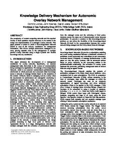
Figure 1. Schematic representation of oxytocin receptor-mediated signaling mechanism in myometrial contractility. Step I shows the inactive oxytocin receptor and the G-protein subunit Ga in its inactive GDP-bound state with high affinity for Gbg. Steps II and III describe the binding of oxytocin to the oxytocin receptor and activation of G-protein, which releases GDP from Ga, allowing it to bind GTP. GTP-bound Ga (Ga-GTP) has a lower affinity toward Gbg, which leads to the dissociation of Ga from Gbg. The G-protein components (Ga-GTP and Gbg) transduce signals to the regulatory molecules downstream. The G-protein signaling is regulated by the hydrolysis of the bound GTP to GDP and further association of resultant Ga-GDP with Gbg. If the oxytocin receptor remains occupied by oxytocin/any other ligand, the G-protein restarts the cyclic reaction by dissociating the bound GDP. Activated G-protein-coupled receptor complex signals the activation of phospholipase C (PLC). The level of phosphatidylinositol 4,5-bisphosphate (PIP2) in the cell is regulated by a balance between hydrolytic activity of PLC and its synthesis by phosphatidylinositol kinase. The PLC activation results in the cleavage of PIP2 into inositol 1, 4, 5-trisphosphate (InsP3) and diacyl glycerol (DAG). InsP3 stimulates the release of Ca2+ from intracellular pools, and this leads to the modulation of calcium voltage channels at the cell membrane, increasing calcium influx. Calcium forms a complex with calmodulin, further activating the myosin light-chain (MLC) kinase. MLC kinase then phosphorylates MLC. In parallel, DAG activates protein kinase C (PKC), leading to increased phosphorylation of downstream targets such mitogen-activated protein (MAP) kinase and stimulates the synthesis of prostaglandins. Together, these cellular events mediate myometrial contraction. GDP indicates guanosine diphosphate; GTP, guanosine-50 -triphosphate.
fluid, crosses the placenta, and is rapidly metabolized in the liver and kidneys, with small amounts of the drug excreted unchanged in urine.
’
Prevention of Postpartum Hemorrhage (PPH)
Oxytocin is the first-line agent in the prevention and treatment of PPH. Active management of the third stage of labor involving www.anesthesiaclinics.com
Oxytocin Protocols for Cesarean Delivery
’
51
prophylactic administration of oxytocin before delivery of the placenta has been shown to reduce PPH by >60%.13,14 In a Cochrane review of 20 trials (n = 10,806), prophylactic oxytocin reduced the risk of blood loss exceeding 500 mL [6 trials; relative risk (RR), 0.53; 95% confidence interval (CI), 0.38-0.74) and the need for therapeutic uterotonic agents (4 trials; RR, 0.56; 95% CI, 0.36-0.87) compared with placebo.13 In 5 trials (n = 2226), prophylactic oxytocin was compared with ergot alkaloids. Oxytocin was associated with fewer side effects, such as nausea and vomiting, and better prevented blood loss >500 mL. There was no difference in the rate of manual removal of the placenta.13 Another review of 6 trials (n = 9332) by McDonald et al15 found a small reduction in the risk of PPH (when defined as an estimated blood loss between 500 and 1000 mL) with a combination of oxytocin 5 IU and ergometrine 0.5 mg, when compared with oxytocin 5 or 10 IU. This difference was greater with the lower dose of oxytocin, but not demonstrated for estimated blood loss of >1000 mL. Vomiting, nausea, and hypertension were more common with the use of the oxytocinergometrine combination. Oxytocin is beneficial as the primary therapy in the management of PPH; however, the literature is inconclusive regarding its most appropriate dose or relative potency when compared with other uterotonic agents. The available studies are limited in number and exhibit heterogeneity in their study populations, oxytocin doses and routes of administration, and the outcomes recorded. As a consequence, the current national and international guidelines for oxytocin administration during cesarean delivery are diverse and mainly empirical, leading to significant variability in global clinical practices. With the rate and total dose unspecified, the World Health Organization suggests an infusion of oxytocin 20 IU/L, and the American College of Obstetricians and Gynecologists practice bulletin indicates a range of oxytocin 10 to 40 IU/L for the prevention of PPH.16,17 The Royal College of Obstetricians and Gynaecologists18 guidelines recommend oxytocin 5 IU by slow IV injection, and the Society of Obstetricians and Gynaecologists of Canada19 recommends carbetocin (an oxytocin analog) 100 mcg IV bolus over 1 minute in lieu of oxytocin for the prevention of PPH during elective cesarean delivery. In addition to these disparities, there is no clear guidance on whether to use oxytocin in a prophylactic or therapeutic manner, or if methods should be altered for women known to be at a high risk for PPH. Inconsistencies in societal guidelines have resulted in significant variation in clinical oxytocin regimens used during cesarean delivery. Routine use of an oxytocin infusion varied greatly (range, 11% to 55%) within Great Britain and Ireland,20 although when used, an oxytocin 5 IU bolus was administered by 88% (85% to 95%) of 391 respondents, with most of the remaining respondents using a 10 IU bolus dose. A similar survey conducted in Australia and New Zealand revealed the use www.anesthesiaclinics.com
52
’
Balki and Tsen
of 68 different regimens during elective cesarean delivery; however, approximately 98% of survey respondents used an oxytocin IV bolus of 5 IU (32%) or 10 IU (67%), as well as an oxytocin maintenance infusion, either routinely or selectively.21
’
Oxytocin Dosing at Cesarean Delivery
The amount of oxytocin required to establish adequate uterine tone in low-risk, nonlaboring women undergoing elective cesarean delivery has been shown to be significantly lower than the commonly used dose of 5 IU (Table 1). In this population of patients, Carvalho et al23 demonstrated that the minimum effective IV bolus dose of oxytocin (ED90) for adequate uterine tone is 0.35 IU (95% CI, 0.18-0.52 IU). Butwick et al,24 in a randomized controlled trial of different doses of oxytocin ranging from 0 to 5 IU, observed a high prevalence of adequate uterine tone with oxytocin doses between 0.5 and 3 IU. Because IV bolus dosing of oxytocin seems to result in greater adverse effects (see below), an infusion regimen has also been examined. George et al25 demonstrated that the infusion dose (ED90) to prevent uterine atony and PPH in nonlaboring women undergoing elective cesarean delivery is 0.29 IU/min (95% CI, 0.15-0.43 IU/min), which correlated with oxytocin 15 IU in 1 L of fluid over a 1-hour period. Together, these 3 studies observed that the use of lower oxytocin doses resulted in a similar estimated blood loss during cesarean delivery when compared with historically higher doses. Sarna et al22 indicated in a randomized controlled trial that the administration of oxytocin doses >5 IU (the lowest evaluated dose), including doses up to 20 IU, did not further improve uterine tone or reduce blood loss. In laboring women induced or augmented with oxytocin, Balki et al27 observed the need for 9-fold greater doses of oxytocin (ED90 = 2.99 IU; 95% CI, 2.32-3.67 IU) to produce adequate uterine contractions during cesarean delivery when compared with the dose effective in nonlaboring women (Table 1). In addition, despite the greater dose of oxytocin used, the blood loss was almost twice that incurred by nonlaboring women [1178 (716) mL vs. 693 (487) mL]. In contrast, Munn at al26 did not find any difference in the amount of blood loss in laboring women undergoing cesarean delivery with oxytocin low-dose (10 IU) versus high-dose (80 IU) regimens; however, the need for additional uterotonic agents was greater in the low-dose group, in women who had experienced labor arrest, and those with an intrapartum clinical diagnosis of chorioamnionitis (Table 1). A metaanalysis of 11 clinical trials on the use of oxytocin for labor induction demonstrated that high-dose or more aggressive oxytocin protocols resulted in more episodes of uterine hyperstimulation, lower rates of spontaneous vaginal delivery, and a higher incidence of PPH compared www.anesthesiaclinics.com
Oxytocin Protocols for Cesarean Delivery
Table 1.
’
53
Oxytocin Dose at CD
References
Study Design
Initial Oxytocin Dose
Elective CD in healthy term nonlaboring women Sarna RCT; n = 40 5, 10, 15, or 20 IU et al22 bolus
Carvalho Biased coin up-andet al23 down method; n = 40
Outcome No difference in uterine tone between groups Mean EBL = 485670 mL ED90 = 0.35 IU; 95% CI, 0.18-0.52 IU EBL = 693 (487) mL
0.5 IU bolus in first patient, dose for next patient based on the response of the previous patient Butwick Double-blinded RCT; 0, 0.5, 1, 3, 5 IU bolus No difference in n = 75 over 15 s adequate uterine et al24 tone at 2 min among groups: 73%, 100%, 93%, 100%, and 93% for 0, 0.5, 1, 3, and 5 IU, respectively Mean EBL = 697836 mL George ED90 = 0.29 IU/min; Biased coin up-and0.4 IU/min dose in et al25 down method; first patient, dose 95% CI, 0.15n = 40 for next patient 0.43 IU/min based on the EBL not mentioned response of the previous patient CD in laboring women 39% required Munn Double-blinded, RCT; 10 IU/500 mL over additional et al26 n = 321 30 min (333 mU/ uterotonics in lowmin) vs. 80 IU/ dose vs. 19% in 500 mL in 30 min high-dose group (2667 mU/min) (P < 0.001) EBL not significantly different [high dose = 957 (148) mL; low dose = 937 (159) mL] Balki ED90 = 2.99 IU; 95% 0.5 IU bolus in first Biased coin up-andet al27 patient, dose for down method; labor CI, 2.32-3.67 IU next patient based augmentation with EBL = 1178 (716) mL on the response of oxytocin for at least the previous patient 2 h; n = 30 CD indicates cesarean delivery; CI, confidence interval; EBL, estimated blood loss; ED90, dose effective in 90% women; RCT, randomized controlled trial.
www.anesthesiaclinics.com
54
’
Balki and Tsen
with low-dose regimens.28 Similarly, a recent study by Grotegut et al29 found a higher incidence of severe PPH secondary to uterine atony when labor augmentation occurred with oxytocin in larger doses and for longer durations. These clinical findings may indicate differences in oxytocin receptor distribution or function, signaling pathways, and sensitivity to oxytocin in laboring and nonlaboring women. Early consideration of uterotonic agents that act through different pathways is therefore advisable in laboring women or those at risk for PPH. Prophylactic administration of a combination of oxytocin-ergometrine has been shown to improve uterine contractility and reduce the need for additional uterotonic agents when compared with oxytocin alone (21% vs. 57%; P = 0.01) in women undergoing cesarean delivery after oxytocin labor augmentation.30 The existing literature is limited regarding the practice of using an oxytocin maintenance infusion after its initial use to achieve adequate uterine tone during cesarean delivery. Because IV oxytocin has a short halflife, there is likely a potential advantage to this practice, particularly in the immediate postpartum period when primary hemorrhage occurs most frequently; this may be especially relevant in laboring women previously exposed to induction or augmentation with oxytocin. A recent study on 2069 nonlaboring women undergoing an elective cesarean delivery compared the efficacy of oxytocin 5 IU given as a slow IV bolus over 1 minute with or without a maintenance infusion of oxytocin 40 IU/500 mL over 4 hours.31 They found no difference in blood loss >1000 mL between the groups (bolus+infusion, 15.7% vs. bolus only 16.0%); however, the need for an additional uterotonic agent in the bolus+infusion group was lower than in the bolus-only group (12.2% vs.18.4%; P < 0.001). In summary, lower oxytocin doses (< 5 IU) are effective for adequate uterine tone after cesarean delivery. The efficacy of these lower doses should not surprise us, as the amount of oxytocin used for labor induction and augmentation is significantly smaller, typically in the range of 0.5 to 30 mIU/min (0.0005 to 0.03 IU/min). It is of interest that even induction or augmentation oxytocin doses may sometimes result in uterine tachysystole.
’
Oxytocin Receptor Desensitization
The entire GPCR family undergoes a desensitization phenomenon, characterized by decreased cellular responsiveness and impaired signal transduction, with continuous or repeated receptor stimulation (ie, prolonged agonist exposure). The phenomenon is intended to protect the tissues from hyperstimulation and likely involves receptor phosphorylation, sequestration, internalization, and either degradation by lysosomes or reincorporation into the cell membrane; ultimately receptor downregulation occurs.32,33 www.anesthesiaclinics.com
Oxytocin Protocols for Cesarean Delivery
’
55
As a GPCR, the oxytocin receptor undergoes rapid molecular desensitization with homologous stimulation. This phenomenon is relevant to oxytocin-induced or augmented labor, especially with the current practice of giving increasingly larger doses over prolonged periods. Robinson et al34 demonstrated this phenomenon in cultured human myocytes through the inhibition of oxytocin-induced calcium efflux after preexposure of the myocytes to oxytocin 10 � 8 M (upper limit of the dose typically used for labor induction and augmentation). This effect was observed within 3 hours of oxytocin exposure, with 50% desensitization in 4.2 hours and complete desensitization in 6 hours. Phaneuf et al32 demonstrated the same phenomenon with myometrium obtained at emergency cesarean delivery; a 50-fold reduction in messenger RNA coding for the oxytocin receptor was observed when the duration of labor was longer than 12 hours. The number of myometrial cell surface oxytocin-binding sites also decreased significantly. The same investigators subsequently demonstrated that in women undergoing cesarean delivery after spontaneous and induced labors of >10 hours duration, messenger RNA levels for the oxytocin receptor decreased by 60and 300-fold, respectively, when compared with nonlaboring women.35 Increasing the duration and dose of oxytocin exposure during labor results in a decrease in oxytocin receptors. This biomolecular desensitization phenomenon has been demonstrated with in vitro studies on isolated term pregnant rat and human myometrial models, which indicate attenuation of oxytocin-induced myometrial contractions after pretreatment with oxytocin.36,37 In human myometrium, the effect was found to be dependent on the concentration and duration of oxytocin preexposure, and was observed with oxytocin 10 � 5 M for at least 2 hours or 10 � 8 M for at least 4 hours of preexposure.37 Similar attenuation of oxytocin-induced myometrial contractions was observed by Balki et al38 with in vitro samples obtained from women with oxytocin-augmented labors, but not in samples obtained from nonlaboring women or those with nonaugmented labors. Of particular clinical importance, this desensitization phenomenon is specific (ie, homologous) for oxytocin; contractions induced by other uterotonic agents (eg, ergonovine and prostaglandin F2) that act through different signaling pathways were unaffected.39 However, despite this desensitization effect on oxytocinexposed myometrial strips, the contractions induced by oxytocin were stronger than those produced by other uterotonic agents39; whether this is also true in vivo is yet to be determined. These findings indicate that women who have received oxytocin induction or augmentation during labor will undergo oxytocin receptor desensitization and downregulation in a dose-related and durationrelated manner. The downregulation of the oxytocin receptor does not affect other uterotonic agents that work through a different mechanism. www.anesthesiaclinics.com
56 ’
’
Balki and Tsen
Cardiovascular Effects of Oxytocin
The Confidential Enquiries into Maternal Deaths (UK 1997 to 1999)40 reported that an IV bolus of oxytocin 10 IU contributed to 2 maternal deaths. One case involved a cesarean delivery patient with spinal anesthesiaassociated hypotension who suffered a cardiac arrest immediately after oxytocin administration; the other case involved a patient with known pulmonary hypertension who collapsed following oxytocin administration after a vaginal delivery. Two additional deaths with an IV bolus of oxytocin 10 IU during cesarean delivery were reported in the Confidential Enquiries into Maternal Deaths in South Africa (2005 to 2007). Similar to the UK report, the first patient expired because of the oxytocin exacerbating the hypotension initially produced by the spinal anesthesia. The other case also involved hypotension, as well as significant respiratory depression, observed after oxytocin administration.41 The UK report recommended the reduction in the initial IV bolus dose of oxytocin from 10 to 5 IU,40 which was adopted into the guidelines from the British National Formulary that now advise the use of “slow IV” oxytocin 5 IU for PPH prevention.42 The hemodynamic effects of oxytocin have been subsequently examined and recorded using intra-arterial blood pressure monitoring, transthoracic bioimpedance, thermodilution recording, beat-to-beat pulse waveform analysis, vectorcardiography, and Holter monitoring. After oxytocin administration, otherwise healthy parturients exhibit peripheral vasodilation, hypotension, tachycardia, increased cardiac output and stroke volume, and increased pulmonary arterial pressure.43 Hypotension is mainly caused by vasodilation mediated by vascular endothelial oxytocin receptors through calcium-dependent stimulation of the nitric oxide pathway.44 Oxytocin also causes the release of atrial natriuretic peptide through the oxytocin receptors within the heart leading to diuresis, natriuresis, and vasodilation,45 and possibly modulates the release of acetylcholine from intrinsic cardiac cholinergic neurons leading to a mild negative inotropic effect.46 Several studies have described the cardiovascular effects of oxytocin during cesarean delivery, which seem to be influenced by the dose and mode of administration (Table 2).
Dose-Dependent Effects
The hemodynamic effects of an IV bolus of oxytocin 10 IU were first studied by Secher et al43 in anesthetized women within their first trimester of pregnancy. Thirty seconds after oxytocin administration, a decrease in systemic resistance (59%), pulmonary resistance (44%), and femoral arterial pressure (40%) was observed. To compensate, an increase in cardiac output (54%) was produced by an increase in heart rate (31%) and stroke volume (17%). Within 150 seconds after oxytocin www.anesthesiaclinics.com
Oxytocin Protocols for Cesarean Delivery
Table 2.
’
57
Oxytocin-induced Hemodynamic Changes During CD
References
Patient Population
Method
Pinder et al47 Double-blinded Oxytocin 5 vs. 10 IU bolus RCT Elective CD in NIBP and thoracic bioimpedance healthy term monitoring women; n = 34 Langesæter et al48
Elective CD in Oxytocin 5 IU bolus healthy term Arterial line and LidCOplus women; monitoring n = 10
Thomas et al49
Double-blinded Oxytocin 5 IU bolus vs. 5 IU infusion over RCT 5 min Elective CD in healthy term Intra-arterial BP monitoring women; n = 30
Svanstrom et al50
Double-blinded Oxytocin 10 IU bolus (n = 20) vs. methylRCT ergometrine 0.2 mg Elective CD in (n = 20) vs. oxytocin healthy term 10 IU bolus in women; nonpregnant, n = 40 nonanesthetized normal controls (n = 10) ECG, vectorcardiography and intra-arterial BP monitoring
Hemodynamic Outcomes Significant decrease in MAP and increase in HR from baseline after 10 IU bolus CO increased 50% and 80% in 5 and 10 IU groups, respectively Median (range) changes from baseline were: 61% (24%-78%) increase in CI; 39% (31%-49%) decrease in SVRi; and 67% (54%72%) decrease in SBP Maximum cardiovascular effects observed 45 s (25-80 s) after oxytocin injection HR increased by 17 (10.7) beats/min in bolus vs. 10 (9.7) beats/ min in the infusion group MAP decreased by 27 (7.6) mm Hg in bolus vs. 8 (8.7) mm Hg in infusion group No differences in the EBL between the 2 groups Oxytocin produced a significant increase in HR by 28 (4) and 52 (3) beats/min (P < 0.001), decreases in MAP by 33 (2) and 30 (3) mm Hg (P < 0.001), and increases in the spatial ST-change vector magnitude by 77 (12) and 114 (8) mV (P < 0.001), in CD patients and controls, respectively Symptoms of chest pain and discomfort were simultaneously present
www.anesthesiaclinics.com
58
’
Balki and Tsen
Table 2. (continued)
References
Patient Population
Sartain et al51 Double-blinded RCT Elective CD in healthy term women; n = 80
Langesæter et al52
RCT Elective CD in healthy term women; n = 80
Jonsson et al53
Double-blinded RCT Elective CD in healthy term women; n = 103
www.anesthesiaclinics.com
Method
Hemodynamic Outcomes
Methylergometrine produced mild hypertension and no significant ECG changes Oxytocin 2 or 5 IU bolus Greater increase in HR with 5 vs. 2 IU group followed by infusion [32 (17) vs. 24 10 IU/h (13) beats/min; P = 0.015] Larger decrease in MAP in 5 vs. 2 IU group [13 (15) vs. 6 (10) mm Hg; P = 0.030] No differences in EBL, uterine tone, or requests for additional uterotonics between groups Maximal change in CO Oxytocin 5 IU bolus after the first and This was followed by a second doses were 94% second bolus of 5 IU in and 42%, respectively 20 patients (P < 0.0001), and for Intra-arterial BP and SBP 31% and 23%, LidCOplus respectively (P = 0.003) monitoring No significant differences in EBL and Hb decrease in these 20 patients compared with the other 60 patients in the study Significantly higher Oxytocin 5 vs. 10 IU occurrence of ST bolus depressions with Holter and NIBP oxytocin 10 IU [11% monitoring (21.6)] than 5 IU [4% Blood sample at 12 h (7.7)] (P < 0.05) postoperatively for The absolute risk troponin levels reduction was 13.9% MAP decreased from baseline by 9 mm Hg in the 5 IU and 17 mm Hg in 10 IU group (P < 0.01) The increase in HR did not differ Troponin I levels were increased in 4 subjects
Oxytocin Protocols for Cesarean Delivery
’
59
Table 2. (continued)
References
Patient Population
Method
McLeod et al54
Elective CD in Oxytocin 5 IU bolus over 3 min and placebo healthy term infusion vs. 5 IU bolus women; over 3 min and 30 IU/ n = 74 4 h infusion Thoracic bioimpedance monitoring
Kim et al55
Oxytocin dose: RCT Elective CD in Group 1 = 0.5 IU/min healthy term Group 2 = 2 IU bolus then 0.25 IU/min women; Group 3 = 5 IU bolus n = 60 then 0.25 IU/min
Langesæter et al56
Oxytocin 5 IU bolus Observational LiDCOplus monitor study Severe preeclampsia undergoing CD; n = 18
Bhattacharya Double-blinded et al57 RCT
Hemodynamic Outcomes (3.9%), but there were no differences in occurrence of symptoms, troponin I levels, or EBL Oxytocin bolus caused rise in CI, LCWi, and HR; decrease in SVRi; and no change in BP Hemodynamic measures returned to normal over 60 min with no adverse effects apparent from the additional oxytocin infusion Maximum decrease in MAP 4.5%, 7.6%, and 11.8% in groups 1, 2, and 3 (P < 0.05), respectively Maximum decrease in HR 7.6%, 17%, and 26.1% in groups 1, 2, and 3 (P < 0.05), respectively Uterine contraction significantly better with 2 and 5 IU boluscontinuous groups than only continuous group EBL not significantly different between groups All patients had an increase in HR 36%, decrease in SVR 52%, and MAP 38% Five patients had a decrease in CO because of an inability to increase SV, suggesting unpredictable response in preeclamptic patients Significant rise in HR and decrease in MAP www.anesthesiaclinics.com
60
’
Balki and Tsen
Table 2. (continued)
References
Patient Population
Method
Elective CD in Oxytocin 3 IU bolus in 15 s vs. infusion over healthy term 5 min women; n = 80
Hemodynamic Outcomes seen in bolus compared with the infusion group ECG changes (ST depression) (n = 3) and chest pain (n = 5) were seen in bolus and not in the infusion group
CD indicates cesarean delivery; CI, cardiac index; CO, cardiac output; EBL, estimated blood loss; HR, heart rate; LCWi, left cardiac work index; MAP, mean arterial pressure; RCT, randomized controlled trial; SBP, systolic blood pressure; SVRi, systemic vascular resistance index.
administration, the pulmonary arterial pressure and wedge pressure increased by 33% and 35%, respectively.43 Several studies have indicated that more profound hemodynamic consequences are observed with oxytocin 10 IU when compared with 5 IU.47,48,50,53,54 Reducing the oxytocin dose from 5 to 2 IU decreases these changes even further without affecting the blood loss or need for additional uterotonic agents.51,55 Although these physiological changes may be tolerated by healthy pregnant women, those with hypovolemia or comorbid conditions may not be able to compensate. Slowing the administration of oxytocin, combined with the coadministration of phenylephrine has been shown to mitigate, but not completely abolish, the hemodynamic effects.58 In contrast with the systemic vasodilation observed with oxytocin, the coronary circulation may respond with vasoconstriction, diminished blood flow, or both. Jonsson et al53 found a significantly higher occurrence of ST depression with IV oxytocin 10 IU than 5 IU [11% (21.6) vs. 4% (7.7); P < 0.05] in women undergoing elective cesarean delivery; no differences in symptoms or troponin I levels were observed. Svanstrom et al50 observed that an IV bolus of oxytocin 10 IU induced chest pain, transient profound tachycardia, hypotension, and concomitant signs of myocardial ischemia on ECG and vectorcardiography. Repeated Oxytocin Dose
Langesæter et al52 observed the hemodynamic effects of a first and second IV bolus dose of oxytocin 5 IU on invasive blood pressure measurements and the lithium dye dilution method of cardiac output. If hypotension is defined as a 20% reduction in systolic blood pressure, then all patients in this study exhibited hypotension after oxytocin www.anesthesiaclinics.com
Oxytocin Protocols for Cesarean Delivery
’
61
administration; a mean decrease in systolic blood pressure (31% and 23%) after both the first and second oxytocin dose, respectively, was observed. The slightly attenuated effects of second-dose oxytocin may have been related to the administration of IV fluids or vasopressors or the concomitant desensitization of oxytocin receptors. Mode of Delivery of Oxytocin
The IV administration of oxytocin 5 IU as a slow versus rapid bolus in healthy term parturients undergoing elective cesarean delivery has been shown to produce less cardiovascular instability, with no difference in total blood loss.49 Similarly, IV oxytocin 3 IU as an infusion, instead of a bolus, was found to cause less cardiovascular instability.57 Oxytocin in Specific Patient Populations
Patients with preeclampsia and cardiac disease are at heightened risk for severe cardiovascular decompensation and unpredictable hemodynamic responses to oxytocin administration.56,59 The response is predominantly mediated by an increase in heart rate, possibly because of the inability to increase stroke volume in the setting of diastolic dysfunction. In a case series by Langesæter et al,60 oxytocin was used in incremental doses of 0.1 to 0.5 IU during cesarean delivery in parturients with advanced cardiac disease that included cardiomyopathy, congenital, and valvular heart disease. There was acceptable hemodynamic stability, although transient changes in blood pressure and cardiac output were observed.60 In view of these hemodynamic effects of oxytocin, it is desirable to administer oxytocin as an infusion in the lowest possible effective dose. ’
Additional Adverse Effects of Oxytocin
Administration of oxytocin can result in nausea, vomiting, headache, and flushing. As a result of structural similarities with vasopressin, large doses of oxytocin may also cause water retention, hyponatremia, seizures, and coma61; however, the antidiuretic effects may be minimized with an infusion rate of <45 mIU/min.62 Consequently, in patients at higher risk for pulmonary edema, such as those with severe cardiac conditions or preeclampsia, a lower rate of oxytocin infusion should be utilized. The cardiovascular and other adverse effects associated with oxytocin have led to its inclusion in a list of “high-alert medications” published by The Institute for Safe Medication Practices (ISMP),63 an independent, nonprofit organization. This designation indicates that the drug represents “a heightened risk of causing significant patient harm when used in error” and may “require special safeguards to reduce the risk of error.” Important medical groups, including the Joint www.anesthesiaclinics.com
62
’
Balki and Tsen
Commission, use the ISMP recommendations when promoting or evaluating medication safety. ’
Rational Plan for Oxytocin Use: The “Rule of Threes”
As a result of the known pharmacokinetic, pharmacodynamic, and clinical responses to oxytocin, we have developed a stepwise, standardized “Rule of Threes” protocol for oxytocin use during cesarean delivery to guide practitioners in a clear and concise manner (Fig. 2).64 Simply stated, the rule indicates: (1) 3 IU oxytocin IV dose over 30 seconds (2) 3-minute intervals before additional oxytocin dosing (3) 3 total doses (initial load+2 rescue doses) (4) 3 IU/h oxytocin infusion for maintenance (30 IU/L at 100 mL/h) (5) 3 pharmacologic options if the 3 doses of oxytocin are ineffective Although the IV dose for oxytocin in low-risk, nonlaboring women has been demonstrated to be lower than for laboring women with previous exposure to oxytocin induction or augmentation (ED90 0.35 vs. 2.99 IU),23,30 the single oxytocin 3 IU dose is effective for the prevention of uterine atony and PPH in both scenarios. Taken together, the “Rule of Threes” protocol emphasizes the following precautions during the administration of oxytocin after cesarean delivery64:
Figure 2. The “Rule of Threes” protocol for oxytocin and uterotonic agent administration during cesarean delivery. Adequate or inadequate refers to the strength of the uterine tone as measured by the obstetric provider at time of cesarean delivery. Cytotec indicates misoprostol; hemabate, carboprost tromethamine; IM, intramuscular administration; IMM, intramyometrial; IV, intravenous administration; methergine, methylergonovine maleate. www.anesthesiaclinics.com
Oxytocin Protocols for Cesarean Delivery
’
63
(1) Oxytocin should not be administered as a rapid IV bolus (eg, <15 s); (2) an initial infusion of oxytocin should be followed by a maintenance infusion; (3) higher initial and infusion doses of oxytocin offer no clinical benefit, may cause more adverse effects, and should be avoided; (4) if oxytocin is not producing effective uterine contractions, other uterotonic agents acting through different pathways [eg, methylergonovine maleate 0.2 mg intramuscular or slow IV over 1 min diluted in 10 mL (relative contraindication: hypertension), carboprost tromethamine 0.25 mg intramuscular or intramyometrial (relative contraindication: asthma or respiratory dysfunction), or misoprostol 800 to 1000 mcg rectal or 600 mcg buccal] should be considered19; (5) oxytocin use in patients with hypovolemia, preeclampsia, and cardiac or pulmonary comorbidities should be closely monitored for hemodynamic and respiratory changes; and (6) vasopressors and resuscitative drugs should be readily available when oxytocin is administered. ’
Conclusions
The isolation and subsequent synthesis of oxytocin has resulted in a greater understanding of its properties and importance to reproductive physiology. Despite over 60 years of use, our understanding of this agent, and certainly its use, deserves further investigation and refinement. An appreciation of the benefits and adverse effects associated with the use of oxytocin to augment uterine tone and prevent PPH indicates the need to administer lower doses at a slower rate and to give earlier consideration to the use of alterative uterotonic agents. ’
Acknowledgment
The authors acknowledge Nivetha Ramchandran, PhD, Department of Anesthesia and Pain Management, Mount Sinai Hospital, Toronto, for creating schematic pathway of the mechanism of action of oxytocin in Figure 1.
The authors have no conflicts of interest to disclose.
’
References
1. Engstrom T. Myometrial receptors in rat parturition. Dan Med Bull. 2002;50: 219–237. 2. Dale HH. “On some physiological actions of ergot”. J Physiol. 1906;34:163–206. 3. du Vigneaud V, Ressler C, Swan JM, et al. The synthesis of an octapeptide amide with the hormonal activity of oxytocin. J Am Chem Soc. 1953;75:4879–4880. www.anesthesiaclinics.com
64
’
Balki and Tsen
4. Caldeyro-Barcia R, Poseiro JJ. Oxytocin and contractility of the pregnant human uterus. Ann N Y Acad Sci. 1959;75:813–830. 5. Caldeyro-Barcia R. Oxytocin in pregnancy and labour. Acta Endocrinol Suppl (Copenh). 1960;34:41–49. 6. Zeeman GG, Khan-Dawood FS, Dawood MY. Oxytocin and its receptor in pregnancy and parturition: current concepts and clinical implications. Obstet Gynecol. 1997; 89:873–883. 7. Inoue T, Kimura T, Azuma C, et al. Structural organization of the human oxytocin receptor gene. J Biol Chem. 1994;269:32451–32456. 8. Michelini S, Urbanek M, Dean M, et al. Polymorphism and genetic mapping of the human oxytocin receptor gene on chromosome 3. Am J Med Genet. 1995;60:183–187. 9. Gimpl G, Fahrenholz F. The oxytocin receptor system: structure, function, and regulation. Physiol Rev. 2001;81:629–683. 10. Kimura T, Takemura M, Nomura S, et al. Expression of oxytocin receptor in human pregnant myometrium. Endocrinology. 1996;137:780–785. 11. Garfield RE. Control of myometrial function in preterm versus term labor. Clin Obstet Gynecol. 1984;27:572–591. 12. Leake RD, Weitzman RE, Fisher DA. Pharmacokinetics of oxytocin in the human subject. Obstet Gynecol. 1980;56:701–704. 13. Westhoff G, Cotter AM, Tolosa JE. Prophylactic oxytocin for the third stage of labour to prevent postpartum haemorrhage. Cochrane Database Syst Rev. 2013;(issue 10): Art. No.: CD001808. 14. Prendiville WJP, Elbourne D, McDonald SJ. Active versus expectant management in the third stage of labour. Cochrane Database Syst Rev. 2000;(issue 3):Art. No.: CD000007. 15. McDonald SJ, Abbott JM, Higgins SP. Prophylactic ergometrine-oxytocin versus oxytocin for the third stage of labour. Cochrane Database Syst Rev. 2004;(issue 1):Art. No.: CD000201. 16. WHO guidelines for the management of postpartum haemorrhage and retained placenta; 2009. Available at: http://whqlibdoc.who.int/publications/2009/9789241598514_ eng.pdf. Accessed July 15, 2013. 17. American College of Obstetricians and Gynecologists. ACOG Practice Bulletin: Clinical Management Guidelines for Obstetrician-Gynecologists Number 76, October 2006: postpartum hemorrhage. Obstet Gynecol. 2006;108:1039–1047. 18. Royal College of Obstetricians and Gynaecologists: prevention and management of postpartum haemorrhage. RCOG Green-top Guideline No. 52; May 2009. Available at: http://www.rcog.org.uk/files/rcog-corp/GT52PostpartumHaemorrhage0411.pdf. Accessed September 24, 2012. 19. Leduc D, Senikas V, Lalonde AB, et al. Clinical Practice Obstetrics Committee; Society of Obstetricians and Gynaecologists of Canada. Active management of the third stage of labour: prevention and treatment of postpartum hemorrhage: no. 235 October 2009 (replaces no. 88, April 2000). J Soc Obstet Gynaecol Can. 2009;31: 980–993. 20. Sheehan SR, Wedisinghe L, Macleod M, et al. Implementation of guidelines on oxytocin use at caesarean section: a survey of practice in Great Britain and Ireland. Eur J Obstet Gynecol Reprod Biol. 2010;148:121–124. 21. Mockler JC, Murphy DJ, Wallace EM. An Australian and New Zealand survey of practice of the use of oxytocin at elective caesarean section. Aust N Z J Obstet Gynaecol. 2010;50:30–35. 22. Sarna MC, Soni AK, Gomez M, et al. Intravenous oxytocin in patients undergoing elective cesarean section. Anesth Analg. 1997;84:753–756. 23. Carvalho JC, Balki M, Kingdom J, et al. Oxytocin requirements at elective cesarean delivery: a dose-finding study. Obstet Gynecol. 2004;104:1005–1010. www.anesthesiaclinics.com
Oxytocin Protocols for Cesarean Delivery
’
65
24. Butwick AJ, Coleman L, Cohen SE, et al. Minimum effective bolus dose of oxytocin during elective Caesarean delivery. Br J Anaesth. 2010;104:338–343. 25. George RB, McKeen D, Chaplin AC, et al. Up-down determination of the ED(90) of oxytocin infusions for the prevention of postpartum uterine atony in parturients undergoing Cesarean delivery. Can J Anaesth. 2010;57:578–582. 26. Munn MB, Owen J, Vincent R, et al. Comparison of two oxytocin regimens to prevent uterine atony at cesarean delivery: a randomized controlled trial. Obstet Gynecol. 2001;98:386–390. 27. Balki M, Ronayne M, Davies S, et al. Minimum oxytocin dose requirement after cesarean delivery for labor arrest. Obstet Gynecol. 2006;107:45–50. 28. Crane JMG, Young DC. Meta-analysis of low dose versus high dose oxytocin for labour induction. J Soc Obstet Gynaecol Can. 1998;20:1215–1223. 29. Grotegut CA, Paglia MJ, Johnson LNC, et al. Oxytocin exposure during labor among women with postpartum hemorrhage secondary to uterine atony. Am J Obstet Gynecol. 2011;204:e1–e6. 30. Balki M, Dhumne S, Kasodekar S, et al. Oxytocin-ergometrine co-administration does not reduce blood loss at caesarean delivery for labour arrest. BJOG. 2008;115:579–584. 31. Sheehan SR, Montgomery AA, Carey M, et al. Oxytocin bolus versus oxytocin bolus and infusion for control of blood loss at elective caesarean section: double blind, placebo controlled, randomised trial. BMJ. 2011;343:d4661. 32. Phaneuf S, Asboth G, Carrasco MP, et al. Desensitization of oxytocin receptors in human myometrium. Hum Reprod Update. 1998;4:625–633. 33. Smith MP, Ayad VJ, Mundell SJ, et al. Internalization and desensitization of the oxytocin receptor is inhibited by dynamin and clathrin mutants in human embryonic kidney 293 cells. Mol Endocrinol. 2006;20:379–388. 34. Robinson C, Schumann R, Zhang P, et al. Oxytocin-induced desensitization of the oxytocin receptor. Am J Obstet Gynecol. 2003;188:497–502. 35. Phaneuf S, Rodriguez Linares B, TambyRaja RL, et al. Loss of myometrial oxytocin receptors during oxytocin-induced and oxytocin-augmented labour. J Reprod Fertil. 2000;120:91–97. 36. Magalhaes JK, Carvalho JC, Parkes RK, et al. Oxytocin pretreatment decreases oxytocin-induced myometrial contractions in pregnant rats in a concentrationdependent but not time-dependent manner. Reprod Sci. 2009;16:501–508. 37. Balki M, Erik-Soussi M, Kingdom J, et al. Oxytocin pretreatment attenuates oxytocininduced contractions in human myometrium in-vitro. Anesthesiology. 2013;119:552–561. 38. Balki M, Erik-Soussi M, Kingdom J, et al. Myometrial contractility with different uterotonic agents in laboring and non-laboring women. Society for Obstetric Anesthesia and Perinatology Meeting, San Antonio, Texas, USA, May 12–18, 2010. 2010:A128. 39. Balki M, Cristian AL, Kingdom J, et al. Oxytocin pretreatment of pregnant rat myometrium reduces the efficacy of oxytocin but not of ergonovine maleate or prostaglandinF2 alpha. Reprod Sci. 2010;17:269–277. 40. Thomas TA, Cooper GM. Maternal deaths from anaesthesia. An extract from why mothers die 1997–1999, the confidential enquiries into maternal deaths in the United Kingdom. Br J Anaesth. 2002;89:499–508. 41. Saving Mothers 2005-2007: fourth report on Confidential Enquiries into Maternal Deaths in South Africa. Available at: http://www.health.gov.za/docs/reports/2011/ saving_b.pdf. Accessed July 20, 2013. 42. British Medical Association and Royal Pharmaceutical Society of Great Britain. British National Formulary. 54th ed. London: BMJ Publishing Group and APS Publishing: 2007;413–416. Available at: http://www.bnf.org/. 43. Secher NJ, Arnsbo P, Wallin L. Haemodynamic effects of oxytocin (syntocinon) and methyl ergometrine (methergin) on the systemic and pulmonary circulations of pregnant anaesthetized women. Acta Obstet Gynecol Scand. 1978;57:97–103. www.anesthesiaclinics.com
66
’
Balki and Tsen
44. Thibonnier M, Conarty DM, Preston JA, et al. Human vascular endothelial cells express oxytocin receptors. Endocrinology. 1999;140:1301–1309. 45. Petersson M. Cardiovascular effects of oxytocin. Prog Brain Res. 2002;139:281–288. 46. Mukaddam-Daher S, Yin YL, Roy J, et al. Negative inotropic and chronotropic effects of oxytocin. Hypertension. 2001;38:292–296. 47. Pinder AJ, Dresner M, Calow C, et al. Haemodynamic changes caused by oxytocin during caesarean section under spinal anaesthesia. Int J Obstet Anesth. 2002;11:156–159. 48. Langesæter E, Rosseland LA, Stubhaug A. Hemodynamic effects of oxytocin during cesarean section. Int J Gynaecol Obstet. 2006;95:46–47. 49. Thomas JS, Koh SH, Cooper GM. Haemodynamic effects of oxytocin given as i.v. bolus or infusion on women undergoing Caesarean section. Br J Anaesth. 2007;98:116–119. 50. Svanstrom MC, Biber B, Hanes M, et al. Signs of myocardial ischaemia after injection of oxytocin: a randomized double-blind comparison of oxytocin and methylergometrine during Caesarean section. Br J Anaesth. 2008;100:683–689. 51. Sartain JB, Barry JJ, Howat PW, et al. Intravenous oxytocin bolus of 2 units is superior to 5 units during elective Caesarean section. Br J Anaesth. 2008;101: 822–826. 52. Langesæter E, Rosseland LA, Stubhaug A. Haemodynamic effects of repeated doses of oxytocin during Caesarean delivery in healthy parturients. Br J Anaesth. 2009;103:260–262. 53. Jonsson M, Hanson U, Lidell C, et al. ST depression at caesarean section and the relation to oxytocin dose. A randomised controlled trial. BJOG. 2010;117:76–83. 54. McLeod G, Munishankar B, MacGregor H, et al. Maternal haemodynamics at elective caesarean section: a randomised comparison of oxytocin 5-unit bolus and placebo infusion with oxytocin 5-unit bolus and 30-unit infusion. Int J Obstet Anesth. 2010;19:155–160. 55. Kim TS, Bae JS, Park JM, et al. Hemodynamic effects of continuous intravenous injection and bolus plus continuous intravenous injection of oxytocin in cesarean section. Korean J Anesthesiol. 2011;61:482–487. 56. Langesæter E, Rosseland LA, Stubhaug A. Haemodynamic effects of oxytocin in women with severe preeclampsia. Int J Obstet Anesth. 2011;20:26–29. 57. Bhattacharya S, Ghosh S, Ray D, et al. Oxytocin administration during cesarean delivery: randomized controlled trial to compare intravenous bolus with intravenous infusion regimen. J Anaesthesiol Clin Pharmacol. 2013;29:32–35. 58. Dyer R, Reed A, van Dyk D, et al. Hemodynamic effects of ephedrine, phenylephrine, and the coadministration of phenylephrine with oxytocin during spinal anesthesia for elective cesarean delivery. Anesthesiology. 2009;111:753–765. 59. Dyer RA, Piercy JL, Reed AR, et al. Hemodynamic changes associated with spinal anesthesia for cesarean delivery in severe preeclampsia. Anesthesiology. 2008;108:802–811. 60. Langesæter E, Dragsund M, Rosseland LA. Regional anaesthesia for Caesarean section in women with cardiac disease: a prospective study. Acta Anaesthesiol Scand. 2010;54:46–54. 61. Bergum D, Lonne H, Hakli TF. Oxytocin infusion: acute hyponatraemia, seizures and coma. Acta Anaesthesiol Scand. 2009;53:826–827. 62. Abdul-Karim R, Assali NS. Renal function in human pregnancy. V. Effects of oxytocin on renal hemodynamics and water and electrolyte excretion. J Lab Clin Med. 1961;57:522–532. 63. Institute for Safe Medical Practices. High alert medications. Available at: http:// www.Ismp.org. Accessed July 20, 2013. 64. Tsen L, Balki M. Oxytocin protocols during cesarean delivery: time to acknowledge the risk/benefit ratio? Int J Obstet Anesth. 2010;19:243–245. www.anesthesiaclinics.com