Prog. Polym. Sci. 31 (2006) 359–397 www.elsevier.com/locate/ppolysci
Polymer–drug conjugates: Progress in polymeric prodrugs Jayant Khandare a, Tamara Minko a,b,c,* a
Department of Pharmaceutics, Ernest Mario School of Pharmacy, Rutgers, The State University of New Jersey, 160 Frelinghuysen Road, Piscataway, NJ 08854-8020, USA b The Cancer Institute of New Jersey, 195 Little Albany Street, New Brunswick, NJ 08903, USA c New Jersey Center for Biomaterials, 610 Taylor Road, Piscataway, NJ 08854, USA Received 13 May 2005; received in revised form 30 August 2005; accepted 12 September 2005
Abstract Polymers are used as carriers for the delivery of drugs, proteins, targeting moieties, and imaging agents. Several polymers, poly(ethylene glycol) (PEG), N-(2-hydroxypropyl)methacrylamide (HPMA), and poly(lactide-co-glycolide) (PLGA) copolymers have been successfully utilized in clinical research. Recently, interest in polymer conjugation with biologically active components has increased remarkably as such conjugates are preferably accumulated in solid tumors and can reduce systemic toxicity. Based on the site and the mode of action, polymer conjugates possess either ‘tuned’ degradable or non-degradable bonds. In order to obtain such bonds, most of the strategies involve incorporation of amino acids, peptides or small chains as spacer molecules through multiple steps to include protections and deprotections. There is a need to design efficient synthetic methods to obtain polymeric conjugates with drugs and other bioactive components. Designs should aim to decrease the steric hindrance exhibited by polymers and the biocomponents. In addition, the reactivity of polymer and drug must be enhanced. This is especially true for the use of high molecular weight linear polymers and bulkier unstable drugs such as steroids and chemotherapeutic agents. Further, it is essential to elucidate the structure activity relationship (SAR) of a drug when it is conjugated with a polymer using different conjugation sites, as this can vary the efficacy and mechanism of action when compared with its free form. This review will discuss the current synthetic advances in polymer-conjugation with different bioactive components of clinical importance. In addition, the review will describe the strategies for reduction of steric hindrance and increase in reactivity of the polymers, drugs and bioactive agents and highlight the requisite structure activity relationship in polymer–drug bioconjugates. Finally, we will focus on passive and active targeting of polymeric drug delivery systems to specific site of drug action. q 2006 Elsevier Ltd. All rights reserved. Keywords: Polymer; Conjugation; Drug delivery; Prodrug; Enhanced permeability and retention; In vivo and in vitro
Contents 1. 2.
Introduction . . . . . . . . . . . . . . . . . . . . . . . . . . . . . . . . . . . . . . . . . . . . . . . . . . . . . . . . . . . . . . . . . . . . . . . . . . Design and synthesis of polymeric prodrugs . . . . . . . . . . . . . . . . . . . . . . . . . . . . . . . . . . . . . . . . . . . . . . . . . . . 2.1. Polymeric drug delivery system (PDDS) . . . . . . . . . . . . . . . . . . . . . . . . . . . . . . . . . . . . . . . . . . . . . . . . 2.2. n-hydroxysuccinimide (NHS) ester and coupling methods . . . . . . . . . . . . . . . . . . . . . . . . . . . . . . . . . . . . 2.3. Incorporation of spacers in prodrug conjugates . . . . . . . . . . . . . . . . . . . . . . . . . . . . . . . . . . . . . . . . . . . .
360 362 364 365 366
* Corresponding author. Address: Department of Pharmaceutics, Ernest Mario School of Pharmacy, Rutgers, The State University of New Jersey, 160 Frelinghuysen Road, Piscataway, NJ 08854-8020, USA. Tel.: C1 732 445 3831x214; fax: C1 732 445 3134. E-mail address:
[email protected] (T. Minko).
0079-6700/$ - see front matter q 2006 Elsevier Ltd. All rights reserved. doi:10.1016/j.progpolymsci.2005.09.004
360
3.
4. 5. 6.
7.
J. Khandare, T. Minko / Prog. Polym. Sci. 31 (2006) 359–397
2.4. Carbodiimide coupling reactions or zero lengths cross-linkers . . . . . . . . . . . . . . . . . . . . . . . . . . . . . . . . . Design and synthesis of polymeric conjugates . . . . . . . . . . . . . . . . . . . . . . . . . . . . . . . . . . . . . . . . . . . . . . . . . 3.1. Dextran . . . . . . . . . . . . . . . . . . . . . . . . . . . . . . . . . . . . . . . . . . . . . . . . . . . . . . . . . . . . . . . . . . . . . . . . 3.2. Poly(ethylene glycol) (PEG) and conjugates . . . . . . . . . . . . . . . . . . . . . . . . . . . . . . . . . . . . . . . . . . . . . . 3.3. PEG–HIV agent conjugates . . . . . . . . . . . . . . . . . . . . . . . . . . . . . . . . . . . . . . . . . . . . . . . . . . . . . . . . . . 3.4. PEG–drug and targeted delivery . . . . . . . . . . . . . . . . . . . . . . . . . . . . . . . . . . . . . . . . . . . . . . . . . . . . . . Poly-amino acid conjugates . . . . . . . . . . . . . . . . . . . . . . . . . . . . . . . . . . . . . . . . . . . . . . . . . . . . . . . . . . . . . . . 4.1. N-(2-hydroxypropyl)methacrylamide (HPMA) copolymer . . . . . . . . . . . . . . . . . . . . . . . . . . . . . . . . . . . . Dendrimer and its conjugates . . . . . . . . . . . . . . . . . . . . . . . . . . . . . . . . . . . . . . . . . . . . . . . . . . . . . . . . . . . . . . Critical aspects of polymer conjugation . . . . . . . . . . . . . . . . . . . . . . . . . . . . . . . . . . . . . . . . . . . . . . . . . . . . . . 6.1. Structure–activity relationship of conjugates (SAR) . . . . . . . . . . . . . . . . . . . . . . . . . . . . . . . . . . . . . . . . 6.2. Steric hindrance . . . . . . . . . . . . . . . . . . . . . . . . . . . . . . . . . . . . . . . . . . . . . . . . . . . . . . . . . . . . . . . . . . 6.3. Enhanced reactivity of polymers by incorporation of spacers . . . . . . . . . . . . . . . . . . . . . . . . . . . . . . . . . 6.4. Targeting of polymeric drugs . . . . . . . . . . . . . . . . . . . . . . . . . . . . . . . . . . . . . . . . . . . . . . . . . . . . . . . . 6.4.1. Passive tumor targeting . . . . . . . . . . . . . . . . . . . . . . . . . . . . . . . . . . . . . . . . . . . . . . . . . . . . . . . . . . . . . 6.4.2. Active targeting . . . . . . . . . . . . . . . . . . . . . . . . . . . . . . . . . . . . . . . . . . . . . . . . . . . . . . . . . . . . . . . . . . Conclusions . . . . . . . . . . . . . . . . . . . . . . . . . . . . . . . . . . . . . . . . . . . . . . . . . . . . . . . . . . . . . . . . . . . . . . . . . . Acknowledgements . . . . . . . . . . . . . . . . . . . . . . . . . . . . . . . . . . . . . . . . . . . . . . . . . . . . . . . . . . . . . . . . . . . . . References . . . . . . . . . . . . . . . . . . . . . . . . . . . . . . . . . . . . . . . . . . . . . . . . . . . . . . . . . . . . . . . . . . . . . . . . . . .
1. Introduction A prodrug is a form of a drug that remains inactive during its delivery to the site of action and is activated by the specific conditions in the targeted site. In other words, a prodrug is an inactive precursor of a drug. Prodrug reconversion (i.e. its conversion into its active form) occurs in the body inside a specific organ, tissue or cell. In most cases, normal metabolic processes such as the cleavage of a bond between a polymer and a drug by specific cellular enzymes are utilized to achieve prodrug reconversion. A conjugation of a drug with a polymer forms so-called ‘polymeric prodrug’. Polymeric conjugates of conventional drugs (polymeric prodrugs) have several advantages over their low molecular weight precursors. The main advantages include: (1) an increase in water solubility of low soluble or insoluble drugs, and therefore, enhancement of drug bioavailability; (2) protection of drug from deactivation and preservation of its activity during circulation, transport to targeted organ or tissue and intracellular trafficking; (3) an improvement in pharmacokinetics; (4) a reduction in antigenic activity of the drug leading to a less pronounced immunological body response; (5) the ability to provide passive or active targeting of the drug specifically to the site of its action; (6) the possibility to form an advanced complex drug delivery system, which, in addition to drug and polymer carrier, may include several other active components that enhance the specific activity of the main drug. Due to these advantages over to free form of a drug, the polymeric prodrug conjugates has lead into a new era of
368 369 369 372 378 378 380 383 385 388 388 388 391 392 392 393 394 394 394
polymeric drug delivery systems (PDDS). The task of obtaining a versatile polymer as an ideal candidate in drug delivery apparently seems to be intricate, as it has to undergo several vigorous clinical barriers. Therefore, many researchers rely on the polymers which have been approved and well established. For decades, the delivery of biomolecules using polymeric materials has attracted considerable attention from polymer chemists, chemical engineers and pharmaceutical scientists [1]. However, the design and synthesis of new polymeric candidates in view of their biological implications has just been initiated. Various architectures of polymers have been used as vehicles to deliver drugs, along with relevant targeting agents [2,3]. Depending on the nature and site of action of a drug, either homopolymers, or graft or block Table 1 Current status of polymeric bioconjugates in cancer therapy Polymer
Drug/biomolecules in conjugate
Current phase
Name of the company
PEGa P-HPMAb
Paclitaxel Doxorubicin (DOX) micelle and Platinate Paclitaxel Camptothecin Camptothecin
I II/III
Pharmacia Access Pharma
I I III
Camptothecin Aspartic acid DOX DOX-Galactosamine
II I II II
Pharmacia Pharmacia Cell Therapeutics Enzon NCI Pharmacia Pharmacia
P-HPMA P-HPMA Poly (glutamate) PEG PEG P-HPMA P-HPMA a b
Polyethylene glycol (PEG). Poly N-(2-Hydroxypropyl) methacrylamide (P-HPMA).
J. Khandare, T. Minko / Prog. Polym. Sci. 31 (2006) 359–397
polymers are being extensively used in bioconjugates. In general, a biologically active component undergoes numerous physio-chemical barriers, which can be further complicated by converting it into a polymeric prodrug. In addition, due to their higher molecular weight, polymers are known to dominate the physical properties of the bioconjugated moiety. It has been well established that polymers can enhance the aqueous solubility of drug molecules by conjugation. Moreover, polymers are being used for the preparation of various formulations such as liposomes, microparticles or nanoparticles [4]. Along with the polymer, the physico-chemical properties of the drug or biomolecule to be conjugated are equally important. The following properties of the drug molecules make it suitable as an ideal candidate to form the polymeric conjugate: (1) lower aqueous solubility, (2) instability at varied physiological pHs, (3) higher systemic toxicity, and (4) reduced cellular entry. Numerous polymeric prodrugs are in clinical phases (Table 1) and several others have been approved (Table 2).
361
Despite progress, the delivery of active components using clinically approved polymers remains a challenge. Methods such as encapsulation, complexation or covalent conjugation are routinely used in drug delivery research [5]. But, the resulting complexes formed are often unstable in a physiological environment. Various bioconjugate methodologies that would form a stable bond are being reported. Covalent conjugation of biomolecules, e.g. protein drugs to synthetic polymers, particularly poly(ethylene glycol) (PEG) does increase their plasma residence, reduces protein immunogenicity and can increase therapeutic index [6]. Successful bioconjugation depends upon the chemical structure, molecular weight, steric hindrance and the reactivity of the biomolecule as well as the polymer. In order to synthesize a bioconjugate, both chemical entities need to posses a reactive or functional groups such as –COOH, – OH, –SH or –NH2. However, the presence of multiple reactive groups makes the task a bit complex. Therefore, the synthetic methodology to form a conjugate involves either protection or deprotection of the groups. There is
Table 2 Polymeric drug delivery systems (PDDS) that have received regulatory approval. [17] Polymer/Formulation
Drug or Active agent
Brand/Trade name/s
Manufacturer
Therapeutic Indication
Approved Year/Country
Liposomal
Amphotericin B
AmBisome
Gilead, Fujisawa
Fungal infection
PEG
Adenosine deaminase
Adagen
Enzon
1990 (Europe), 1997 2000 1990
Styrene maleic acid and neocarzinostatin copolymer in Ethiodol Stealth (PEG-stabilized) liposomal
Zinostatin stimalamer
SMANCS/ Lipiodol
Yamanouchi
Doxorubicin
Doxil/Caelyx
Schering Plough, Alza
Liposomal
Doxorubicin
Myocet
Elan
Liposomal cytosine
Arabinoside
DepoCyt
SkyePharma
PEG
Interferon a-2b
PEG-Intron
Liposomal
Verteporfin
Visudyne
Enzon, ScheringPlough QLT, Novartis
PEG
Neulasta
Amgen
PEG
Granulocyte colony stimulating factor or pegfilgrastim L-asparaginase
PEG
Adenosine deaminase.
Oncaspar/ pegaspargase Adagen
Leishmaniasis Severe combined immunodeficiency Hepatocellular carcinoma
1993 1996 (Japan)
Kaposi’s sarcoma
1995
Refractory ovarian cancer Refractory breast cancer
1999 2003 (Europe, Canada) 2000 (Europe)
Metastatic breast cancer in combination with cyclophosphamide Lymphomatous meningitis Neoplastic meningitis Hepatitis C
1999 Phase IV 2001
Enzon
Wet macular degeneration in conjunction with laser treatment Reduction of febrile neutropenia associated with chemotherapy Antineoplastic
2000 2001 (Japan)
1997
Enzon
immunodeficiency disease
1993
2002
362
J. Khandare, T. Minko / Prog. Polym. Sci. 31 (2006) 359–397
further need to design simple and yet appropriate polymeric-conjugation methodology. Many of the most commonly used strategies involve use of both coupling agents such as dicyclohexyl carbodiimide (DCC) and 1-ethyl-3-(3-dimethylaminopropyl)carbodiimide or use of N-hydroxysuccinimide esters. Chemical conjugation of drugs or other biomolecules to polymers and its modifications can form stable bonds such as ester, amide, and disulphide. The resulting bond linkage should be relatively stable to prevent drug release during its transport before the cellular localization of the drug. Covalent bonds (e.g. ester or amide) are stable and could deliver the drug at the targeted site, but such bonds may not easily release targeting agents and peptides under the influence of acceptable environmental changes [7]. In the past, most of the polymeric prodrugs have been developed for the delivery of anticancer agents. High molecular weight prodrugs containing cytotoxic components have been developed to decrease peripheral side effects and to obtain a more specific administration of the drugs to the cancerous tissues [8,9]. Favorably, a macromolecular antitumor prodrug is expected to be stable in circulation and should degrade only after reaching the targeted cells or tissues. Polymer–drug conjugates can therefore be tailored for activation by extra- or intracellular enzymes releasing the parent drug in situ. In this review, our focus is on the most promising polymer candidates that are being used to form bioconjugates. There is a necessity to decrease the steric hindrance and increase the reactivity of polymers as well as biomolecules to be chemically conjugated. In this regard, we will discuss the current progress and advances with various methodologies to obtain polymeric prodrugs.
type of prodrug—the drug delivery system (DDS). Such a system can be constructed not only to target a desired organ as a whole, its cells or specific organelles inside certain cells but also to release a specified amount of the drug at specific times. The polymeric prodrug conjugate can also increase aqueous solubility, enhance biodistribution and retain the inherent pharmacological properties of the drug intact [11]. Three major types of polymeric prodrugs are currently being used [12]. Prodrugs of the first type are broken down inside cells to form active substance or substances. The second type of prodrug is usually the combination of two or more substances. Under specific intracellular conditions, these substances react forming an active drug. The third type of prodrug, targeted drug delivery systems, usually includes three components: a targeting moiety, a carrier, and one or more active component(s). The targeting ability of the delivery system depends on the several variables including: receptor expression; ligands internalization; choice of antibody, antibody fragments or non-antibody ligands; and binding affinity of the ligand [13]. Therefore, the selection of a suitable polymer and a targeting moiety is vital to the effectiveness of prodrugs.
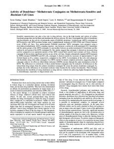
2. Design and synthesis of polymeric prodrugs Over the last decade, polymer chemists have been actively involved in designing polymeric materials for biomedical applications. One particular approach towards an improved use of drugs for therapeutic applications is to design polymeric prodrugs. A ‘prodrug’ is a chemical entity of an active parent drug with altered physico-chemical properties [10]. In a prodrug, a drug precursor remains inactive during delivery to the site of action and is specifically activated at the target site. The utilization of prodrugs, to a certain extent, allows for the preservation of specific activity of a drug and targets its release to certain cells or their organelles. The most complete realization of the prodrug approach is possible by the use of an advanced
Fig. 1. Schematic presentation for (a) polymeric prodrug with targeting agent and (b) hyperbranched polymer conjugate with targeting and imaging agent.
J. Khandare, T. Minko / Prog. Polym. Sci. 31 (2006) 359–397
363
Fig. 2. Schemes of various polymer architectures showing steric hindrance and crowding effect for chemical conjugation of drug molecules (a) linear polymer, (b) copolymer, (c) branched polymer, and (d) hyperbranched polymer.
364
J. Khandare, T. Minko / Prog. Polym. Sci. 31 (2006) 359–397
Numerous polymeric conjugation methods have been attempted since 1950s. In 1955, Jatzkewitz reported peptamin–polyvinylpyrrolidone conjugates improve the efficacy of the drug [14]. Subsequential biological aspects of polymeric prodrugs were not previously taken into consideration. However, the conjugation methodology would be more applied in the forthcoming years. In 1975, a rational model for pharmacologically active polymers was proposed by Ringsdorf—considered to be the pioneer of polymeric prodrug research [15]. In general, an ideal polymeric prodrug model consists mainly of a combination of one or more components: (a) a polymeric backbone as a vehicle, (b) one or more drugs of the biological active components, (c) spacer(s) for hydrolysis of the biomolecule and versatility for conjugation, (d) an imaging agent and (e) targeting moiety (Fig. 1a and b). The drug delivery carrier can be either biocompatible or an inert biodegradable polymer. The drug is coupled directly or via a spacer arm onto the polymer backbone. Selection of the spacer arm is critical as it opens the possibility of controlling the site and the rate of release of the active drug from the conjugates either by hydrolysis or by enzymatic degradation. The most challenging aspect of this protocol is the possibility of altering the body distribution and the cellular uptake by cell-specific or non-specific uptake enhancers. Due to current interdisciplinary research, molecular biologists and organic, polymer chemists can now design tailor-made polymeric carriers with different structural architecture. Soluble polymers as potential drug carriers have been reviewed in detail [16]. The polymers selected for preparing macromolecular prodrugs can be categorized according to: (a) chemical nature (vinylic or acrylic polymers, polysaccharides, poly(a-amino acids), etc., (b) biodegradability, (c) origin (either natural polymers or synthetic polymers) and (d) molecular weight (oligomers, macromers and polymers). Below are the schematic polymeric architectural structures in conjugated form with their bioactive components. Most of the polymers possess crowding effect for chemical conjugation (Fig. 2). Further synthetic strategies will need to be designed to reduce such effects that will improve the conjugation ratio and payload of component with the polymer. 2.1. Polymeric drug delivery system (PDDS) During the last decade, polymer chemistry was dedicated to synthesis, derivatization, degradation, characterization, application, and evaluation, for newer biocompatible and biodegradable polymers, all
of which were used as carriers for polymeric drug delivery systems. These systems possess unique roles in enhanced physiological drug distribution, bioavailability, drug targeting, time-controlled release, sensorresponsive release, etc. The emergence of polymer therapeutics has instigated research interfacing polymer chemistry and the biomedical sciences including initiation of nanosized (5–100 nm) polymer-based pharmaceuticals [6]. Polymer therapeutics include: rationally designed macromolecular drugs, polymer– drug and polymer–protein conjugates, polymeric micelles containing covalently bound drug, and polyplexes for DNA delivery. The clinical applications of polymer–protein conjugates and their resulting outcomes seem to be promising, especially for polymer–anticancer-drug therapeutics [6]. Many of the pharmacological properties of conventional free drugs can be improved through the use of polymeric drug delivery systems (PDDS), which include particulate carriers composed primarily of lipids and/or polymers, and their associated therapeutics [17]. Several PDDS have already reached the market (Table 2). The majority of the PDDS approved are mainly targeted for parenteral administration and include either liposomal or therapeutic molecules linked to poly(ethylene glycol) (PEG) polymers. The pegylation process is being used extensively in prodrug conjugates and the synthesis approach involves attachment of repeating units of poly(ethylene glycol) to a polypeptide drug. More than 30 years, scientists have developed various techniques to build PEG polymers and attach them to a drug of choice. Recently, pegylated drug forms in therapeutics of hepatitis C, acromegaly, rheumatoid arthritis, neutropenia, various cancers, wound healing, and other disorders either have been approved or are undergoing clinical trials [18]. Proteins and peptides hold great promise as therapeutic agents; however, they are prone for degradation by proteolytic enzymes. These bioactives can be rapidly cleared by the kidneys, generate neutralizing antibodies, and have a short circulating half-life. Therefore, pegylation of such drugs can overcome these and other shortfalls. In addition, increased molecular mass of proteins and peptides shields them from proteolytic enzymes and enhances pharmacokinetics [18]. Modification of a polymer to form a conjugate with a biomolecule depends upon two interrelated chemical reactions: (1) reactive functional groups present in the polymer and (2) functional groups present on the biological component. In general, most of the biomolecules such as ligands, peptides, proteins, carbohydrates, lipids, polymers, nucleic acid and
J. Khandare, T. Minko / Prog. Polym. Sci. 31 (2006) 359–397
oligonucleotide possess combinations of these functional groups. Selection of a suitable method, process, and reagents are crucial for successful chemical conjugation. Polymers may undergo several structural changes with solvents, coupling agents, and reactants. Peptide and protein PEGylation problems and solutions have been reviewed [19]. A detail of bioconjugation strategies involving PEG and other polymers with various biomolecules has been discussed briefly in Section 3.2. Most of the bioconjugation strategies involve coupling reactive nucleophiles with the following order of reactivity: thiol, a-amino groups, epsilone amino group, carboxyl and hydroxyl. The order of reactivity depends upon the pH in the reaction and presence of steric hindrance on the coupling moiety. Recent advances in bioconjugation use homobifunctional amine or heterobifunctional coupling reagents. A number of electrophilic groups are capable of reacting with amines and other nucleophiles, e.g. epoxides, vinylsulphones, and aziridines. In addition, sulphonyl chlorides also react with amines; however, they are generally water-insoluble and may get hydrolyzed. Homobifunctional compounds can form protein–protein linkages such as N,N 00 -ethyleneiminoyl-1-6-diaminohexane, bis-aziridin, divinyl sulphone (DVS), nitrogen mustard and bis-sulphonyl chloride. On the other hand, heterobifunctional reagents are useful to couple amines with other functional groups. The most important amine reacting heterobifunctional compounds are used in protein chemistry; e.g. photoactivation, biotinylation and thiolation reactions. Reactive groups in protein–carboxyl functions offer alternating thiol reactions as a site for heterobifunctional coupling with amines. The linkages between these two functions are formed without incorporation of additional atoms by dehydration to an amide. A class of such reagents contain compounds with an amine at one end to allow a reaction with an activated carboxyl function and an amine reactive moiety at the other. However, such reactions may undergo cyclization or
R-NH2
+
365
cross-linking which will lead to an unstabilized form and undesired products. Modification of the polymer or its bioconjugate can provide increased biocompatibility, reduced immune response, enhanced in vivo stability, and passive tumor targeting for anticancer DDS. In addition, modification can significantly increase the water solubility of the insoluble component. The following are common strategies adapted to obtain a polymeric drug delivery system as biologically active prodrug conjugates. 2.2. N-hydroxysuccinimide (NHS) ester and coupling methods Due to their higher reactivity at physiological pH makes NHS a choice for amine coupling reactions in bioconjugation synthesis [20]. NHS ester compounds react with nucleophiles to release the NHS leaving group and form an acylated product (Fig. 3). NHS ester is the most common activation chemical agent used to form reactive acylating agent. Carboxyl groups activated with NHS esters are highly reactive with amine nucleophiles. Polymers containing –hydroxyl groups (e.g. PEG) can be modified to obtain anhydride compounds. PEG or mPEG can be acetylated with anhydrides to form an ester terminating to free carboxylate groups (Fig. 4). PEG and its succinimidyl succinate and succinimidyl glutarate derivative can be further used for conjugation with drugs or proteins. The differences in the mode of action of conjugates have created awareness of the importance in coupling protocols appropriate for different components. The successful application of conjugates for therapeutic implication requires conjugates of defined composition and of low molecular weight using heterobifuntional coupling reagents under controlled and optimized conditions [21]. Among the commonly used coupling procedures, a heterobifunctional reagent is used to couple modified lysine residues on one protein to sulphydryl groups on the second. The modification of lysine residues involves use of a heterobifunctional reagent comprising of a N-hydroxysuccinimide functional group, together with a maleimide or
R'
NHR
+
=O
H
=O R'
Amine component
NHS ester derivative
Amide bond
NHS leaving group
Fig. 3. NHS esters compounds react with nucleophiles to release the NHS leaving group and form an acetylated product. PEG can be succiniylated to form –COOH group, which can further form amide or ester bond with biomolecules.
366
J. Khandare, T. Minko / Prog. Polym. Sci. 31 (2006) 359–397 O
O
O
+
H 3C
O
O
O
O
n
EDC
O
Succinylated mPEG
+
H 3C
O R-OH DCC or EDC.HCl Organic Solvent
O
O
O
n
R
=
n
OH
=
O
=
O
O
=
H 3C
o
mPEG with amidebond
O (b)
R
O
R-NH 2 Succinylated mPEG
NH =
n
OH
=
O
O
=
H 3C
=
(a)
O
mPEG with ester bond
Fig. 4. Succinylated mPEG coupled to amine terminated component using a carbodiimide as coupling agent to form (a) amide bond and (b) ester bond conjugate.
protected sulphydryl group. The linkage formed is one of two basic types, a disulphide bridge or a thioether bond (Fig. 5); the difference depends on whether the introduced group is a sulphydryl or maleimide, respectively. The thiol group on the second protein may be an endogenous free sulphydryl or chemically introduced by modification of lysine residues. NHS is widely used as an acylating agent and is preferred for conjugation with amine terminal compounds. To optimize PEGylation, scFvs have been recombinantly developed in a vector that adds an unpaired cysteine (c) near the scFv carboxy terminus (scFv-c), to provide a specific site for thiol conjugation (Fig. 6). Random PEG conjugation (PEGylation) of small molecules via amine groups demonstrated variations in structural conformation and binding affinity [22]. The authors evaluated applicability of unpaired cysteine for PEGylation of scFv-c. Conjugation efficiency was determined for four different scFvs along with several PEG molecules having thiol reactive groups. ScFvs produced as scFv-c and purified by anti-E-TAG affinity chromatography were conjugated using PEG molecules with maleimide (Mal) or o-pyridyl disulfide (OPSS). Conjugations were carried out at pH 7.0, with 2 M excess tris(2-carboxyethyl)phosphine hydrochloride (TCEP)/scFv and PEG–Mal or PEG–OPSS, using 5:1 (PEG:scFv). PEG–Mal conjugation efficiency was further evaluated with 1:5 (PEG:scFv). PEGylation efficiency was determined for each reaction by quantitation of the products on SDS-PAGE. ScFv-c conjugation with unifunctional maleimide PEGs resulted in PEG conjugates incorporating 30–80% of the scFv-c with a consistent average above 50%. The efficiency of scFv-c conjugation to both functional groups of the bifunctional PEG–(Mal)2 varied between the PEG and scFv-c molecules studied. A maximum of
45% of scFv-c protein was conjugated as PEG–(scFv-c)2 using the smallest PEG–(Mal)2 (2 kDa). However, no significant increase in scFv-c conjugation was observed by use of a greater than 5 M excess of PEG/scFv-c. Specific examples related to the coupling reactions have been described in Section 3. 2.3. Incorporation of spacers in prodrug conjugates Various spacers have been incorporated along with the polymers and copolymers to decrease the crowding effect and steric hindrance [23]. The incorporation of a spacer arm can enhance ligand–protein binding and has application in prodrug conjugates and in biotechnology [24]. Ideal linkers possess the following characteristics: (1) stability in the physiological pH if the drug is to be O NH-C-X-
O
N
O
S
(a) Thioether linkage (x = Spacer) Antibody Enzyme Toxin- NH-C-CH2-CH2-S-SO
(b) Disulphide linkage Fig. 5. Types of protein–protein linkages used for synthesis of antibody–enzyme or antibody–toxin conjugates. The positions of the proteins with respect to the linkage may be varied (modified from Ref. [21]).
J. Khandare, T. Minko / Prog. Polym. Sci. 31 (2006) 359–397
367
delivered to the tumor vasculature and (2) they release the bioactive agent at an appropriate site of action. For example, amino acid spacers such as glycine, alanine, and small peptides are preferred due to their chemical versatility for covalent conjugation and biodegradability. Heterobifunctional coupling agents containing succinimidyl have also been used extensively as spacers (Fig. 7). Therapeutic potential of a carboxypeptidase monoclonal antibody conjugate were reported using N-succinimidyl anhydrides [25–29]. The higher conjugation ratio of an antibody with a drug can result in a decrease in the ability of the antibody to bind to its specific receptor. This could be overcome by introducing a polymer spacer between the targeting antibody and the drug. The use of an intermediate polymer with drug molecules carried in
Fig. 6. PEG–maleimide (PEG–Mal) structures and the maleimide (Mal) thiol conjugation reaction with scFv-c (scFv–SH): (a) methoxyPEG–Mal; (b) PEG–(Mal)2; (c) branched methoxy-PEG–Mal; (d) thioether bond formation between maleimide and cysteine of scFv-c (reproduced from Ref. [22]).
Fig. 7. Structures of commonly used heterobifunctional coupling agents with spacer lengths N-succinimidyl-4-(N-maleimidomethyl)cyclohexane-l-carboxylate (SMCC), N-succinimidyl-4-(p-maleimidophenyl)-butyric acid (SMPB), N-maleimidobenzoyl-N-hydroxysuccinimide (MBS) (reproduced from Ref. [29]).
Fig. 8. Structure of (a) acid-sensitive bifunctional reagents used for coupling of anthracycline drugs with polymers or antibodies and (b) antibody–drug and antibody–polymer–drug conjugates (reproduced from Ref. [28]).
368
J. Khandare, T. Minko / Prog. Polym. Sci. 31 (2006) 359–397
Fig. 9. Scheme for protein coupling using N-hydroxysuccinimide ester/maleimide heterobifunctional agents. X represents the spacer groups of varying chain lengths (reproduced from Ref. [29]).
its side chains increases the potential number of drug molecules able to attach to that antibody by modification of only a minimum amount of existing amino acid residues (Fig. 8a and b) [29]. In most of the bioconjugates, the NHS ester anhydride is reacted with primary –NH2 of the peptide at slightly higher pH (7.5) to form an amide bond which links the maleimide group to the protein and releases NHS (Figs. 9 and 10). N-hydroxysuccinimide released from the protein can be easily removed either by dialysis or by gel filtration using Sephadex columns such as G10 or G25. Thereafter, the maleimide group can be further reacted with the thiol containing moieties or proteins to form a thioether bond in the presence of a slightly acidic or neutral pH [29]. The applications of spacers to form a polymer conjugate are discussed in the polymer conjugate text.
2.4. Carbodiimide coupling reactions or zero lengths cross-linkers Coupling and condensation reactions are unique to obtain chemical conjugates involving drugs or other biocomponents with polymers. The smallest possible reagents for bioconjugate synthesis are called zero length cross-linkers [30]. Modification of these conjugates depends upon the following two interrelated chemical reactions; the reactive functional groups on the different cross-linking or deriavatizing reagents and the functional groups present on the target biomolecule to be modified. Coupling agents mediate the conjugation of the two molecules by forming a bond with no additional spacer atom. Therefore, one atom of the molecule is covalently linked to an atom of the second molecule with no additional linker or spacer needed.
Fig. 10. Schemes for two N-hydroxysuccinimide-based reagents commonly used for insertion of thiol residues into proteins (reproduced from Ref. [29]).
J. Khandare, T. Minko / Prog. Polym. Sci. 31 (2006) 359–397
peptides, and even polymer–polymer conjugates. Usually, a byproduct is formed; which is mostly insoluble in the solvent medium and facilitates easier purification (Fig. 12). Biomacromolecules containing phosphate groups such as the 5 0 phosphate of oligonucleotide can be conjugated to amine containing molecules by using a carbodiimide mediated reaction (e.g. EDC). Carbodiimide activates the phosphate to an intermediate phosphate ester, identical to its reaction with carboxylates. Further, in the presence of an amine on a polymer containing –NH2 terminal groups, carbodiimide can be conjugated to form a stable phosphoramidate bond (Fig. 13). The application of carbodiimide as a coupling agent to form a polymeric bioconjugate, with examples, has been discussed in the subsequent polymeric text.
CH3 – N + Cl H CH 3
N CH3
C N
369
Fig. 11. Structure of 1-ethyl-3-(3-dimethylaminopropyl)carbodiimide hydrochloride (EDC) coupling agent.
Carbodiimides (Figs. 11 and 12a and b) are most commonly used as coupling reagents to obtain amide linkage between a carboxylate and an amine or phosphoramidate linkage between a phosphate and an amine. They are unique due to their efficiency and versatility to form a conjugate between two polymers, between protein molecules, between a peptide and a drug molecule, or between a peptide and a protein plus any combination of these small molecules. The carbodiimides are either water-soluble or waterinsoluble. The water-soluble carbodiimides are used for biomacromolecular conjugation with variations of buffer solutions. Water-insoluble carbodiimides are used in presence of organic solvents for conjugation of polymers with drugs and imaging agents, polymers and
3. Design and synthesis of polymeric conjugates 3.1. Dextran Several natural and synthetic polymers have attracted the attention as prodrugs in biomedical
R
(a)
CH3
N
C
CH3
O
O
o-acylisourea active intermediate
N + Cl H CH 3
N
CH3
R-COOH +
N + Cl H CH 3
N CH3
N
C
R'-NH2
R'-OH
EDC.HCl
R-COO-NHR' Amide bond formation
R-COOH +
R-COO-R' Esterbondformation
O o-acylisourea intermediate
(b)
NH-C-NH R-COOH +
N N
R'-NH2
C
R-COOH + R'-OH
DCC R-COO-NHR'
R-COO-R'
Amide bond formation
Esterbondformation
Fig. 12. Formation of amide or ester bond. EDC$HCl (a) and DCC (b) activates carboxylic acid and further reacts with amine or hydroxyl group to form amide or ester bond, respectively.
370
J. Khandare, T. Minko / Prog. Polym. Sci. 31 (2006) 359–397
OO
O-
P OH
+ R'-NH2 EDC
O
O
O
5' phosphate oligonucleotide
Glu O R'
P
O
N H
Phosphoramidate bond
O
HO O
applications. In particular, natural biopolymers, e.g. dextran and chitosan have been used extensively for prodrug conjugation research. Dextran is a natural polysaccharide containing monomers of simple sugar glucose (Fig. 14). This polyglucose biopolymer is characterized by a-1,6 linkages, with hydroxylated cyclohexyl units and generally produced by enzymes from certain strains of Leuconostoc or Streptococcus. However, dextran has more compact structure than any other polymers. The chemical properties of dextran can be modified chemically for the specific applications. Dextran possesses multiple primary and secondary hydroxyl groups and therefore can be easily conjugated with drugs and proteins with reactive groups either by direct conjugation or by incorporation of a spacer arm. After oral administration, the polymer is not significantly absorbed. Therefore, most of the effective applications of dextran as polymeric carriers are through injections. A few studies have reported the potential of dextran in colon-specific delivery of drugs via the oral route [31]. On the other hand by systemic administration, the pharmacokinetics of the dextran conjugates along with therapeutic agents are significantly affected by the kinetics of the dextran. Animal and human studies have shown that both the distribution and elimination of dextrans are dependent on molecular weight and the net charge. Dextran has been extensively evaluated as a polymeric vehicle for delivery of anticancer drugs to the tumor tissue through a passive accumulation of the dextran–anticancer conjugate [32]. Conjugates of dextrans with corticosteroids have been evaluated previously for the local delivery of steroids in colon as anti-inflammatory agents [33]. A macromolecular O
HO HO
HO
O
OH
O OH
OH O
Fig. 14. Structure of dextran.
O
Glu
O
OH R'~
Fig. 13. Phosphate oligonucleotide-polymer conjugation using EDC as couplng agent.
CH2
Dextran
CH2
C
O H 2 H2 C C C
O
Succinate linker H2C C=O OH
HO
O
Methylprednisolone CH3
Fig. 15. Dextran–methylprednisolone conjugate. MP-succinate (MPS) conjugate was prepared using 1,1 0 -carbonyldidmidazole as a coupling agent with succinic acid as a spacer (reproduced from Ref. [34]).
prodrug of methylprednisolone (MP) was synthesized by conjugating MP with dextran using succinic acid (S) as a spacer [34]. MP-succinate (MPS) conjugate was prepared using 1,1 0 -carbonyldidmidazole as a coupling agent with succinic acid as a spacer (Fig. 15). The hydrolysis of dextran MP (DMP) conjugate in rat blood was achieved with a half-life of w25 h. The hydrolysis of MPS to MP was proved to occur in the liver lysosomal fraction, but not in the control samples lacking lysosomes. The hydrolysis rate constants for DMP conjugate over to MP and MPS in the lysosomal fraction were not significantly different from those in the control samples. The authors also delineated the applications of dextrans for targeted and sustained delivery of therapeutic and imaging agents [35]. The dextran was chemically conjugated with the amine containing drugs or proteins using the periodate method (Fig. 16). The method to obtain b-(4-hydroxy-3,5-di-tertbutylphenyl) propionic acid (PA)–dextran conjugates (PADC) was reported [36]. The reaction was carried out using dicyclohexyl carbodiimide (DCC) as a coupling agent (Fig. 17). In most of the coupling reactions, either pyridine or p-dimethylaminopyridine (DMAP) was used as a catalyst. Dicyclohexylurea was formed as a byproduct in the form of precipitate during the reaction, easily removed by filtration. A macromolecular prodrug of Tacrolimus (FK506), FK506–dextran conjugate, was developed and its physico-chemical, biological and pharmacokinetic
J. Khandare, T. Minko / Prog. Polym. Sci. 31 (2006) 359–397
371
Fig. 16. Dextran was chemically conjugated with the amine containing drugs or proteins using the periodate method (reproduced from Ref. [35]).
characteristics were studied [37]. The conjugate was estimated to have 0.45% of Tacrolimus (FK506) agent and the coupling molar ratio was approximately 1:1 (dextran to FK506) (Fig. 18). Low molecular weight radioactive compound(s) which eluted in the same fractions as [ 3H]FK506 were released from [3H]FK506–dextran conjugate by chemical hydrolysis, with a half-life of 150 h in phosphate buffer. The dextran–FK506 conjugate was synthesized using a solution of carboxy-n-pentyl-dextran (C6D-ED) in phosphate buffer with activated ester of FK506 in dioxane. Potent anticancer agents, such as camptothecin have been conjugated to dextran to form prodrugs (Fig. 19). Carrier and dose effects on the pharmacokinetics of T-0128, a camptothecin analogue-carboxymethyl dextran conjugate, were reported in control and tumor bearing rats [38]. Conjugation of drugs with dextrans has exhibited prolonged effect, reduced toxicity, and immunogenicity. Most of the studies have been carried out in animals, however, with only a few experiments being performed on humans [35]. The multiple –OH groups
on the polymeric dextran backbone provides possible functional sites for drug conjugation. Dextran of Mw 70,000 Da was conjugated to doxorubicin via an acidlabile linkage for intratumoral delivery [39]. Dextran is considered as a polymeric carrier because of its biocompatibility and biodegradability [40]. An example of tumor-targeted dextran-based drug delivery system has been described by Chau et al. [41]. A dextran–peptide–methotrexate conjugate for tumortargeted delivery of chemotherapeutics contains ProVal-Gly-Leu-Ile-Gly peptide linker cleavable by matrix metalloproteinase II (MMP-2) and IX (MMP-9). Both enzymes are over expressed in tumors. The linker chemistry and the backbone charge were optimized to allow high sensitivity of the conjugates toward the targeted enzymes. In the presence of the targeted enzymes, the peptide linker was cleaved and peptidyl methotrexate (anticancer drug) was released. Satisfactory stability of the new conjugates was demonstrated in serum containing conditions, suggesting that the conjugates can remain intact in systemic circulation. Apart from natural polymers such as dextran, other synthetic polymers such as PEG conjugates have
Fig. 17. Synthetic scheme for sterically hindered phenol–dextran conjugate (reproduced from Ref. [36]).
372
J. Khandare, T. Minko / Prog. Polym. Sci. 31 (2006) 359–397
Fig. 18. Scheme for FK506–dextran conjugate. The dextran–FK506 conjugate was synthesized using a solution of carboxy-n-pentyl-dextran (C6DED) in phosphate buffer with activated ester of FK506 in di-oxane (reproduced from Ref. [37]).
gained interest for numerous therapeutic applications and will be discussed in Section 3.2. 3.2. Poly(ethylene glycol) (PEG) and conjugates PEG conjugates are classical prodrugs with an enhanced permeability and retention (EPR) effect, and which can accumulate significantly into tumor mass and cross the cell membranes by endocytosis to reach intracellular targets. Covalent conjugation of synthetic polymers—particularly poly(ethylene glycol) (PEG)— to bioactive components increases plasma residence
time and the therapeutic index yet protein immunogenicity is reduced. Several PEGylated enzymes (adenosine deaminase, L-asparaginase) and cytokines (including interferon a and G-CSF) have now entered routine clinical use. PEG-modified adenosine deaminase (ADAGEN w) and PEG- L -asparaginase (ONCASPARw) were the first PEG modified enzymes being used in early 1990s. Most commonly used, monomethoxy poly(ethylene glycol) (mPEG–OH), can be functionalized and conjugated with drug and other biological components. Having only one or two terminal functional groups at
Fig. 19. Synthesis of FITC-labeled CPT (T-2513)–dextran conjugate (reproduced from Ref. [38]).
J. Khandare, T. Minko / Prog. Polym. Sci. 31 (2006) 359–397
(a) H3C
the end of polymer chain, PEG has a limitation with a poor loading capacity. PEG has a linear or branched polyether terminated with hydroxyl groups and is synthesized by an anionic ring opening polymerization of ethylene oxide initiated by a nucleophilic attack of a hydroxide ion on the epoxide ring. Most used PEGs for prodrug modification are either monomethoxy PEG (Fig. 20a) or di-hydroxyl PEG (Fig. 20b). High aqueous solubility makes PEG polymer a versatile candidate for the prodrug conjugation. PEG is also considered to be somewhat hydrophobic due to its solubility in many organic solvents.
O O
OH
n
(b) HO
OH O
n
PEG Fig. 20. Structures for (a) monomethoxy-poly(ethylene glycol) and (b) di hydroxyl terminated poly(ethylene glycol).
(a)
H2 C
C
X
mPEG
3 x =O,n=2 4 x =O, n =2 5 x = NH, n = 2
R N
O
n C
O
Su
O O O
mPEG
N
O 6 O
Cl C
Su
O
O
13
AA
7 R = Imidazole 8 R = O-TCP 9 R = O-pNP 10 R = O-Su
mPEG-OH
O
O
mPEG
1 R = Cl 2 R = mPEG-O
mPEG
Su
C
N
mPEG O
373
O
Su
mPEG
C
O
Su
C O
O O 2HC
mPEG
S
11 AA=Gly,Ala etc
OF3
O
O 12
R
(b) N O
mPEG
N N
N H
Protein
R = Cl or mPEG-O
1 or 2 3 -11 H2N
12
Protein
13 and NaCN BH3
mPEG-HN
mPEG
X
C
HN
Protein
O x = O2C (CH2)n , OCH 2, O, or an amino acid residue
Protein
Fig. 21. (a) mPEG-based protein-modifying methods. Protein modification with all of these agents results in acylated amine-containing linkages: amides, derived from active esters 3–6 and 11, or carbamates, derived from 7 to 10. Alkylating reagents (12 and 13) both react with proteins forming secondary amine conjugation with amino-containing residues. As represented in (b) tresylate (12) alkylates directly, while acetaldehyde (13) is used in reductive alkylation reactions. The numbering (1–13) represent to the order in which these activated polymers were introduced (reproduced from Ref. [42]).
374
J. Khandare, T. Minko / Prog. Polym. Sci. 31 (2006) 359–397
Fig. 22. PEG-carbamate-paclitaxel (PCT) conjugate (reproduced from Ref. [44]).
Coupling reactions between –NH 2 groups of proteins and mPEG with an electrophilic functional group have been used in most cases for preparation of PEG–protein conjugates [42]. Such reactions usually result in formation of conjugates composed of a globular protein in its core to which several polymer chains are covalently linked. The composition of such a graft copolymeric system is dependent on the number of available attachment sites (–NH2 and other nucleophilic groups) on the reactivity of the mPEG, and reaction conditions. Fig. 21a and b illustrates the most commonly used methods of mPEG-based protein modifying reagents. Figure represents multiple components of mPEG–OH polymer conjugates. Derivatives 1 and 2 contain a reactive aryl chloride residue, which is displaced by a nucleophilic amino group by a reaction with peptides or proteins, as shown in Fig. 21b. Derivatives (1 and 2) are acylating reagents, whereas derivatives (3–11) contain reactive acyl groups referenced as acylating agents. Protein modification with all of these agents results in acylated amine-containing linkages: amides—derived from active esters (3–6 and 11)—or carbamates—derived from (7–10). Alkylating reagents (12 and 13) both react with proteins forming secondary amine conjugation with amino-containing residues. As represented in Fig. 21b, tresylate (12) alkylates directly, while acetaldehyde (13) is used in reductive alkylation reactions. Numbers (1–13) represent the order in which these activated polymers were introduced [42]. It has been established that PEGylation greatly enhances water solubility and decreases immunogenicity [43]. However, bioactive compounds conjugated with PEG polymers must be chemically stable in its conjugated form until released. Successful applications of covalently bonded PEG with proteins and anticancer agents have been
reported [44]. The authors designed and evaluated water-soluble mPEG 5000 paclitaxel-7-carbamates conjugates (Fig. 22). PEG has severe limited conjugation capacity since only two terminal functional groups exist at the end of the polymer chain (or just one in the case of the most used monomethoxy poly(ethylene glycol) (mPEG– OH)), which can be functionalized and conjugated to the biocomponents. Recently, this limitation of PEG was overcome by coupling amino acids, such as bicarboxylic amino acid and aspartic acid, to the PEG [45,46]. Such derivatization doubled the number of active groups of the original molecule of PEG. Using the same method with recursive derivatization, dendrimeric structures were achieved at each PEG’s extremity. However, the authors encountered some problems in this study, namely the lower reactivity of the bicarboxylic acids groups towards Ara-C binding. It was inferred that low reactivity is a result of steric hindrance between two molecules of Ara-C when they are conjugated to neighboring carboxylic moieties. It was suggested that this effect might be overcome by incorporating the dendrimer arms with an amino alcohol (H2N–[CH2–CH2–O]2–H). PEG polymers with –hydroxyl terminals can be modified easily using small amino acids or other aliphatic chains molecules. For example, linear or branched PEG of varying molecular weight PEG–AraC conjugates for controlled release were reported [47]. The antitumor agent 1-b-D-arabinofuranosilcytosyne (Ara-C) was covalently linked to varying molecular weight –OH terminal PEGs through an amino acid spacer in order to improve the in vivo stability and blood residence time. Conjugation was carried out with one or two available hydroxyl groups at the polymer’s terminals. Furthermore, to increase the drug loading of the polymer, the –hydroxyl of PEG was functionalized
J. Khandare, T. Minko / Prog. Polym. Sci. 31 (2006) 359–397 O
(a) +
HO-PEG-OH
Cl
375
O O
C
NO 2
PEG-O
Et3N
CO
NO2 2
CH2Cl2
MW 10000 Da
13 1) Amino adipic acid
H2O/CH2CN
2) Et3N
CONHS
O C
PEG- O
O
EDC / NHS
H N CH
COOH
H N CH
C
PEG-O CH2Cl2
CH2-CH2-CH2-COOH
CH2-CH 2-CH2-CONHS
2
2
Ara C Pyridine
15
14
AD= Amino adipic acid
PEG-AD2-Ara-C 7
COOH
(b) PEG -O
C
Et3N Amino adipic acid
CONHS
O H N CH
CONH
O
PEG-O
C
CH
H N CH
CH 2-CH2-CH2-COOH
H2O/CH2CN CH2-CH2-CH2- CONH
CH2-CH2-CH2-CONHS 2
CH
15
COOH
16
CH 2-CH2-CH2-COOH EDC / NHS
2
CH2Cl2
CONHS CONH
O PEG-O
C
CH
H N CH
CH2-CH2-CH2-CONHS
CH2-CH2-CH2- CONH CH 17
CONHS CH2-CH2-CH2-CONHS
Ara-C
2
Pyridine AD= Amino adapic acid PEG-A2-AD4-Ara-C8 8
Fig. 23. Synthetic schemes for PEG10000–AD2–Ara-C4 (7) (a) and PEG10000–AD2–AD4–Ara-C8 (8) conjugates (b). The antitumour agent 1-b-Darabinofuranosilcytosyne (Ara-C) was covalently linked to varying molecular weight –OH terminal PEGs through an amino acid spacer in order to improve the in vivo stability and blood residence time (reproduced from Ref. [47]).
376
J. Khandare, T. Minko / Prog. Polym. Sci. 31 (2006) 359–397 CO-Ara-C
(a) CONH
O PEG-O
C
CH
H N CH
CH2-CH2-CH2-COOH
CH2-CH2-CH-
CONH HC
COOH CH 2-CH2-CH2-CO Ara- C
CO-Ara-C
(b) CONH
O PEG-O
C
CH
H N CH -
H2C
CH2-COOH CONH HC
COOH CH2-CO - Ara -C
Fig. 24. (a) Most stable molecular structure of PEG–(aminoadipic acid)–(Ara-C)2 conjugate. The two carboxylic acid functions are ˚) emphasized by a space filling representation, and the distance (A between them is reported. (b) Most stable conformation of PEG– (aspartic acid)–(Ara-C)2 conjugate. The two carboxylic acid functions ˚ ) between them is are emphasized by space filling, and the distance (A reported (reproduced from Ref. [47]).
with a bicarboxylic amino acid to form a tetrafunctional derivative. Finally, the conjugates with four or eight Ara-C molecules for each PEG chain were prepared (Fig. 23). The authors investigated steric hindrance in
PEG–Ara-C conjugates using molecular modeling to investigate the most suitable bicarboxylic amino acid with the least steric hindrance (Fig. 24a and b). Computer aided design suggested that aminoadipic acid was most suitable, because the carboxylic groups are sufficiently separated to accommodate Ara-C without the necessity to incorporate spacer arms. The theoretical findings were confirmed and supported by the experimental conjugation results. PEG conjugates with Ara-C were prepared through an amino acid spacer (e.g. non-leucine or lysine). Hydroxyl groups of PEG were activated by p-nitrophenyl chloroformate to form a stable carbamate linkage between PEG and the amino acid. The degree of PEG hydroxyl group activation with p-nitrophenyl chloroformate was determined by UV analysis of the p-nitrophenol released from PEG–pnitrophenyl carbonate after alkaline hydrolysis. Activated PEG was further coupled with amino acid and the intermediate PEG–amino acid was linked to Ara-C by EDC/NHS activation. Design and synthesis of non-targeted or antibodytargeted biodegradable PEG multi-block coupled with N2,N5-diglutamyllysine tripeptide with doxorubicin (Dox) attached through acid-sensitive hydrazone bond was reported [48–51]. PEG activated with phosgene and N-hydroxysuccinimide was reacted with –NH2 groups of triethyl ester of tripeptide N2,N6-diglutamyllysine to obtain a degradable multi-block polymer. The polymer was then converted to the corresponding polyhydrazide by hydrazinolysis of the ethyl ester with hydrazine hydrate. The non-targeted conjugate was prepared by direct coupling of Dox with the hydrazide PEG multi-block polymer (Fig. 25). Whereas the antibody-targeted conjugates, a part of the polymerbound hydrazide group was modified with succinimidyl 3-(2-pyridyldisulfanyl) propanoate to introduce a pyridyldisulfanyl group for subsequent conjugation with a modified antibody. Dox was coupled to the remaining hydrazide groups using acid-labile hydrazone bonds to obtain a polymer precursor. In addition, human immunoglobulin IgG modified with 2-iminothiolane was conjugated to the polymer by substitution of the 2-pyridylsulfanyl groups of the polymer with –SH groups of the antibody. It was demonstrated that Dox was rapidly released from the conjugates when incubated in phosphate buffer at lysosomal pH 5 and 7.4 (blood). PEG has been used to modify a number of therapeutically interesting proteins [52]. Conjugates of adriamycin with PEG–poly(aspartamide) block copolymers forms micelles [53]. Adriamycin (ADR), an anthracycline anticancer drug, was bound to the
J. Khandare, T. Minko / Prog. Polym. Sci. 31 (2006) 359–397
377
Fig. 25. Scheme for multi-block PEG–Dox (hydrazone) with antibody conjugate (reproduced from Ref. [50]).
poly(aspartic acid) chain of poly(ethylene glycol)– poly(aspartic acid) block copolymer by amide bond formation between an amino group of adriamycin and the carboxyl groups of the poly(aspartic acid) chain (Fig. 26). The hydrophilic PEG chains form the outer shell and the hydrophobic poly(aspartic acid)–doxorubicin components form the inner core. It was demonstrated that these systems have very high in vivo antitumor activity and show a reduced nonspecific accumulation in the heart, lungs and liver. Water-soluble PEG polymer–drug conjugates are considered to be most promising with unique delivery systems [54–59]; especially PEG polymer which is the most utilized carrier to deliver drugs in cancer therapy. In tumor tissue, spacers are necessary to be incorportated between the drug and its carrier in order to enable the release drug from that carrier either in slightly acidic extracellular fluids or, after endocytosis, in endosomes or lysosomes of cancer cells [60]. Acidsensitive hydrazone bond was formed between the C13 carbonyl group of anthracyclines (Dox, daunomycin (Dau) and polymer hydrazides or amide bond of a cisaconityl residue containing spacer. Structure of Dox bound to the polymer via pH-sensitive trityl spacer is shown in Fig. 27. It is worthwhile to note that even the larger proteins or peptides have been successfully conjugated with linear polymers. Various amounts of a releasable PEG linker (rPEG) were conjugated to the protein lysozyme (Fig. 28). rPEG–lysozyme conjugate is relatively stable
in pH 7.4 buffer for over 24 h. However, regeneration of native protein from the rPEG conjugates occurred, as expected, in a higher pH buffer of rat plasma [61]. Lysozyme is released more rapidly from the monosubstituted conjugate than from the di-substituted conjugate, suggesting possibility of steric hindrance for the enzyme cleavage. PEG, being a linear polymer, is first activated at the –OH terminal of either diol or mono methoxy PEG. Modification of a branched PEG
Fig. 26. Adriamycin-conjugated poly(ethylene glycol)–poly(aspartic acid) block copolymer. Adriamycin (ADR), an anthracycline anticancer drug, was bound to the poly(aspartic acid) chain of poly(ethylene glycol)–poly(aspartic acid) block copolymer by amide bond formation between an amino group of adriamycin and the carboxyl groups of the poly(aspartic acid) chain (reproduced from Ref. [53]).
378
J. Khandare, T. Minko / Prog. Polym. Sci. 31 (2006) 359–397
(R.I.CK-Tat9) [a cationic retro-inverso-cysteinelysine-Tat nonapeptide]–PEG3400, and SQV–cysteine (R.I.CK(stearate)-Tat9)–PEG3400. In all the conjugates, SQV was linked with cysteine to form a degradable SQV–cysteine ester bond. In addition, the amino group of the cysteine moiety provided an attachment site for degradation of the amide bond with N-hydroxysuccinimide-activated forms of PEG and PEG–biotin (Fig. 29). 3.4. PEG–drug and targeted delivery
Fig. 27. Structure of Dox–polymer conjugate with a pH-sensitive trityl spacer (reproduced from Ref. [60]).
chain can provide an umbrella-like covering (U-PEG, PEG 2) and has demonstrated applications in protein or peptide conjugation [62,63]. 3.3. PEG–HIV agent conjugates PEGylation significantly enhances biopharmaceutical characteristics such as circulation half-life and aqueous solubility of drugs [64–66]. Recently, HIV prodrug conjugates were designed, synthesized, and evaluated to overcome several biopharmaceutical challenges associated with HIV-1 protease inhibitor (PIs) and prodrugs [67]. Various PEG-based prodrug conjugates of the HIV-1 (PI) saquinavir (SQV) were synthesized using different chemical groups having the capability to modify pharmacokinetic properties. The prodrug conjugates included SQV–cysteine–PEG3400, SQV–cysteine–PEG3400–biotin, SQV–cysteine
Fig. 28. Scheme for PEGylation of lysozyme with PEG-BE linker (reproduced from Ref. [61]).
Targeting of the cancer drug and other antineoplastic agents is critical to the specific sites as it can maximize ‘cell-death effect’ during the tumor growth phase during which majority of the cells remain sensitive to pharmacotherapy. Also, healthy cells are protected from exposure to the cytotoxic agent [68]. Recently, a two-tier approach was demonstrated using the drug, camptothecin (CPT), and two different targeting agents—luteinizing hormone-releasing hormone (LHRH) and BCL2 homology 3 (BH3) peptide. LHRH peptide was targeted to extra-cellular LHRH receptors overexpressed in several cancer cells in order to: increase the cancer specificity of the drug, reduce adverse drug side effects, and enhance drug uptake by cancer cells. BH3 peptide was targeted to intracellular controlling mechanisms of apoptosis used to suppress the cellular anti-apoptotic defense to enhance drug anticancer activity [69]. CPT was first coupled to an amino acid via a biodegradable ester bond to the hydroxyl group at position 20, using Boc-Cys (Trt) amino acid. Diisopropylcarbodiimide, was used as a coupling agent and protecting groups were removed using trifluoroacetic acid in methylene chloride. The prodrug conjugate, CPT–cysteine ester, had two potential, orthogonal conjugation sites—the amino group and the thiol group (Fig. 30a). CPT-Gly ester was first reacted with bifunctional reagent, NHS–PEG–Vinyl sulphone (VS), where the amino group formed an amide bond with the active ester (N-hydroxysuccinimide ester of PEG (Fig. 30b). An analog of BH3 (Ac-Met-Gly-Gln-Val-Gly-Arg-GlnLeu-Ala-Ile-Ile-Gly-Asp-Asp-Ile-Asn-Arg-Arg-TyrCys–NH2), containing an extra residue of cysteine at the C-terminus, was synthesized by the solid phase peptide method. The previous reaction mixture was coupled to the thiol group of BH3, which formed a thioether bond with the VS group on the PEG. The LHRH analogLHRH–Lys6–des-Gly10–Pro9-ethylamide (Gln-HisTrp-Ser-Tyr-DLys-Leu-Arg-Pro-NH-Et), which had a reactive amino group only on the side chain of the lysine
J. Khandare, T. Minko / Prog. Polym. Sci. 31 (2006) 359–397
379
Fig. 29. Scheme to prepare saquinavir (SQV) conjugates. SQV was linked with cysteine to form a degradable SQV–cysteine ester bond. In addition, the amino group of the cysteine moiety provided an attachment site for steady sate degrading amide bond with N-hydroxysuccinimide-activated forms of PEG and PEG–biotin (reproduced from Ref. [67]).
at position 6, was first reacted with one equivalent of NHS–PEG–VS in DMF. CPT-Cys was then added to obtain the thioether bond formation between the VS group and the thiol group. The efficacy of the targeted CPT–PEG–BH3 and CPT–PEG–LHRH conjugates was higher than the non-targeted PEG–CPT conjugate. The feasibility of a two tier targeting of CPT– PEG conjugates to LHRH receptors and cellular antiapoptotic defense using LHRH and BH3 peptides, respectively, was investigated [69]. In this study, human ovarian carcinoma cells were incubated with free CPT, CPT–PEG, CPT–PEG–BH3 or CPT–PEG– LHRH conjugates and the mixture of CPT–PEG– BH3 or CPT–PEG–LHRH conjugates. Two
approaches were used to assess the induction of apoptosis: (1) measurement of the enrichment of cell cytoplasm by histone-associated DNA fragments (mono- and oligonucleosomes) and (2) the detection of single- and double-stranded DNA breaks (nicks) using terminal deoxynucleotidyl transferase mediated dUTP-fluorescein nick end labeling (TUNEL) method. The results obtained in these experiments demonstrated that conjugation of CPT to PEG increased its proapoptotic activity (Fig. 31). Further enhancement was achieved by using BH3 peptide in a CPT–PEG–BH3 and LHRH peptide in a CPT– PEG–LHRH conjugates and their mixture. These results were consistent with the cytotoxicity and
380
J. Khandare, T. Minko / Prog. Polym. Sci. 31 (2006) 359–397
Fig. 30. Synthetic schemes for (a) CPT–PEG–BH3 conjugate. CPT was first coupled to an amino acid via a biodegradable ester bond to the hydroxyl group at position 20, using Boc-Cys (Trt) amino acid and (b) LHRH–PEG–CPT conjugate (reproduced from Ref. [69]).
gene expression analysis results and showed that simultaneous targeting and suppression of cellular anti-apoptotic defense substantially increased anticancer activity of camptothecin. Recent development has lead to the use of higher molecular weight PEG (O20,000 Da); especially the use of PEG 40,000 Da which is estimated to have a plasma circulating half-life of approximately 8–9 h in a mouse. Conjugation of small organic molecules with high molecular weight PEG conjugates to form a prodrug has proven to be a successful polymeric candidate [70]. 4. Poly-amino acid conjugates Due to demand of new biomaterials for a variety of biomedical application, new potential candidates are being designed and synthesized. Various amino acid based polymers and their complex forming nature are
being explored to deliver drugs to genetic materials. Since poly(amino acid)s are structurally related to natural proteins, the synthesis of amino acid-based polymers is explored as a potential source of new biomaterials [71]. In the past, various reviews have been published predicting poly(amino acids) as promising polymeric candidates [72–74]. However, only few polymers such as poly(g-substituted glutamates) and copolymers have been screened as promising materials for biomedical applications. Cationic polymers such as poly(L-lysine) and their complexes with DNA have increased attention as synthetic vectors for the delivery of genes. Synthetic poly(a-amino acids) like poly(L-lysine), poly(L-glutamic acid), and poly((N-hydroxyalkyl) glutamines) can be synthesized by ring-opening polymerization of the N-carboxyanhydride monomers. These polymers have functionalities in their side groups (amine, hydroxyl, and carboxyl) that allow covalent coupling with drug
J. Khandare, T. Minko / Prog. Polym. Sci. 31 (2006) 359–397
381
Fig. 31. Typical fluorescence microscopy images of TUNEL labeled A2780 human ovarian carcinoma cells after exposure to CPT, CPT–PEG, CPT–PEG–BH3, CPT–PEG–LHRH and the mixture of CPT–PEG–BH3 and CPT–PEG–LHRH. Cells were incubated 48 h with equivalent CPT concentration of 3 nM (modified from Ref. [69]).
molecules (Fig. 32a and b). In general, poly(L-amino acids) are biocompatible and biodegradable. Poly-N-(2-hydroxyethyl)-L-glutamine (PHEG) prodrugs of the antitumor antibiotic mitomycin C (MMC) were synthesized using peptidyl spacers along with tri and tetrapeptides to link drugs to a macromolecular carrier (Fig. 33) [75]. Conjugates having a terminal glycine in the spacer are less stable to hydrolysis than those with a terminal hydrophobic amino acid both in buffer and in serum. The Gly-Phe-Ala-Leu conjugate released MMC very rapidly in the presence of both lysosomal enzymes and collagenase IV. Biological experiments indicate that PHEG–MMC conjugates act as prodrugs of MMC. Cytotoxicity was observed after hydrolytic release of the active compound in vitro. MMC was coupled to the Fmoc-protected oligopeptide pentafluorophenyl ester. After deprotection, the amine-containing spacer-MMC derivatives and tyrosinamide were coupled with 4-nitrophenyl carbonate containing PHEG (Fig. 33). Delivery of genetic materials is a long-term goal for which researchers continually strive. Vectors for gene delivery formed by self-assembly of DNA with poly (L-lysine) grafted with hydrophilic polymers was reported [76]. Poly(L-lysine) (PLL) grafted with range of hydrophilic polymer blocks, including poly(ethylene glycol) (PEG), dextran and poly[N-(2-hydroxypropyl)methacrylamide] (PHPMA) showed efficient binding to DNA. PEG-containing complexes increased transfection activity against cells in vitro. Complexes formed with all polymer conjugates exhibit greater aqueous solubility than simple PLL/DNA complexes, particularly at charge
neutrality. a-Methoxy-u-carboxy–poly(ethylene oxide) (PEG–COOH) was prepared by the reaction of PEG–NH2 with succinic anhydride (Fig. 34). Conversely, PLL-gPEG copolymers were synthesized by partially coupling amino functions of PLL with a-methoxy-u-carboxypoly(ethyleneoxide) using EDC as a coupling agent. Further, dextran-grafted PLL was prepared by reductive coupling of –NH2 groups of PLL with the terminal aldehyde groups of dextran using NaCNBH3 for selective reduction of the Schiff base. In addition, a(a)
n
(b)
O [-NH-CH-C ]
n
(CH2)2 O=C-NHCH2CH2OH Fig. 32. Structures of (a) polylysine and (b) poly-N-(2-hydroxyethylL-glutamine).
382
J. Khandare, T. Minko / Prog. Polym. Sci. 31 (2006) 359–397
Fig. 33. Mitomycin-C (MMC) poly-N-(2-hydroxyethyl)-L-glutamine) (PHEG) conjugate. MMC was coupled to the Fmoc-protected oligopeptide pentafluorophenyl ester. After deprotections the aminecontaining spacer-MMC derivatives and tyrosinamide were coupled with 4-nitrophenyl carbonate containing PHEG (reproduced from Ref. [75]).
(carboxyethylenethio)–poly(HPMA) (PHPMA–COOH) was prepared by radical solution polymerization in the presence of 3-mercaptopropionic acid (MPA) as a chain transfer agent. The PLL and PHPMA–COOH were dissolved in water and adjusted to pH 5.0 by dropwise addition of HCl (0.5%). 1-Ethyl-3-[(dimethylamino)propyl]carbodiimide hydrochloride (EDC) was added as coupling agent and the pH was maintained at 5. Despite the advances in the field of therapeutics, the delivery of nucleic acids and other genetic materials remains a challenge [77,78]. Due to their intrinsic ability, viral vectors introduce exogenous DNA into host cells and have had limited success in delivery of therapeutic genes due to immunogenicity. In this regard, synthetic cationic polymers are a promising alternative to viral vectors, e.g. polycations [79]. Whereas certain polycations can transfect mammalian cells, these vectors can be cytotoxic and are much less efficient than their viral counterparts [80]. A polycation gene delivery vector, polylysine-graftimidazoleacetic acid (Fig. 35), has recently been shown to deliver genes in vitro with low cytotoxicity [81]. The polycation imidazole groups were conjugated to the
Fig. 34. Structures of the grafted copolymers (a) PLL-g-PEG, (b) PLL-g-dextran and (c) PLL-g-PHPMA (reproduced from Ref. [76]).
3-amines of Mw 34,300 Da poly-L-lysine in three different molar ratios. It was observed that the reporter gene expression increased non-linearly with the increasing imidazole content in polycations, providing an example of the structure–activity relationships of these polymers. Amidation of the 3-amine of poly-Llysine with 4-imidazoleacetic acid was achieved using a EDAC/NHS coupling conjugation method.
Fig. 35. Polylysine-graft-imidazoleacetic acid conjugates (reproduced from Ref. [81]).
J. Khandare, T. Minko / Prog. Polym. Sci. 31 (2006) 359–397 CH3 H2 C
C
O
C HN
CH3 H2 C O
C C NH
CH2 CH2 HO
CH CH3
C=O NH
H3C
CH3 CH CH2
CH2-CH-CONH-CH-CONH-CH 2-CO
NH O
OH CH3
O OH
O
OCH 3
HO HOH2CC HO
O
O
Fig. 36. Structure of PHMPA copolymer–adriamycin conjugate (reproduced from Ref. [83]).
4.1. N-(2-hydroxypropyl)methacrylamide (HPMA) copolymer Since 1973, HPMA is the most investigated and advanced polymer used in therapeutics due to its versatility as a vehicle. Other clinically established polymers in terms of biocompatibility and biodegradation kinetics are linear poly(ethylene glycol) and PLGA copolymers. HPMA homopolymer was designed and synthesized in Czechoslovakia as a plasma expander [82]. HPMA being hydrophilic, increases water solubility of the drugs and has proven to be non-toxic in rats. Currently, antitumor agent HPMA–Dox conjugate is under clinical trials. An HPMA copolymer with adriamycin conjugated with the peptidyl linker Gly-Phe-Leu-Gly (PK1) (Fig. 36), has been developed [83]. It was demonstrated that HPMA–adriamycin conjugates are less toxic than the free drug and can accumulate inside solid tumor models. HPMA drug conjugates are known to induce endocytosis for the delivery of drugs into the cells. PHPMA hydrazides were synthesized by modifying it with N-succinimidyl 3-(2-pyridyldisulfanyl) propanoate (SPDP) to introduce the pyridyldisulfanyl groups
383
for subsequent conjugation with a modified antibody (Fig. 37). Dox was bound to the remaining hydrazide groups via an acid-labile hydrazone bond [84,85]. Human immunoglobulin IgG was modified with 2-iminothiolane by conjugating to the HPMA polymer by substitution of the 2-pyridylsulfanyl groups of the polymer with –SH groups of the antibody. Another type of the conjugate used a hydrazone linkage formed by direct coupling of the periodate-oxidized antibody with hydrazide groups remaining in the PHPMA-hydrazide polymer after Dox attachment. Anticancer drug, Paclitaxel, due to its low water solubility, has been an ideal candidate for preparing water-soluble prodrugs. It has been established that the 2 0 - or 7-hydroxy group of Paclitaxel is suitable for structure modification. Many attempts have been made to couple low-molecular-weight solubilizing moieties at the C2 0 or C7 position. These prodrugs are mainly ester derivatives including succinate, sulfonic acid, and amino acid/phosphate derivatives [86,87]. Polymer–drug conjugates have demonstrated high potential for targeting drugs to a cancerous tumor [88]. Apart from the selection of the ideal polymer to form a prodrug, there are several other important aspects that govern the success of polymeric conjugation. Most of the carriers selected for covalent conjugation of drugs with polymers must be water-soluble, non-toxic, and possess a degree of compatability for chemical conjugation. Advanced study of the acid-sensitive Dox–HPMA copolymer conjugates was reported [89–91]. Polymer–Dox conjugates containing side chains of hydrazone-bound Dox moieties were attached via single-amino-acid or the longer oligopeptide spacers. Enzymatically degradable Gly-Phe-Leu-Gly or non-degradable Gly, Gly-Gly, b-Ala, 6-aminohexanoyl (AH) or 4-aminobenzoyl (AB) spacers were used. Also HPMA-based conjugates with Dox attached through Gly-Phe-Leu-Gly, Gly-Gly, and AH spacers—containing cis-aconityl residue at the spacer end—were synthesized and studied (Fig. 38). It was shown that the rate of Dox released from all the conjugates under study was pH-dependent, with highest rates obtained at pH 5. A detailed investigation showed that mechanisms of anticancer action of HPMA-copolymer bound anticancer drug, Doxorubicin, differ substantially from those of free drugs (Fig. 39) [92–96]. It was shown that high molecular weight HPMA-copolymer bound drugs accumulated preferentially in solid tumors, with only a trace amount of drugs detected in healthy organs. In contrast, significant amount of free drugs were also found in the liver, heart, lungs, spleen and kidneys.
384
J. Khandare, T. Minko / Prog. Polym. Sci. 31 (2006) 359–397 CH3 H2 C
C
O
C
CH3 H2 C O
HN
CH3
CH3
C
(H2C
C
H2 C
C
C
O
C
O
C
X
X
NH
NH
O
(H2C
C
C
O
C
X
X
CH2
N
CH3 O
CCH2OH
H3C HO
CH2 OCH3 O
OH S
OH CH2
O S
O
C=O
OH CH2
O
NH
OH
OH OCH3 O
NH
CH
C=O
OH CCH2OH
C
NH HO
N
O
CH3
CH2
CH CH3
H2 C
HN
CH2 HO
CH3
H3C
NH2 . HCl N
HO
O
S
NH2 .HCl HN
C
(CH2)3 S
NH
+
NH HN
C
(CH2)3 SH
S NH
Fig. 37. Scheme of the synthesis of HPMA copolymer–Dox-antibody conjugates. PHPMA hydrazides were synthesized by modifying it with N-succinimidyl 3-(2-pyridyldisulfanyl) propanoate (SPDP) to introduce the pyridyldisulfanyl groups for subsequent conjugation with a modified antibody (reproduced from Ref. [84]).
Therefore, conjugation of the anticancer drug to HPMA copolymer substantially limited adverse side effects to healthy organs imposed upon by free drugs. In addition, the distribution of polymeric drugs within the tumor was considerably more uniform when compared with that of free drugs, which accumulated mainly in the tumor regions with maximal permeability of the vascular endothelium. HPMA-copolymer bound anticancer drugs overcame existing drug efflux pumps located in the plasma membrane of cancer cells and prevented de novo development of multi-drug resistance during repeated chemotherapy. In contrast, free low molecular weight drugs activated existing multidrug resistance and initiated its de novo development after prolonged treatment. The polymeric drugs internalized into the cancer cells by endocytosis were transported through the cellular cytoplasm in
membrane limited organelles. This protected the drugs from cellular detoxification enzymes and preserved their high anticancer activity. Moreover, HPMA-copolymer bound drugs suppressed the activity of cellular detoxification enzymes. In contrast, free drugs—in most cases—activated cellular detoxification mechanisms, antioxidant defense systems, and other non-specific cellular defensive mechanisms. Finally, the polymeric drugs activated caspase-dependent cell death signaling pathways as they induced both apoptosis and necrosis in tumor cells. Simultaneously, HPMA-polymer bound drugs suppressed cell death dependent mechanisms. In contrast, free low molecular weight anticancer drugs activated cellular cell death defensive mechanisms. As a result, cell death induction by polymeric drugs was more pronounced when compared with the free drugs. These data justify that
J. Khandare, T. Minko / Prog. Polym. Sci. 31 (2006) 359–397 CH3
(a) H2 C
C
O
C
CH3 H2 C O
HN
(H2C
C
C
O
C
O
X
NH2
C
O
CH2
NH
N
C
CH3 H2 C
HN
HO
CH3 O
CH3
C
NH
CH
(b) H2 C
X
CH2 HO
CH3
385
CH
CH3
C
(H2C
C
C
O
C
X
X
NH
NH
(CH2)2
CH3
NH
OH CCH2OH
NH
(CH2)2
OH C=O OCH3 O
CH2
O H3C HO
NH2
OH
HC
O NH2 . HCl
COOH
C=O NH OH O
CH3
O OH O
OCH3
HO HOH2COC
OH
O
Fig. 38. Structure of HPMA copolymer–Dox conjugates: (a) hyrazone bond-containing spacer; (b) cis-aconityl group-containing spacer. Polymer– Dox conjugates containing side chains of hydrazone-bound Dox moieties were attached via single-amino-acid or longer oligopeptide spacers (reproduced from Ref. [89]).
conjugation of low molecular weight drugs to HPMA copolymer carrier substantially increased its antitumor activity. 5. Dendrimer and its conjugates Dendritic polymers are emerging as potentially ideal drug delivery vehicles because they are easily manipulated and they provide a large density of functional groups [2,97–99]. Dendrimers possess unique properties and are considered to be most promising candidates for delivery of drug and bioactives [100–104]. The following characteristics of dendrimers makes them ideal candidates for conjugation with biomolecules. Dendrimers are (1) monodisperse (w1.0), (2) nanosize (w20 nm), (3) have multiple functional groups at the terminal (generation 2–8), and (4) possess end group ‘tailorability’ for conjugation. Due to these unique properties, dendrimers can be modified to obtain a ‘high payload’ of
bioactive components. So far they have been primarily functionalized to deliver anticancer agents along with targeting moieties. Recently, polyether dendritic compounds with folate residues on their surface were prepared as model drug carriers with potential tumor cell specificity [2]. Although the dendrimers have capability to conjugate high amounts of drug molecules, most of the reports imply conjugation of only 4– 5 molecules. In general, the low conjugation ratio could be a net effect of all of the following: (1) lower reactivity, (2) higher steric hindrance exhibited by the biomolecule as well as the dendrimer, (3) small radius of gyration (Rh) for chemical conjugation, and (4) crowding effect of the functional groups at the terminals. Recently, nanoscale dendritic drug delivery was targeted to the tumor cells by using the folate receptor [102]. The nanodevice was ethylenediamine core polyamidoamine (PAMAM) dendrimer of generation 5. Folic acid, fluorescein, and methotrexate were covalently attached to the surface to
386
J. Khandare, T. Minko / Prog. Polym. Sci. 31 (2006) 359–397
Fig. 39. Comparison of main mechanisms of antitumor action of high molecular weight HPMA-copolymer bound and low molecular weight free anticancer drugs.
provide targeting, imaging, and intracellular drug delivery capabilities. In other research, PAMAM dendrimer generation 3 (G3) with 32 amino terminal groups and 5-acetyl salicylic acid (ASA) pro-moiety containing two different azo linkers PABA or PAH were synthesized [105]. The release of drug from the conjugates in vitro was compared to sulfasalazine (Fig. 40). Various applications of dendrimers have yet to be explored for a very broad range of therapeutic areas. Simultaneous conjugation of drug and targeting moiety on dendrimers has been popular. Ester-terminated polyether hydrazide groups were introduced on to the surface of the dendrimers by reacting with the hydrazine moiety (Fig. 41). In addition, folate residues were conjugated to the hydrazide chain ends of the dendrimers by direct condensation with folic acid in the presence of a condensing agent or by reaction with an active ester derivative of folic acid. Complete fictionalization of the terminal hydrazide groups was achieved for both the first and the second generation dendrimers with four and eight hydrazide groups. These functionalized dendrimer conjugates were prepared as model drug carriers with potential tumor cell specificity. One of the key features of the dendrimer is the nanosize vehicle, which can be conjugated to the drug
or bioactive agents to obtain a high payload per mole of the dendrimer. This created device has the potential to ‘bombard’ multiple drug molecules and other agents simultaneously inside the cell. Recently, the high payload of an average of w50 molecules of ibuprofen were conjugated per dendrimer. In addition, the pharmacological activity was measured by inhibition of prostaglandin secretion [106]. In spite of the high payload capacity of conjugation, dendrimers seem to conjugate with only 4–5 drug molecules. As previously discussed, lower drug conjugation ratios can also be obtained with most of the drug molecules [107]. As many as 12 steroid molecules of methylprednisolone (MP) per molecule of generation 4 polyamidoamine-hydroxyl terminated dendrimer (PAMAM) were obtained (Fig. 42). In the first experiment, glutaric acid was coupled to MP to enhance the reactivity of the drug and as a spacer molecule. This modified MP was further conjugated with –hydroxyl terminal dendrimer using DCC coupling agent. By lowering the steric hindrance and using a spacer payload of the drug conjugation was increased. Even at short intervals of time, this conjugate demonstrated comparable therapeutic activity as to that of the free drugs.
J. Khandare, T. Minko / Prog. Polym. Sci. 31 (2006) 359–397
387
(a)
COOH
(b) O PAMAM
H N
C
R=
R
N
N
OH
PAMAM-PABA-SA
b)
PAMAM-PAH-SA
c)
O H2CHN-C
Fig. 40. Structures of PAMAM dendrimer G3 (a), PAMAM–PABA–SA conjugate (b), and PAMAM–PAH–SA conjugate (c) (reproduced from Ref. [105]).
388
J. Khandare, T. Minko / Prog. Polym. Sci. 31 (2006) 359–397
Fig. 41. Scheme for conjugation of folic acid to hydrazide-terminated dendritic macromolecules (reproduced from Ref. [2]).
Dendrimers have also been considered to be prevalent nanovehicles for the delivery of bioactives. Further research is needed to design, synthesize and evaluate these nanovehicles to measure their biodegradable ability, cellular uptake, release response, hemolytic effect, and protein interactions. 6. Critical aspects of polymer conjugation 6.1. Structure–activity relationship of conjugates (SAR) The structure–activity relationship (SAR) means the effect of a drug, in its conjugated form, on an animal, plant or the environment as it relates to its molecular structure. This type of relationship may be assessed by considering a series of molecules, making gradual changes to them, then noting the effect on the biological activity of each change. Alternatively, it may be possible to evaluate a large body of toxicity data using in vitro and in vivo assays. Very few reports suggest the differences in SAR due to variations of the conjugated sites of a drug with the polymer. Such studies are possible if a drug candidate has different sites for conjugation and their activity mechanisms are established. The drug, Methotrexate (MTX), is an ideal candidate for these studies, as it has two –COOH groups available for the covalent linkage with the polymeric carrier. The drug activity is relatively
maintained when the gamma-carboxyl is chemically modified, whereas the alpha-carboxyl has much less bulk tolerance [108]. Recently, design and synthesis of dextran–peptide–MTX conjugates for tumor-targeted delivery of chemotherapeutics via the mediation of matrix metalloproteinase II (MMP-2) and matrix metalloproteinase IX (MMP-9) was reported [109]. The linker chemistry and the backbone charge were optimized to allow high sensitivity of the conjugates toward the targeted enzymes (Fig. 43). The Pro-ValGly-Leu-Ile-Gly conjugate carriers were used as the peptide linker. The charge on the dextran backbone was neutralized. In the presence of enzymes, the peptide was cleaved and peptidyl methotrexate was released. Cytotoxicity assays concluded that the released product was effective in inhibiting the proliferation of tumor cells but the potency was lower than the free MTX. It was assumed that the drug was not released in the original free form. Further study is necessary for the SAR of drugs in conjugated form with polymers. 6.2. Steric hindrance Steric hindrance describes how molecular groups interfere with other groups in the structure or other molecules during chemical conjugation. This effect is
J. Khandare, T. Minko / Prog. Polym. Sci. 31 (2006) 359–397
389
Fig. 42. Schematic for dendrimer–glutaric acid–methylprednisolone conjugate. As much as 12 steroid molecules of methylprednisolone (MP) per molecule of generation 4 polyamidoamine–OH terminated dendrimer (PAMAM) were obtained (reproduced from Ref. [107]).
390
J. Khandare, T. Minko / Prog. Polym. Sci. 31 (2006) 359–397
Fig. 43. Schematic synthesis procedure of dextran–peptide–methotrexate (MTX) conjugate (i) methotrexate with alpha carboxyl protected MTX (OtBu); (ii) peptide on O-bis-(aminoethyl)ethylene glycol trityl resin; (iii) jeffamine–peptide–MTX (OtBu); (iv) carboxymethyl dextran with degree of substitution of CM groups at w50% and nominal Mw 70,000; (v) ethanolamine; (vi) modified dextran–peptide–MTX (OtBu) with degree of modification at w50% and degree of drug loading; (vii) modified dextran–peptide–MTX with EDC in H2O; 2B: charge neutralization by EDC/ethanolamine in excess [109].
J. Khandare, T. Minko / Prog. Polym. Sci. 31 (2006) 359–397
due to the interaction of molecules as dictated by their shape and/or spatial relationships. For example, molecular atoms that have affinity for one another may not be at an appropriate distance to attract each other due to their shape, or they may have other atoms blocking them. The macroscale architecture of polymers causes steric hindrance for covalent conjugation with drugs in general, and large peptides molecules in particular. Steric hindrance drives chemical conformations and may affect the chemical conjugation with bulkier, unstable molecules. Therefore, a conjugation reaction involving polymers, peptides and unstable molecules requires methodologies to reduce this effect. The most preferred method to decrease steric hindrance has been to alter the synthesis approach either by incorporating a spacer arm or by increasing the reactivity of the polymer or biomolecules [107], including examples cited in the following. An efficient method for synthetic carbohydrate conjugate vaccine was reported for the synthesis of sialyl Tn (STn)– keyhole limpet hemocyanin (KLH) conjugate using a 4(4-N-maleimidomethyl) cyclohexane-1-carboxyl hydrazide (MMCCH) as a spacer arm to decrease the steric hindrance during conjugation [110]. It was observed by the authors that STn(c) conjugation by direct reductive amination, only 30–100 STn(c) haptens molecules were conjugated per KLH. This was attributed to the steric hindrance caused by the large STn(c) molecules. To over come this problem the authors introduced an MMCCH spacer arm using the bifunctional cross-linker molecule MMCCH. The STn(c)–MMCCH–KLH conjugate resulted to form a higher epitope density than was possible with STn(c)– KLH without the spacer arm. When the two conjugates were used to immunize mice, the STn(c)–MMCCH– KLH conjugate was found to elicit a higher titer antibody response than STn(c)–KLH. The higher antibody titer was probably due to the higher epitope ratio but may also be due to qualitative issues related to the carbohydrate epitopes available to the immune system [110]. During bioconjugation, high molecular weight biomolecules and polymers exhibit steric hindrance for the reactions. This is especially true for the linear polymers, in general, and dendrimers in particular. Therefore, the hindrance must be reduced either by incorporation of the spacer molecule or by increasing the reactivity of the bioconjugating moiety. Instead of conjugating two large molecules directly, one may be reacted first with small, reactive spacer arm moiety to increase the final reactivity. Further, the resultant conjugate can be coupled with the second molecule
391
[107]. Schematic steric hindrance exhibited by different architectures of polymers is represented by Fig. 2a–d. Few researchers have studied the effect of steric hindrance in conjugation of biomolecules to polymers. Several studies do suggest the elucidation of spacer effects in ligand–protein binding [23]. Crowding of functional groups and steric hindrance may lead to lower conjugate ratios with unreacted polymers. There is further need to understand the role of steric hindrance in bioconjugation to enhance the efficiency of conjugation 6.3. Enhanced reactivity of polymers by incorporation of spacers The reactivity of functional polymers to couple with other biomolecules, which may be low, could be enhanced by first conjugating the polymer with reactive bis functional molecules. The resulting polymer–spacer conjugate moiety often enhances the reactivity and decreases steric hindrance for further coupling with drugs or biomolecules [107]. Commonly used as spacers for conjugating polymers with drugs and other biomolecules include a-amino acids such as glycine, alanine, and serine. The a-amino acids in peptides and proteins (excluding proline) consist of a carboxylic acid (–COOH) and an amino (–NH2) functional group attached to the same tetrahedral carbon atom which extends diversity for conjugation with hydroxyl, carboxyl or amino groups of polymer or biomolecule. Moreover, amino acid based spacers are short-chained, reactive, biocompatible and may release the active agent from the conjugate. Di-functional amino acids such as 6-amino caproic acid (6-ACA) or 4 amino butyric acids (4 ABA) have been used as spacer arms between the polymers and the ligands for applications in biotechnology [23]. Polymer carriers used for conjugation with anticancer drugs are often linked by polypeptides [111,112]. The authors reported conjugates of alkylating agent mitomycin C (MMC) with poly[N5-(2-hydroxyethyl)-L-glutamine] (PHEG) using oligopeptide spacers designed predominantly for enzymatic degradation. Hydrolytic stability studies carried out in buffers at pH 5.5 and 7.4, in serum, demonstrated that MMC was released from the conjugates with a rate dependant on the detailed structure of the spacer (Fig. 44). Most of the conjugation methods involve the use of spacers, which provide chemical flexibility for coupling biological compounds to the polymers.
392
J. Khandare, T. Minko / Prog. Polym. Sci. 31 (2006) 359–397
Fig. 44. Structure of the biodegradable PHEG carrier bearing mitocin (MMC) bound via an oligopeptide spacer (reproduced from Ref. [112]).
6.4. Targeting of polymeric drugs Conjugation of low molecular weight drugs to high molecular weight carriers results in high molecular weight prodrugs. Such conjugation substantially changes the mechanisms of cellular drug entrance. While small molecular weight drugs enter cells primarily by diffusion, high molecular weight drugs are internalized mainly by endocytosis. Endocytosis is a much slower internalization process when compared with simple diffusion and generally requires much higher drug concentration outside the cell to produce the same cellular effect as corresponding low molecular weight drug. Therefore in most cases, excluding those when conjugation of a drug to a polymer improves its bioavailability (i.e. increasing drug solubility), high molecular weight drugs displayed lower specific activity, compared to low molecular weight drugs, yet required a higher dose. For example, polymeric anticancer drugs are generally less toxic when compared with free drugs yet require substantially higher concentrations inside the tumor to kill the same amount of cancer cells as low molecular weight drugs. Compensation for this decrease in drug efficacy can be achieved by targeting a polymeric drug to the specific organ, tissue and/or cell. The main advantages of such targeting are the prevention of negative side effects of regular drugs with an enhancement of the drug uptake by the targeted cells. Targeting a drug specifically to certain type of cells and/or specific organelles inside the cells permits the internalization of
substances with low cellular permeability by endocytosis and drug release in targeted organelles (e.g. lysosomes, mitochondria, nucleus, etc.). As a result, the targeted drug acts like a ‘magic bullet’ selectively killing the villain and sparing the innocent. The targeting of a drug delivery system is especially important for cancer chemotherapy, mainly because of the high toxicity of anticancer drugs currently being used. Two approaches are generally used to target polymeric anticancer drugs to the tumor or cancer cells: (1) passive and (2) active targeting [113,114]. Ligandtargeted therapeutics (LTTs) is a considered to be a successful mean for improving the selective toxicity of anticancer therapeutics. A radioimmunotherapy, an immunotoxin and an immunoconjugate have each received clinical approval and over 100 ligand-targeted therapeutics are currently in clinical trials [13]. 6.4.1. Passive tumor targeting Passive targeting approaches include: (1) the enhanced permeability and retention (EPR) effect, (2) the use of special conditions in tumor or tumor-bearing organ and (3) topical delivery directly to the tumor. The EPR effect was first described by Maeda and coworkers [115–118]. The EPR effect is the result of the increased permeability of the tumor vascular endothelium to circulating macromolecules combined with limited lymphatic drainage from the tumor interstitium. Low molecular drugs coupled with high molecular weight carriers are inefficiently removed by lymphatic drainage and therefore accumulate in tumors.
J. Khandare, T. Minko / Prog. Polym. Sci. 31 (2006) 359–397
393
Theoretically, any high molecular water-soluble drug carrier should show such an effect, including watersoluble polymers, liposomes, etc. The existence of the EPR effect was experimentally confirmed for the many types of macromolecular anticancer drug delivery systems [12]. Passive targeting increases the concentration of the conjugate in the tumor environment and therefore ‘passively’ forces the polymeric drug to enter the cells by means of the concentration gradient between the intracellular and extracellular spaces and therefore is not very efficient. The more efficient way to provide targeting is so-called ‘active targeting’ [12].
Fig. 45. Main stages of receptor-mediated endocytosis: (1) interaction of targeting moiety with corresponding receptor and internalization of drug delivery system (DDS); (2) transport of DDS in membranelimited organelles; (3) fusion with lysosomes; (4) degradation of a spacer and release of the drug into cellular cytoplasm.
10
2
10
1
10
0
*
10
–1
10
–2
10
–3
10
–4
*† 1
2
Increase in cytotoxicity
from the plasma membrane and forms an endocytic vesicle and endosomes-membrane-limited transport vesicles with a polymeric delivery system inside. Transport inside the membrane-coated endosome prevents drugs from degradation by cellular detoxification enzymes and therefore preserves its activity. Endosomes move deep inside the cell and fuse with lysosomes forming secondary lysosomes. If the bond
IC50, nM
6.4.2. Active targeting Active targeting of a drug delivery system is usually achieved by adding, to DDS, a targeting component that provides preferential accumulation of the whole system or drug in a targeted organ, tissue, cells, intracellular organelles or certain molecules in specific cells. The active targeting approach is based on the interactions between a ligand and a receptor or between a specific biological pair (e.g. avidin–biotin, antibody–antigen, lectin–carbohydrate, etc.). In most cases, a targeting moiety in a polymeric drug delivery system is focused on the specific receptor or antigen overexpressed in the plasma membrane or intracellular membrane of the targeted cells. The targeted anticancer LHRH–PEG–CPT conjugate described above is an example of such targeted anticancer drug delivery system [69]. In this system, LHRH peptide is used as a targeting moiety to the corresponding receptors overexpressed in several cancer cells, PEG polymer—as a carrier and CPT— as an anticancer drug. The use of a targeting moiety not only provides targeting of the whole system to the targeted cells limiting adverse side effects, but also facilitates cellular uptake of the whole conjugate by receptor-mediated endocytosis. The interaction of such molecules with their receptor initiates receptormediated endocytosis; an active process that requires a significantly lower gradient of internalized substance across the plasma membrane when compared with simple endocytosis. By conjugating the drug delivery system with molecules that are recognized by extracellular plasma membrane receptors, it is possible to enhance the influx of whole conjugate by receptor-mediated endocytosis. This process can be divided into several distinct steps as schematically presented in Fig. 45. Interaction of a targeted carrier with a corresponding receptor leads to the engulfing of the plasma membrane inside the cells and the formation of a coated pit. The pit then pinches off
3
Fig. 46. Cytotoxicity of CPT (1), CPT–PEG (2), CPT–PEG–LHRH (3) in A2780 human ovarian carcinoma cells. IC50 doses were measured using a modified MTT assay. Cells were incubated 48 h with 45 different equivalent CPT concentrations. MeansGSD are shown (modified from Ref. [69]). *P!0.05 when compared with CPT. †P!0.05 when compared with CPT–PEG.
394
J. Khandare, T. Minko / Prog. Polym. Sci. 31 (2006) 359–397
between the targeting moiety (or targeted carrier) or spacer is designed in such a way that lysosomal enzymes are capable of breaking it, the drug is released from the drug delivery complex and might exit a lysosome by diffusion. As a result of receptormediated endocytosis and transport inside cells in membrane-limited organelles, targeted polymeric drugs no longer require a higher concentration gradient across the plasma membrane. They are protected from degradation inside the cell and therefore the specific efficacy is substantially higher than non-targeted conjugates and free drugs. For instance, targeted LHRH–PEG–CPT conjugate exhibited several order of magnitudes higher cytotoxicity against cancer cells when compared with non-targeted CPT–PEG conjugate and free Camptothecin (Fig. 46) [69]. Analysis of receptor mediated endocytosis showed two main limiting critical points of this process, namely interaction of receptors with corresponding ligands and drug release from drug delivery system and lysosomes. Both of them should be addressed when developing a targeted drug delivery system for effective treatment.
7. Conclusions Use of polymeric materials and their implications in delivering biocomponents seems to be crucial in therapeutics. It is well accepted that the bioactive components can be delivered more efficiently by being converted into a prodrug form. However, such polymeric bioconjugates need extensive structural and clinical studies. To date, poly(ethylene glycol) continues to be a highly investigated polymer for the covalent modification of biological macromolecules and has evolved as a successful candidate for many pharmaceutical and biotechnical applications. Modification of biological macromolecules, anticancer drugs, peptides, and proteins in the form of prodrugs are of extreme importance in therapeutics. Selection of a suitable polymer and methodology to conjugate the same with different bioactive components is a critical step. The prodrug conjugation approach is a fascinating field and appears to have a bright future in therapeutics. Acknowledgements This research was supported in part by National Institute of Health grant CA100098 from the National Cancer Institute.
References [1] Lewis DH. Controlled release of bioactive agents from lactide/glycolide polymers. In: Chasin M, Langer R, editors. Biodegradable polymers as drug delivery systems. New York: Marcel Dekker; 1990. p. 1–2. [2] Kono K, Liu M, Frechet JM. Design of dendritic macromolecules containing folate or methotrexate residues. Bioconjugate Chem 1999;10(6):1115–21. [3] Thomas AC, Campbell JH. Conjugation of an antibody to cross-linked fibrin for targeted delivery of anti-restenotic drugs. J Control Release 2004;100:357–77. [4] Charrois GJR, Allen TM. Drug release rate influences the pharmacokinetics, biodistribution, therapeutic activity, and toxicity of pegylated liposomal doxorubicin formulations in murine breast cancer. Biochim Biophys Acta Biomembr 2004; 1663:167–77. [5] Maeda H, Seymour LW, Miyamoto Y. Conjugates of anticancer agents and polymers: advantages of macromolecular therapeutics in vivo. Bioconjugate Chem 1992;5:351–62. [6] Duncan R. The dawning era of polymer therapeutics. Nat Rev Drug Discov 2003;2:347–60. [7] Satchi-Fainaro R, Wrasidlo W, Lode HN, Shabat D. Synthesis and characterization of a catalytic antibody-HPMA copolymerconjugate as a tool for tumor selective prodrug activation. Bioorg Med Chem 2002;10:3023–9. [8] Kopecek J. Synthesis of tailor-made soluble polymeric drug carriers. In: Anderson JM, Kim SW, editors. Recent advances in drug delivery systems. New York, NY: Plenum Press; 1984. p. 41–62. [9] Duncan R. Sustained and controlled drug delivery systems. In: Robinson JR, Lee V, editors. Controlled drug delivery: fundamentals and applications. New York, NY: Marcel Dekker; 1987. p. 581–621. [10] Hoste K, Winne KD, Schacht E. Polymeric prodrugs. Int J Pharm 2004;277:119–31. [11] Oliyai R, Stella VJ. Prodrugs of peptides and proteins for improved formulation and delivery. Annu Rev Pharmacol Toxicol 1993;32:521–44. [12] David A, Kopeckova P, Minko T, Rubinstein A, Kopecek J. Design of a multivalent galactoside ligand for selective targeting of HPMA copolymer–doxorubicin conjugates to human colon cancer cells. Eur J Cancer 2004;40:148–57. [13] Allen TM. Ligand-targeted therapeutics in anticancer therapy. Nat Rev Cancer 2002;2:750–63. [14] Jatzkewitz H. Peptamin (glycyl-L-leucyl-mescaline) bound to blood plasma expander (polyvinylpyrrolidone) as a new depot form of a biologically active primary amine (mescaline). Z Naturforsch 1955;10b:27–31. [15] Ringsdorf H. Structure and properties of pharmacologically active polymers. J Polym Sci Symp 1975;51:135–53. [16] Sezaki H, Takakura Y, Hashida M. Soluble macromolecular carriers for the delivery of antitumour drugs. Adv Drug Deliv Rev 1989;3:247–56. [17] Allen TM, Cullis PR. Drug delivery systems: entering the mainstream. Science 2004;303:1818–22. [18] Harris JM, Chess RB. Effect of pegylation on pharmaceuticals. Nat Rev Drug Discov 2003;2:214–21. [19] Veronese FM. Peptide and protein PEGylation: a review of problems and solutions. Biomaterials 2001;22:405–17.
J. Khandare, T. Minko / Prog. Polym. Sci. 31 (2006) 359–397 [20] Hermanson G. Bioconjugate techniques. San Diego, CA/Amsterdam: Academic Press/Elsevier; 1996. [21] Cruz LJ, Iglesias E, Aguilar JC, Quintana D, Garay HE, Duarte C, et al. Study of different coupling agents in the conjugation of a V3-based synthetic MAP to carrier proteins. J Pept Sci 2001;7(9):511–8. [22] Natarajan A, Xiong C-Y, Albrecht H, DeNardo GL, DeNardo SJ. Characterization of site-specific ScFv PEGylation for tumor-targeting pharmaceuticals. Bioconjugate Chem 2005;16(1):113–21. [23] Vaidya AA, Lele BS, Kulkarni MG, Mashelkar RA. Enhancing ligand-protein binding in affinity thermoprecipitation: elucidation of spacer effects. Biotechnol Bioeng 1999;64:418–25. [24] Khandare JJ, Kulkarni M G. Polymerizable macromers and preparation thereof. US patent (Council of Scientific and Industrial Research, New Delhi, IN 6,822,064.)2004. [25] Esswein A, Hanseler E, Montejano Y, Vitols KS, Huennekens FM. Construction and therapeutic potential of a carboxypeptidase A/monoclonal antibody conjugate. Adv Enzyme Regul 1991;31:3–12. [26] Melton RG, Boyle JMB, Rogers GT, Burke P, Bagshawe KD, Sherwood RF. Optimization of small-scale coupling of A5B7 monoclonal antibody to carboxypeptidase-G. J Immunol Methods 1993;158:49–56. [27] Searle F, Bier C, Buckley RG, Newman S, Pedley RB, Bagshawe KD, et al. The potential of carboxypeptidase G,antibody conjugates as anti-tumor agents. I. Preparation of antihuman chorionic gonadotrophin-carboxypeptidase G 2 and cytotoxicity of the conjugate against JAR choriocar-cinema cells in vitro. Br J Cancer 1986;53:377–84. [28] Ulbrich K, Subr V. Polymeric anticancer drugs with pHcontrolled activation. Adv Drug Deliv Rev 2004;56:1023–50. [29] Melton RG. Preparation and purification of antibody–enzyme conjugates for therapeutic applications. Adv Drug Deliv Rev 1996;22:289–301. [30] Hermanson G. Bioconjugate techniques. San Diego: Academic Press/Elsevier; 1996. p. 169–86. [31] Sund M, Xu LL, Rahman A, Qian BF, Hammarstrom ML, Danielsson A. Reduced susceptibility to dextran sulphate sodium-induced colitis in the interleukin-2 heterozygous (IL-2) mouse. Immunology 2005;14:54–64. [32] Takakura Y, Hashida M. Macromolecular carrier systems for targeted drug delivery: pharmacokinetic considerations on biodistribution. Pharm Res 1996;13:820–31. [33] McLeod AD, Friend DR, Tozer TN. Synthesis and chemical stability of glucocorticoid-dextran esters: potential prodrugs for colon-specific delivery. Int J Pharm 1993;92:105–14. [34] Mehvar R, Dann RO, Hoganson DA. Kinetics of hydrolysis of dextran-methylprednisolone succinate, a macromolecular prodrug of methylprednisolone, in rat blood and liver lysosomes. J Control Release 2000;68:53–61. [35] Mehvar R. Dextrans for targeted and sustained delivery of therapeutic and imaging agents. J Control Release 2000;69: 1–25. [36] Arefjev DV, Domnina NS, Komarova EA, Bilibin AY. Sterically hindered phenol–dextran conjugates: synthesis and radical scavenging activity. Eur Polym J 1999;35:279–84. [37] Yura H, Yoshimura N, Hamashima T, Akamatsu K, Nishikawa M, Takakura Y, et al. Synthesis and pharmacokinetics of a novel macromolecular prodrug of Tacrolimus (FK506), FK506-dextran conjugate. J Control Release 1999; 57:87–99.
395
[38] Harada M, Murata J, Sakamura Y, Sakakibara H, Okuno S, Suzuki T. Carrier and dose effects on the pharmacokinetics of T-0128, a camptothecin analogue-carboxymethyl dextran conjugate, in non-tumor- and tumor-bearing rats. J Control Release 2001;71:71–86. [39] Munechika K, Sogame Y, Kishi N, Kawabata Y, Ueda Y, Yamanouchi K, et al. Tissue distribution of macromolecular conjugate, adriamycin linked to oxidized dextran, in rat and mouse bearing tumor-cells. Biol Pharm Bull 1994;17: 1193–8. [40] Vercauteren R, Schacht E, Duncan R. Effect of the chemical modification of dextran on the degradation by rat-liver lysosomal-enzymes. J Bioact Compat Polym 1992;7:346–57. [41] Chau Y, Tan FE, Langer R. Synthesis and characterization of dextran–peptide–methotrexate conjugates for tumor targeting via mediation by matrix metalloproteinase II and matrix metalloproteinase. IX. Bioconjugate Chem 2004;15:931–41. [42] Zalipsky S. Chemistry of polyethylene glycol conjugates with biologically active molecules. Adv Drug Deliv Rev 1995;16: 157–82. [43] Greenwald RB, Choe YH, McGuire J, Conover CD. Effective drug delivery by PEGylated drug conjugates. Adv Drug Deliv Rev 2003;55:217–50. [44] Greenwald RB, Gilbert CW, Pendri A, Conover CD, Xia J, Martinez A. Drug delivery systems: water soluble taxol 2 0 poly(ethylene glycol) ester prodrugs–design and in vivo effectiveness. J Med Chem 1996;39:424–31. [45] Choe YH, Conover CD, Wu D, Royzen M, Greenwald RB. Anticancer drug delivery systems: N4-acyl poly(ethyleneglycol) prodrugs of ara-C. I. Efficacy in solid tumors. J Control Release 2002;79:41–53. [46] Choe YH, Conover CD, Wu D, Royzen M, Greenwald RB. Anticancer drug delivery systems: multi-loaded N4-acyl poly(ethylene glycol) prodrugs of ara-C. II. Efficacy in ascites and solid tumors. J Control Release 2002;79:55–70. [47] Schiavon O, Pasut G, Moro S, Orsolini P, Guiotto A, Veronese FM. PEG-Ara-C conjugates for controlled release. Eur J Med Chem 2004;39:123–33. [48] Pechar M, Ulbrich K, Jelı´nkova´ M, Rı´hova´ B. Enzymatically degradable PEG multiblock copolymers with hydrazoneattached doxorubicin in cancer therapy. In: Sixth European symposium on controlled drug delivery, Noordwijkaan Zee (Neederlands); 2000. p. 155–6. [49] Rı´hova´ B, Etrych T, Pechar M, Jelı´nkova´ MM, Stastny´ M, Hovorka O, et al. Doxorubicin bound to a HPMA copolymer carrier through hydrazone bond is effective also in a cancer cell line with a limited content of lysosomes. J Control Release 2001;74:225–32. [50] Ulbrich K, Etrych T, Chytil P, Pechar M, Jelı´nkova´ M, Rı´hova´ B. Polymeric anticancer drugs with pH-controlled activation. Int J Pharm 2004;277:63–72. [51] Ulbrich K, Etrych T, Chytil P, Jelı´nkova´ M, Rı´hova´ B. Novel generation of polymer–drug-carrier systems for site-specific therapy. In: Seventh European symposium on controlled drug delivery systems, Noordwijkaan Zee, Netherlands; 2002. p. 3–15. [52] Abuchowski A, Es TV, Palczuk NC, Davis FF. Alteration of immunological properties of bovine serum albumin by covalent attachment of polyethylene glycol. J Biol Chem 1977;252:578–3581. [53] Yokoyama M, Miyauchi M, Yamada N, Okano T, Sakurai Y, Kataoka K, et al. Characterization and anticancer activity of the
396
[54]
[55]
[56] [57]
[58]
[59]
[60] [61]
[62]
[63]
[64]
[65]
[66]
[67]
[68]
J. Khandare, T. Minko / Prog. Polym. Sci. 31 (2006) 359–397 micelle forming polymeric anti-cancer drug adriamycinconjugated poly(ethylene glycol)–poly(aspartic acid) block copolymer. Cancer Res 1990;50:1693–700. Ulbrich K, Subr V, Strohalm J, Plocova´ D, Jelı´nkova´ M, Rı´hova´ B. Polymeric drugs based on conjugates of synthetic and natural macromolecules: I. Synthesis and physicochemical characterisation. J Control Release 2004;64:63–79. Rı´hova´ B, Jelı´nkova´ M, Strohalm J, Subr V, Plocova´ D, Hovorka O, et al. Polymeric drugs based on conjugates of synthetic and natural macromolecules: II. Anti-cancer activity of antibody or (Fab’) 2-targeted conjugates and combined therapy with immunomodulators. J Control Release 2000;64:241–61. Garnett MC. Targeted drug conjugates: principles and progress. Adv Drug Deliv Rev 2001;53:171–216. Hashida M, Takakura Y, Matsumoto S, Sasaki H, Kato H, Kojima T, et al. Regeneration characteristics of mitomycin C-dextran conjugate in relation to its activity. Chem Pharm Bull 1983;31:2055–63. Duncan R, Spreafico F. Polymer conjugates-pharmacokinetic considerations for design and development. Clin Phamacokinet 1994;27:290–306. Hoste K, Schacht EH, Seymour LW. New derivatives of polyglutamic acid as drug carrier systems. J Control Release 2000;64:53–61. Ulbrich K, Subr V. Polymeric anticancer drugs with pHcontrolled activation. Adv Drug Deliv Rev 2004;56:1023–50. Lee S, Greenwald RB, McGuire J, Yang K, Shi C. Drug delivery systems employing 1,6-elimination: releasable poly(ethylene glycol) conjugates of proteins. Bioconjugate Chem 2001;12:163–9. Lee LS, Conover CD, Shi C, Whitlow M, Filpula D. Prolonged circulating lives of single-chain Fv proteins conjugated with polyethylene glycol: a comparison of conjugation chemistries of conjugation chemistries and compounds. Bioconjugate Chem 1999;10:973–81. Somack R, Saifek MGP, Williams LD. Preparation of long acting superoxide dismutase using high molecular weight polyethylene glycol (41,000–72,000 Daltons). Free Radic Res Commun 1991;12:553–62. Conover CD, Pendri A, Lee C, Gilbert CW, Shum KL, Greenwald RB. Camptothecin deliverys: the antitumor activity of a camptothecin-polyethylene glycol ester transport form. Anticancer Res 1997;17:3361–8. Minko T, Paranjpe PV, Qiu B, Lalloo A, Won R, Stein S, et al. Enhancing the anticancer efficacy of camptothecin using biotinylated poly(ethylene glycol) conjugates in sensitive and multidrug-resistant human ovarian carcinoma cells. Cancer Chemother Pharmacol 2002;50:143–50. Davis S, Abuchowski A, Park YK, Davis FF. Alteration of the circulating life and antigenic properties of bovine adenosine deaminase in mice by attachment of polyethylene glycol. Clin Exp Immunol 1981;46:649–52. Gunaseelan S, Debrah O, Li W, Leibowitz MJ, Rabson AB, Stein S, et al. Synthesis of poly(ethylene glycol)-based saquinavir prodrug conjugates and assessment of release and anti-HIV-1 bioactivity using a novel protease inhibition assay. Bioconjugate Chem 2004;15:1322–33. Shenoy DB, Amiji MM. Poly(ethylene oxide)-modified poly(3caprolactone) nanoparticles for targeted delivery of tamoxifen in breast cancer. Int J Pharm 2005;293:261–720.
[69] Dharap SS, Qiu B, Williams GC, Sinko P, Stein S, Minko T. Molecular targeting of drug delivery systems to ovarian cancer by BH3 and LHRH peptides. J Control Release 2003;91:61–73. [70] Greenwald RB. PEG drugs: an overview. J Control Release 2001;74:159–71. [71] Bourke SL, Kohn J. Polymers derived from the amino acid L-tyrosine: polycarbonates, polyarylates and copolymers with poly(ethylene glycol). Adv Drug Deliv Rev 2003;55:447–66. [72] Katchalski E. Poly(amino acids): achievements and prospects. In: Blout ER, Bovey FA, Goodman M, Lotan N, editors. Peptides, polypeptides, and proteins—proceedings of the Rehovot symposium on poly(amino acids), polypeptides, and proteins and their biological implications. New York: Wiley; 1974. p. 1–13. [73] Lotan N, Berger A, Katchalski E. Conformation and conformational transitions of poly-a-amino acids in solution. In: Snell EE, Boyer PD, Meister A, Sinsheimer RL, editors. Annual review of biochemistry, vol. 41. Palo Alto: Annual Reviews, Inc.; 1971. p. 869–901. [74] Fasman GD. The road from poly(a-amino acids) to the prediction of protein conformation. Biopolymers 1987;26: S59–S79. [75] Marre AD, Soyez H, Schacht E, Shoaibi MA, Seymour LW, Rihova B. Synthesis and evaluation of macromolecular prodrugs of mitomycin C. J Control Release 1995;36:87–97. [76] Toncheva V, Wolfert MA, Dash PR, Oupicky D, Ulbrich K, Seymour LW, et al. Novel vectors for gene delivery formed by self-assembly of DNA with poly(L-lysine) grafted with hydrophilic polymers. Biochim Biophys Acta 1998;1380: 354–68. [77] Crystal G. Transfer of genes to humans: early lessons and obstacles to success. Science 1995;270:404–10. [78] Verma LM, Somia N. Gene therapy—promises, problems and prospects. Nature 1997;389:239–42. [79] Luo D, Saltzman WM. Synthetic DNA delivery systems. Nat Biotechnol 2000;18:33–7. [80] Smedt SED, Demeester J, Hennink WE. Cationic polymer gene delivery systems. Pharm Res 2000;17:113–26. [81] Putnam D, Gentry CA, Pack DW, Langer R. Polymer-based gene delivery with low cytotoxicity by a unique balance of side-chain termini. Proc Natl Acad Sci USA 2001;98:1200–5. [82] Kopecek J, Bazilova H. Poly(N-(hydroxypropyl)-methacrylamide). 1. Radical polymerization and copolymerization. Eur Polym J 1973;9:7–14. [83] Duncan R, Cable R, Lloyd HC, Rejmanova P, Kopecek J. Degradation of side-chains of N-(2-hydroxypropyl)methacrylamide copolymers by lysosomal thiol-proteinases. Biosci Rep 1982;2(12):1041–6. [84] Ulbrich K, Etrych T, Chytil P, Jelı´nkova´ M, Rı´hova´ B. HPMA copolymers with pH-controlled release of doxorubicin. In vitro cytotoxicity and in vivo antitumor activity. J Control Release 2003;87:33–47. [85] Hovorka O, Stastny´ M, Etrych T, Subr V, Strohalm J, Ulbrich K, et al. Differences in the intracellular fate of free and polymerbound doxorubicin. J Control Release 2002;80:101–17. [86] Deutsch HM, Glinski JA, Hermandez M, Haugwitz RD, Narayanan LV, Suffness M, et al. Synthesis of congeners and prodrugs. 3. Water-soluble prodrugs of taxol with potent antitumor activity. J Med Chem 1989;32:788–92. [87] Zhao Z, Kingston DG. Preparation of water-soluble prodrugs of taxol. J Nat Prod 1991;54:1607–11.
J. Khandare, T. Minko / Prog. Polym. Sci. 31 (2006) 359–397 [88] Duncan R, Seymour LW, O’Hare KB, Flanagan PA, Wedge S, Hume IC, et al. Preclinical evaluation of polymer-bound doxorubicin. J Control Release 1992;19:331–46. [89] Etrych T, Chytil P, Jelı´nkova´ M, Rı´hova´ B, Ulbrich K. Synthesis of HPMA copolymers containing doxorubicin bound via a hydrazone linkage. Effect of spacer on drug release and in vitro cytotoxicity. Macromol Biosci 2002;2:43–52. [90] Etrych T, Jelı´nkova´ M, Rı´hova´ B, Ulbrich K. New HPMA copolymers containing doxorubicin bound via pH-sensitive linkage: synthesis and preliminary in vitro and in vivo biological properties. J Control Release 2001;73:89–102. [91] Rı´hova´ B, Etrych T, Pechar M, Jelı´nkova´ M, St’astny´ M, Hovorka O, et al. Doxorubicin bound to a HPMA copolymer carrier through hydrazone bond is effective also in a cancer cell line with a limited content of lysosomes. J Control Release 2001;74:225–32. [92] Minko T, Kopeckova P, Pozharov V, Kopecek J. HPMA copolymer bound adriamycin overcomes MDR1 gene encoded resistance in a human ovarian carcinoma cell line. J Control Release 1998;54:223–33. [93] Minko T, Kopeckova P, Kopecek J. Chronic exposure to HPMA copolymer-bound adriamycin does not induce multidrug resistance in a human ovarian carcinoma cell line. J Control Release 1999;59:133–48. [94] Kopecek J, Kopeckova P, Minko T, Lu Z. HPMA copolymeranticancer drug conjugates: design, activity, and mechanism of action. Eur J Pharm Biopharm 2000;50:61–81. [95] Kunath K, Kopeckova P, Minko T, Kopecek J. HPMA copolymer-anticancer drug-OV-TL16 antibody conjugates. 3. The effect of free and polymer-bound adriamycin on the expression of some genes in the OVCAR-3 human ovarian carcinoma cell line. Eur J Pharm Biopharm 2000;49:11–15. [96] Minko T, Kopeckova P, Kopecek J. Preliminary evaluation of caspases-dependent apoptosis signaling pathways of free and HPMA copolymer-bound doxorubicin in human ovarian carcinoma cells. J Control Release 2001;71:227–37. [97] Tomalia DA, Fre´chet JM. Introduction to dendrimers and dendritic polymers. Prog Polym Sci 2005;30:294–324. [98] Grainger DW, Okano T. Biomedical micro- and nanotechnology. Adv Drug Deliv Rev 2003;55:311–3. [99] Newcome GR, Moorefield CN, Keith JN, Baker GR, Escamilla GH. Chemistry within a unimolecular micelle precursor: Boron superclusters by site- and depth-specific transformations of dendrimers. Angew Chem Int Ed 1994;33: 2413–20. [100] Malik N, Wiwattanapatapee R, Klopsch R, Lorenz K, Frey H, Weener JW, et al. Dendrimers: relationship between structure and biocompatibility in vitro, and preliminary studies on the biodistribution of I-l polyamidoamine dendrimers in vivo. J Control Release 2002;65:133–48. [101] Kannan S, Kolhe P, Raykova V, Glibatec M, Kannan RM, Lieh-Lai M, et al. Dynamics of cellular entry and drug delivery by dendritic polymers into human lung epithelial carcinoma cells. J Biomater Sci Polym Ed 2004;15:311–30. [102] Quintana E, Raczka L, Piehler I, Lee A, Myc I, Majoros AK, et al. Design and function of a dendrimer-based therapeutic nanodevice targeted to tumor cells through the folate receptor. Pharm Res 2002;19:1310–6.
397
[103] Jevprasesphant R, Penny J, Attwood D, McKeown NB, Emanuele AD. Engineering of dendrimer surfaces to enhance transepithelial transport and reduce cytotoxicity. Pharm Res 2003;20:1543–50. [104] Tajarobi F, El-Sayed M, Rege BD, Polli JE, Ghandehari H. Transport of polyamidoamine dendrimers across madin-D canine kidney cells. Int J Pharm 2001;215:263–7. [105] Wiwattanapatapee R, Lomlim L, Saramunee K. Dendrimers conjugates for colonic delivery of 5-aminosalicylic acid. J Control Release 2003;88:1–9. [106] Kolhe P, Khandare J, Pillai O, Kannan RM, Kannan S, LiehLai M. Hyperbranched polymer drug conjugates with high drug payload for enhanced drug delivery. Pharm Res 2004;21: 2185–95. [107] Khandare J, Kolhe P, Pillai O, Kannan S, Lieh-Lai M, Kannan RM. Synthesis, cellular transport, and activity of polyamidoamine dendrimer–methylprednisolone conjugates. Bioconjugate Chem 2005;16:330–7. [108] Rosowsky A, Forsch R, Uren J, Wick M. Methotrexate analogues. Synthesis of new gamma substituted derivatives as dihydrofolate-reductase inhibitors and potential anticancer agents. J Med Chem 1981;24:1450–5. [109] Chau Y, Tan FE, Langer R. Synthesis and characterization of dextran-peptide-methotrexate conjugates for tumor targeting via mediation by matrix metalloproteinase II and matrix metalloproteinase IX. Bioconjugate Chem 2004;15:931–41. [110] Ragupathi G, Koganty RR, Qiu D, Lloyd KO, Livingston PO. A novel and efficient method for synthetic carbohydrate conjugate vaccine preparation: synthesis of sialyl Tn-KLH conjugate using a 4-(4-N-maleimidomethyl) cyclohexane-1carboxyl hydrazide (MMCCH) linker arm. Glycoconjugate J 1998;15:217–21. [111] Li C. Poly(L-glutamic acid)-anticancer drug conjugates. Adv Drug Deliv Rev 2002;54:695–713. [112] Soyez H, Schacht E, Vanderkerken S. The crucial role of spacer groups in macromolecular prodrug design. Adv Drug Deliv Rev 1996;21:81–106. [113] Minko T. Drug targeting to the colon with lectins and neoglycoconjugates. Adv Drug Deliv Rev 2004;56:491–509. [114] Dharap SS, Wang Y, Chandna P, Khandare JJ, Qiu B, Gunaseelan S, et al. Tumor-specific targeting of an anticancer drug delivery system by LHRH peptide. Proc Natl Acad Sci USA 2005;102:12962–7. [115] Fang J, Sawa T, Maeda H. Factors and mechanism of ‘EPR’ effect and the enhanced antitumor effects of macromolecular drugs including SMANCS. Adv Exp Med Biol 2003;519: 29–49. [116] Greish K, Fang J, Inutsuka T, Nagamitsu A, Maeda H. Macromolecular therapeutics: advantages and prospects with special emphasis on solid tumour targeting. Clin Pharmacokinet 2003;42:1089–105. [117] Maeda H. The enhanced permeability and retention (EPR) effect in tumor vasculature: the key role of tumor-selective macromolecular drug targeting. Adv Enzyme Regul 2001;41: 189–207. [118] Matsumura Y, Maeda H. A new concept for macromolecular therapeutics in cancer chemotherapy: mechanism of tumoritropic accumulation of proteins and the antitumor agents. Cancer Res 1986;46:6387–92.