SEPARC Information Sheet # 16
DERMOCYSTIDIUM (AMPHIBIOCYSTIDIUM) INFECTIONS IN AMPHIBIANS By: Amanda L. J. Duffus*, Christie M. Jackson and Abby M. Andrews Pathogen Characteristics: Dermocystidium infections are also called Amphibiocystidium and Ichthyosporus infections, depending on the specific organism that is responsible for the infection (1). This group of pathogens is not new to amphibians with the first reported cases being seen in Europe in the early 1900s (2). However, reports of mortality of animals that are infected with Dermocystidium spp. parasites appear to be on the rise (see 3, 4, 5) and have been described in several North American species including American Bullfrogs (6), American Toads (7), Eastern Spotted Newts (4). Over the years, the exact classification of Dermocystidium and Amphibiocystidium species has been a source of considerable debate. The organisms that are responsible for the infection are quite unique and ancient, having diverged from the fungus-animal line shortly after they split from each other (8). Currently these organisms belong to the class Mesomycetozoea, order Dermocystidia, and genus Amphibiocystidium (9, 10). Signs of Disease and Potential Impacts: Infections typically begin with a cyst developing in the subcutaneous tissue, as they become mature, they move towards the surface of the skin (11). As they move through the layers of the skin, the shape of the cyst changes from being flat and disk-like to a more U-shape (11). Typically, when the cysts reach the surface of the skin, they are visible to the naked eye, looking like small glass beads that are clear or pearly in color, and full of microscopic spores (11, 6). However, cysts can also appear in the internal organs (5), therefore proper post mortem examinations should be performed. Infection with these types of organisms is thought to be self-limiting and non-lethal (1); however, mortality events have been associated with Dermocystidium infections in North America (see 3). No investigations have been performed into potential secondary infections which may affect an animal whose skin has been broken by Dermocystidium infection.
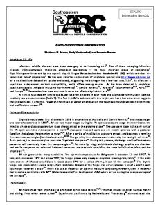
Figure 1: Image of skin with the typical presentation of dermocystidium cysts from Pascolini, R., Daszak, P., Cunningham, A.A., Tei, S., Vagnetti, D., Bucci, S., Fagotti, A. and I. Di Rosa (2003). Parasitism by Dermocystidium ranae in a population of Rana esculenta complex in Central Italy and description of Amphibiocystidium n. gen.. Diseases of Aquatic Organisms, 56:65-74.
References: 1. Densmore, C. L., and D. E. Green (2007). Diseases of Amphibians. Institute for Laboratory Animal Research Journal, 48:235254. 2. Pérez, C. (1907). Dermocystis pusula, organisme nouveau parasite de la peau des tritons. Reunion Biologique de Bordeaux, Nov 5, 1907:445-446. 3. Green, D. E., Converse, K. A., and A. K. Schrader (2002). Epizootiology of sixty-four amphibian morbidity and mortality events in the USA, 1996-2001. Annals of the New York Academy of Sciences, 969:323-339. 4. Raffel, T.R., Bommarito, T., Barry, D. S., Witiak, S. M., and L. A. Shackelton (2008). Widespread infection of the Eastern spotted newt (Notophthalmus viridescens) by a new species of Amphibiocystidium, a genus of fungus-like mesomycetozoan parasites not previously reported in North America. Parasitology, 135:203-215. 5. González-Hernádez, M., Denoël, M., Duffus, A. L. J., Garner, T. W. J., Cunningham, A. A., and K. Acevedo-Whitehouse (2010). Dermocystid infections and associated skin lesions in free living palmate newts (Lissotrition helveticus) from Southern France. Parasitology International, 59:344-350. 6. Goodchild, C. G. (1953). A subcutaneous cyst-parasite of bullfrogs: Hystocydium ranae, n.g., n. sp.. The Journal of Parasitology, 39:395-405 7. Jay, J. M., and W. J. Pohley (1981). Dermocystidium penneri sp.n. from the skin of the American toad, Bufo americanus (Amphibia: Bufonidae). Journal of Parasitology, 67:108-110. 8. Baldauf, S. L. (2008). An overview of the phylogeny and diversity of eukaryotes. Journal of Systematics and Evolution, 46:263-273. 9. Pascolini, R., Daszak, P., Cunningham, A. A., Tei, S., Vagnettie, D., Bucci, S., Fagotti, A., and I. Di Rosa (2003). Parasitism by Dermocystidium ranae in a population of Rana esculenta complex in Central Italy and description of Amphibiocystidum n.gen.. Diseases of Aquatic Organisms, 56:65-74. 10. Pereira, C. N., Di Rosa, I., Simoncelli, F., Pascolini, R., and L. Mendoza (2005). The pathogen of frogs Amphibiocystidium ranae is a member of the order Dermocystidia in the class Mesomycetozoea. Journal of Clinical Microbiology, 43:192-198. 11. Broz, O. and M. Privora (1952). Two skin parasites of Rana temporaria: Dermocystidium ranae Guyénot & Naville and Dermosporidium granulosum n.sp.. Parasitology, 42:65-69.
Author’s Affiliation and Contact Information Department of Biology Gordon College 419 College Drive Barnesville, GA, 30204 *Email Amanda Duffus:
[email protected] Recommended Citation: Duffus, A.L.J, C.M. Jackson, and A.M. Andrews. 2013. Dermocystidium (Amphibiocystidium) Infections in Amphibians. Southeastern Partners in Amphibian and Reptile Conservation, Disease, Pathogens and Parasites Task Team, Information Sheet #16