GABAA receptor facilitation reduces firing of neuronal assemblies in a computational cortical model Kingsley Storer, George Reeke Weill Cornell Medical College & The Rockefeller University, New York, New York
20%
Baseline average density 38 neurons/mm2
excitatory neuron
v = 0.15 m/s
r
mm 8 =
Cl-
r = 2mm
50
local
axonal
and Crick and Koch’s model of
b
B D
E
G
F H
I
20
40
60
time (ms)
c
300 ms
neuronal group
firing
in
connections, are integral to the conscious state.
What effect
would anesthesia have on such group formation?
Baseline
600
γ rhythm
24
3
sedation 50
general anesthesia 25
θ rhythm 1
2
3
4
5
time (hours)
40
60
frequency (Hz)
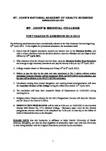
Figure 5. Left panel is a simulated EEG spectrogram demonstrating the development of γ and θ rhythms over time under the influence of synaptic plasticity. Colors denote power of EEG at a particular frequency, red indicating the highest power and violet the lowest. Right panel shows the reduction in the amplitude and frequency of the underlying γ rhythm that results from combined GABAA facilitation and input drive reduction. The parameters for each clinical state correspond to those from equivalent clinical states in figure 4.
Conclusions • modeling has permitted description of neural network phenomena that are not amenable to study using existing in vivo and in vitro techniques • the computational model spontaneously forms neuronal groups • direct and indirect GABAA effects of propofol prevent the model from forming neuronal groups. This occurs concurrently with slowing and weakening of the γ rhythm • the findings are consistent with theories that emphasize the importance of neuronal groups as substrates of consciousness
400
References 200
300%/ 25.5% 500%/ 41.6% (sedation) (general anesthesia)
GABAA facilitation / input drive reduction
1. Alkire MT, Hudetz AG, Tononi G. Consciousness and anesthesia. Science 2008;322:876-880 2. Crick F, Koch C. A framework for consciousness. Nat Neurosci 2003;6:119-126 3. Edelman GM. Neural Darwinism: selection and reentrant signaling in higher brain function. Neuron 1993;10:115-125 4. Greenfield SA, Collins TF. A neuroscientific approach to consciousness. Prog Brain Res 2005;150:11-23 5. Seth AK, Izhikevich E, Reeke GN, Edelman GM. Theories and measures of consciousness: an extended framework. Proc Natl Acad Sci U S A 2006;103:10799-0804 6. Izhikevich EM, Gally JA, Edelman GM. Spike-timing dynamics of neuronal groups. Cereb Cortex 2004;14:933-944 7. Adodra S, Hales TG. Potentiation, activation and blockade of GABAA receptors of clonal murine hypothalamic GT1-7 neurones by propofol. Br J Pharmacol 1995;115:953-960 8. McCarthy MM, Brown EN, Kopell N. Potential network mechanisms mediating electroencephalographic beta rhythm changes during propofol-induced paradoxical excitation. J Neurosci 2008;28:13488-3504 9. Izhikevich EM. Polychronization: computation with spikes. Neural Comput 2006;18:245-282
10 ms
specific patterns and formed by large numbers of re-entrant
GABAA facilitation (general anesthesia)
Baseline
to these is the idea that groups,
48
75
1 Hz random input drive to each neuron
A
0
96
12
r = 1.5mm
neuronal coalitions. Common neuronal
activation frequency
To see animation of figure 4 use QR or go to http://tinyurl.com/7jhjpmd
Finding neuronal groups • Certain groups of neurons fire with persistent spike timing patterns • These groups are termed polychronous (meaning multiple timing9)
group
neuronal assembly hypothesis
group identity determined
Figure 2. Schematic of properties of the model.
C
baseline 192
multiple active groups
150
from
selection, Greenfield’s transient
6
Direct and indirect effects of GABAA facilitation prevent activation of polychronous groups
GABAA receptor
A to I
neuronal
Propofol Cl-
neurons
of
5
100
384
6
160
neuroscience include Edelman’s theory
synaptic weights stabilizing
Polychronous group activation per second
arising
3 time (hrs)
inhibitory neuron
Major theories of
consciousness
0
80%
a Figure 1.
test
baseline
Results
Connections
The exact mechanisms by which general anesthetic agents induce unconsciousness remain unclear1. Considerable effort has been directed at identifying the cellular effects of anesthetics and determining the anatomic sites of action. What is lacking is a coherent explanation of how anesthetic effects on individual cells scale to the whole-brain level to abolish consciousness. Progress towards this goal is hampered by the spatial and temporal limitations of available modalities including fMRI and EEG. This mystery is part of a larger problem – the underpinnings of consciousness. Prominent theories for the neural correlates of consciousness share the idea that dynamically created groups of neurons, formed by extensive re-entrant connections between cells are integral to production of the conscious state2-5. In the absence of the ability to directly observe neuronal group formation, computer modeling has become a valuable adjunct in the study of this process. We have examined a model of a cortical neuronal network subjected to varying degrees of GABAA facilitation as would occur with propofol administration to study the effects of anesthesia on neuronal group formation.
run-in
7680 cells total
r = 1.5mm
Introduction
± input drive reduction
Computational model Details of the structure and function of the model are summarized in figure 2. Features that are incorporated include: • biologically realistic Izhikevich spiking model6 • synaptic strengths modified by spike timing dependent plasticity • activation of AMPA and NMDA receptor channels by excitatory cell synapses • activation of GABAA and GABAB receptor channels by inhibitory cell synapses • propofol parameters consistent with in vitro data covering a clinically relevant range of plasma concentrations7,8
on ax /s ted lina = 1 m mye v
Background Our understanding of how general anesthetics act on individual cells and on global brain function has increased significantly over the last decade. What remains poorly understood is how anesthetics act at intermediate scales. Several major theories emphasize the importance of neuronal groups in the phenomenon of consciousness. We have undertaken computer modeling to determine how GABAA facilitating agents such as propofol may influence the dynamics of neuronal group formation. Methods A computer model of cortical neurons with connections modified by synaptic plasticity was examined. At baseline the model spontaneously formed neuronal collections. Direct effects of GABAA facilitation and indirect effects on input drive were examined to study their effects on this process. Results Facilitation of GABAA and input drive reduction reduced the firing frequency of both inhibitory and excitatory neurons in a dose-dependent manner. The diminution in spiking rates led to dramatic reductions in the firing frequency of neuronal groups. Simulated EEG output from the model at baseline exhibits γ and θ rhythmicity. The direct and indirect GABAA effects reduce the amplitude of these underlying rhythms and modestly slow the γ rhythm. Conclusions GABAA facilitation both directly and indirectly inhibits the ability of neurons to form spontaneous interacting collections. A lack of group formation supports explanations of anesthetic induced loss of memory and consciousness arising from theories of neuronal group selection and transient neuronal assembly formation.
± GABAA facilitation
% of baseline gamma amplitude
Methods
GABAA facilitation reduces the frequency and amplitude of spontaneously forming γ rhythm
frequency (Hz)
Abstract
Time course of simulation
Figure 3. Panels a and b. Example of a polychronous group found in a subset of neurons. Lines indicate strong synaptic connections and rectangles represent firing of excitatory (green) or inhibitory (red) neurons. Panel c. The precise spatio-temporal firing pattern of the group is searched for in the spiking output of the model. On two occasions during the depicted time period, the majority of neurons fire in the precise pattern identified in panel a. When neurons fire in this manner, the group is said to be activated.
Figure 4. The effect of a combined increase of GABAA receptor channel conductance and decreased input drive reduction on the activation frequency of polychronous groups is shown at left. The film strips depict five corresponding time points at 10 seconds apart for the baseline condition and combined GABAA receptor facilitation and input drive reduction. The images show the distribution of activated polychronous groups on the simulated cortical surface. Lines of the same color connect neurons that are firing as part of the same activated polychronous group.
Further information Corresponding author: Kingsley Storer (
[email protected]) Link to poster and further information by using QR reader on your mobile device to read the QR code at right or by going to http://tinyurl.com/7h4dv9l