Title:
Spike-timing-dependent plasticity of inhibitory synapses in the entorhinal cortex
Authors:
Julie S. Haas1, Thomas Nowotny1, and H. D. I. Abarbanel1,2
Affiliation:
1. Institute for Nonlinear Science University of California, San Diego La Jolla, CA 92093-0402 2. Department of Physics and Marine Physical Laboratory (Scripps Institution of Oceanography) University of California, San Diego La Jolla, CA 92093-0402
Running Head: Spike-timing-dependent plasticity of inhibitory synapses Contact:
Julie S. Haas, Ph.D. 9500 Gilman Drive MC0402 La Jolla, CA 92093-0402 USA Phone: 858-534-4511 FAX: 858-534-7664 Email:
[email protected]
Spike-timing-dependent plasticity of inhibitory synapses
JN-00551-2006.R1
Abstract Actions of inhibitory interneurons organize and modulate many neuronal processes, yet the mechanisms and consequences of plasticity of inhibitory synapses remains poorly understood. We report on spike-timing-dependent plasticity of inhibitory synapses in the entorhinal cortex. After pairing presynaptic stimulations at time t pre with evoked postsynaptic spikes at time t post under pharmacological blockade of excitation we found, via whole-cell recordings, an asymmetrical timing rule for plasticity of the remaining inhibitory responses. Strength of response varied as a function of the time interval
∆t = t post − t pre : for ∆t > 0 inhibitory responses potentiated, peaking at a delay of 10 ms. For ∆t < 0 the synaptic coupling depressed, again with a maximal effect near 10 ms of delay. We also show that changes in synaptic strength depend on changes in intracellular calcium concentrations, and demonstrate that the calcium enters the postsynaptic cell through voltage-gated channels. Using network models, we demonstrate how this novel form of plasticity can sculpt network behavior efficiently and with remarkable flexibility. Introduction Experience dependent forms of synaptic plasticity have been measured in many areas and for many types of synapses in the brain (Bliss and Collingridge 1993; Malenka and Bear 2004) and are widely thought to form the cellular basis of learning and memory. One form of plasticity induction uses tightly controlled precise temporal relationships between presynaptic and postsynaptic activations. The resulting longlasting change in synaptic strength, or spike-timing-dependent plasticity (STDP) is then expressed as a function of that precise timing relationship. Most investigations have explored STDP in excitatory synapses (Bi and Poo 1998; Dan and Poo 2004; Egger et
Spike-timing-dependent plasticity of inhibitory synapses
JN-00551-2006.R1
al. 1999; Feldman 2000; Levy and Steward 1983; Malenka and Bear 2004), though a small subset have addressed the issue of plasticity at inhibitory synapses, reviewed in Gaiarsa (2002). In the neocortex, coincidence-dependent potentiation of inhibitory synapses is calcium-dependent and can be induced by trains or bursts of postsynaptic spikes paired to a single presynaptic spike (Holmgren and Zilberter 2001). In cultured hippocampal neurons and hippocampal slices, repetitive postsynaptic spikes within 20 ms in either direction of presynaptic activation of inhibitory synapses led to a symmetrical window of potentiation of inhibitory synapses, an effect which is also calcium-dependent and dependent on chloride reversal potential modulation via the K-Cl cotransporter KCC2 (Fiumelli et al. 2005b; Woodin et al. 2003). In immediately postnatal CA1 pyramidal cells, long depolarizing postsynaptic pulses increase both amplitude and frequency of spontaneous inhibitory events, but this effect tapered off by postnatal day 12 (Gubellini et al. 2001, 2005). Also in CA1 pyramidal cells, repetitive firing of a presynaptic interneuron decreased the probability of synaptic failures in a postsynaptic neuron (Kang et al. 1998). Finally, Huang et al. (2005) showed that presynaptic stimulation at 3 Hz combined with prolonged postsynaptic depolarization of CA1 pyramidal cells resulted in long-term potentiation of slow, metabotropic IPSCs, a process dependent on postsynaptic NMDA-R activation, Ca2+ increase, and CaMKII activity. In the present work we report experimental results of STDP at inhibitory synapses in the entorhinal cortex (EC). The EC serves as an anatomical signaling gateway for the hippocampus (Kloosterman et al. 2003; Witter 1993). Signals passing through layer II are transformed by both the intrinsic and synaptic dynamics of the principal excitatory stellate cells (SCs) (Alonso and Klink 1993; Haas and White 2002; Klink and Alonso 1993; White et al. 1998) and by the regional theta rhythm, a 4-12 Hz pattern of oscillatory behavior linked to learning and memory processes (Jensen and Lisman 2005; Kahana et al. 2001; Raghavachari et al. 2001). Layer II of the EC is thought to be more
Spike-timing-dependent plasticity of inhibitory synapses
JN-00551-2006.R1
seizure-resistant than Layer V through effects of inhibition (Bailey et al. 2004; Bradford 1995), and inhibition is likely to modulate the activation of SCs by incoming synaptic input as well, as in other cortical areas (Hasenstaub et al. 2005). Synaptic plasticity is largely unexplored in the EC, but a handful of groups have begun to explore LTP and LTD in this area (Cheong et al. 2001; Solger et al. 2004; Yang et al. 2004; Yun et al. 2002). Understanding how SCs process their synaptic inputs, and how processing changes with those inputs, is vital to understanding how we learn and remember information. In this work, we demonstrate a novel form of STDP at inhibitory synapses onto SCs in the entorhinal cortex. We show that this form of plasticity is dependent on a rise in intracellular calcium levels, mediated by L-type voltage-gated channels. In addition to demonstrating STDP of inhibitory synapses, we also explore the possible consequences of the observed plasticity. We construct a one-dimensional chain and a 2-dimesional layer model and show that the inhibitory plasticity leads to efficient and flexible control of network activity in both cases. We hypothesize that the observed plasticity of inhibitory synapses is a mechanism to control inappropriate “run-away”, seizure-like activity. Our results provide strong evidence for the importance of inhibitory inputs in maintaining an appropriate balance of synaptic signaling in the brain. Methods All experiments were conducted as approved by the UCSD IACUC. Young (14- to 21-day-old) Long-Evans rats were anesthetized by overexposure to CO2 and decapitated. The brain was quickly removed and immersed in cold (0° C) oxygenated artificial cerebral spinal fluid (ACSF) (in mM: NaCl 126, KCl 3, NaH2PO4 1.25, MgSO4 2, NaHCO3 26, Glucose 10, CaCl2 2, buffered to pH 7.4 with 95/5 % O2/CO2). Horizontal slices were prepared using a Vibratome cutter (TPI). Slices were allowed to
Spike-timing-dependent plasticity of inhibitory synapses
JN-00551-2006.R1
recover for 1 hour prior to recording in a holding chamber at room temperature, continuously bathed in oxygenated ACSF. Slices were transferred to an immersion chamber (RC-27L, Warner Instruments) and visualized with IR-DIC optics (Zeiss Axioskop 2FS Plus, Dage CCD100), maintained at 34°C (TC-344B, Warner). Electrodes of resistance 4-6 MΩ were pulled on a horizontal puller (Sutter Instruments) and filled with a recording solution (in mM: KGluconate 135, KCl 4, NaCl 2, HEPES 10, EGTA 0.2, MgATP 4, GTP-tris 0.3, phosphocreatine-tris 10). Intracellular signals were amplified (Axoclamp 2B, Molecular Devices), lowpass filtered (8-pole Butterworth at 5 kHz), and digitized at 10 kHz with a DAQ card (NI PCI-6035E) controlled by lab-made software created in LabView (National Instruments). In most experiments, excitatory synaptic transmission was blocked by CNQX (10µM), and D(−)-APV (50 µM), obtained from Sigma (St. Louis, MO). In some experiments, 10 mM BAPTA (Sigma) was added to the internal solution and 15 µM nimodipine (Tocris; from stock solution of 10 mM in DMSO) was added to the bath. We obtained whole-cell recordings from superficial EC layer II neurons. We selected SCs by their superficial-most position in layer II and oblong cell bodies, as well as particular characteristics of their electrophysiological responses to long current steps: a prominent (>30%) sag in response to both depolarizing and hyperpolarizing current injections (Alonso and Klink 1993; Haas and White 2002), as well as an early first spike in response to suprathreshold stimuli (Figure 1A). From a total of 68 neurons, average rest potential was –61.2 ± 4.8 mV, without correction for junction potential. Neurons were recorded in current clamp mode with no extra holding current. Presynaptic, extracellular stimulation was delivered as 1 ms, 10-50 µA current pulses via 125 µm concentric bipolar electrodes (FHC) in layer II, within 100-200 µm of the recording electrode. We paired synaptic responses to spikes by delivering extracellular
Spike-timing-dependent plasticity of inhibitory synapses
JN-00551-2006.R1
stimulations and forcing intracellular spikes using1 ms, 2-3 nA current injections through the recording electrode in current clamp mode at fixed time delays. We used 500 ms intervals between pairings for a total of 3-5 minutes resulting in about 320 to 600 pairings. Baseline and post-pairing synaptic responses were collected as sets of 30 postsynaptic responses to presynaptic stimulation collected at 0.5 Hz, in 5 minute intervals. Series and input resistances, and resting potentials, were monitored throughout each experiment; data from cells with variations greater than 25% in those parameters were discarded. Offline analysis was performed in Matlab (Mathworks). Numerical methods and modeling details are described in online supplemental material. Values are reported as mean ± SEM; statistical differences were measured with Anova unless indicated otherwise. Results Bidirectional STDP of inhibitory synpases In control solution, synaptic responses of SCs to intra-layer stimulations are a mix of excitatory and inhibitory responses (Figure 1). The excitatory effects can be blocked by addition of the antagonists CNQX (10 µM, blocking AMPA receptors) and D(−)-APV (50 µM, blocking NMDA receptors). The inhibitory responses can be blocked by addition of bicuculline (10 µM, blocking GABAA receptors) to the bath solution. To focus on the inhibitory portion of the response, all recordings reported here were made in the presence of CNQX and D(−)-APV. In each experiment, sets of 30 baseline responses, recorded at 0.5 Hz, were monitored every 5 minutes over a period of 10-15 minutes to ensure a stable synaptic response. We paired presynaptic stimulations with single induced postsynaptic spikes (Figure 2, arrow), and varied the interval between those stimuli, ∆t = t post − t pre , between –25 ms
Spike-timing-dependent plasticity of inhibitory synapses
JN-00551-2006.R1
and +25 ms. Pairings were repeated at a rate of 2 Hz, for 5 minutes. Following the pairings we monitored synaptic strength for up to an hour, recording sets of 30 postsynaptic responses at 0.5 Hz, at 5 minute intervals. We quantified IPSPs by their initial slopes (the slope of a linear fit to the first 40% of the IPSP rise, 11.6 ± 3.5 ms), and we normalized all responses to baseline. We quantified the effective plasticity as the mean IPSP slope between 20 and 30 min. following pairings, normalized to the mean of the slopes for 15 minutes preceding pairings. We recorded IPSPs before and after pairings from a total of 78 neurons. IPSPs had initial sizes of 1.5 ± 0.9 mV. Examples of these results are shown in Figure 2. In the top panels, we show the evolution of changes in IPSP initial slope following pairings with ∆t > 0 (A) and ∆t < 0 (B). Representative IPSPs for ∆t < 0 and ∆t > 0 are shown in the lower panels. We found that for pairings in which presynaptic stimulation preceded postsynaptic stimulation ( ∆t = t post − t pre > 0 ), IPSP initial slopes potentiated. This effect was maximal for delays close to 10 ms and appears to be a very precise effect: delays of less than 5 ms or greater than 20 ms were less effective in inducing potentiation. For all pairings with ∆t between +5 and +15 ms, IPSP slope was enhanced on average to 134.3% ± 5.9% (n=26, p<.02) of control values. Potentiation tended to evolve slowly in time following pairings. We found that for pairings in which ∆t = t post − t pre < 0 , IPSP initial slopes were diminished. As for potentiation, this effect is also very precise in its temporal requirements: for delays less than 5 ms or greater than 15 ms, no substantial effect was found. For all pairings with ∆t between -15 and -5 ms, IPSP slope was diminished on average to 83.2% ± 5.8% (n=19, p<.05) of control values. In contrast to potentiation, depression was usually much faster, and was usually expressed immediately following pairings.
Spike-timing-dependent plasticity of inhibitory synapses
JN-00551-2006.R1
In contrast to STDP of excitatory synpases, for ∆t near zero we observed very little change in synaptic strength. For all pairings with ∆t between -5 and +5 ms, IPSPs were on average 103.6% ± 3.3% (n=11, p>.3) of control values. Neither presynaptic stimulation alone nor postsynaptic spiking alone affected synaptic response at our stimulation rates. As an experimental control, we delivered isolated pre- or post-synaptic stimulation at the same interval and duration as in pairing experiments; IPSPs were not significantly different following pairings in both of these cases (n=4, p>0.2). In Figure 3A we show a summary plot of normalized change in IPSP slope as a function of the pairing interval ∆t. For both ∆t < 0 and ∆t > 0, significant changes in IPSPs were maximal near |∆t|=10 ms, and were restricted to relatively narrow temporal windows. The general trends (potentiation, depression, and temporal windows) do not depend on the details of evaluation; using IPSP slope, integral, or amplitude to evaluate the net change in synaptic strength yields the same overall effect. Pooled timecourse data is also shown in Figure 3B, and shows on average that potentiation is expressed more slowly than depression. For responses that potentiated significantly (p<.01) following pairings between +5 and +15 ms, the first set of IPSPs had a mean slope of 109.6% ± 3.9% (n= 22). In comparison, the first set of IPSPs that depressed to significant levels (p<.01) had a mean slope of 82.2% ± 7.7% (n=15).
Mechanisms of inhibitory STDP To initiate the investigation of mechanisms for this bi-directional plasticity, we repeated the pairings at delays of +10 and –10 ms with 10 mM BAPTA added to the intracellular medium. With intracellular calcium concentrations buffered, no significant potentiation was observed for pairings with ∆t > 0 (99.5% ± 1.5%, n=5; p>0.7). Potentiation, but no significant depression, was observed for pairings with ∆t < 0
Spike-timing-dependent plasticity of inhibitory synapses
JN-00551-2006.R1
(115.3% ± 3.3%, n=5, p<0.01). Results of these experiments are shown in Figure 4, along with representative IPSPs. Our results confirm previous reports (Gaiarsa et al. 2002; Woodin et al. 2003) that plasticity of inhibitory synapses is a process dependent on intracellular calcium dynamics. In our initial experiments, we blocked both AMPA/ kainate and NMDA receptors, leaving voltage-gated calcium channels (VGCCs) as one possible site of calcium entry into the postsynaptic cell. To investigate the role of VGCCs, we repeated the pairings at delays of +10 and –10 ms, with 15 µM nimodipine, to block L-type calcium channels, added to the bath solution. Under this recording condition, we saw no significant potentiation for pairings with ∆t > 0 (101.89% ± 1.9%, n=5, p>0.4). A small but insignificant depression was observed for pairings with ∆t < 0 (95.1% ± 3.9%, n=4, p>0.2). Results of these experiments are shown in Figure 5, along with representative IPSPs. As an experimental control, we repeated pairings in DMSO, the solvent for nimodipine, and as in control conditions, observed depression (to 80.6% ± 4.8 % of control, n=5, p<.001) for pairings with ∆t = -10. We observed potentiation for ∆t > 0 (to 136.0 ± 8.3% of control, n=3, p< .01). We also recorded paired-pulse responses before and after IPSP-spike pairings to investigate possible dependence of STDP on changes in presynaptic transmission. In control conditions, inhibitory synapses onto SCs exhibit paired-pulse depression, measured as average of the second response divided by average of the first response (Kim and Alger 2001), shown in Figure 6. Paired-pulse ratios did not depend on IPSP size for our dataset. After induction of either LTP or LTD, paired-pulse ratios were not significantly different from their pre-pairing values (n=10, p>.4), providing one indication that presynaptic release mechanisms remain unaffected by IPSP-spike pairings.
Spike-timing-dependent plasticity of inhibitory synapses
JN-00551-2006.R1
Possible functions for inhibitory plasticity in the brain To investigate functions of the observed plasticity rule we first created a model of a unidirectional chain of fifteen model SCs, each coupled reciprocally to a single model interneuron (Figure 7A). A pool of background neurons, firing with sinusoidally modulated Poisson rates, provides background activity to the fifteen SCs and to the interneuron. We chose initial values of excitatory couplings that would allow propagation of single spikes along the chain of SCs for most inputs. The simplicity of this chain model offers us insight into the effects of the observed inhibitory STDP rule on neuronal signal transmission. As an output of the model, we determined the amount of inhibition necessary to terminate the propagation of spikes along the chain of SCs. We compared simulations in which the inhibitory synapses were set to a constant conductance, to simulations in which each inhibitory synapse was allowed to change according to the plasticity rule derived from a fit to the experimental data. For constant inhibitory synapses, two prominent changes occurred as we increased excitatory coupling between SCs along the chain in different simulations. The amount of inhibitory synaptic conductance required to terminate propagation of activity along the chain increased, and the neuron at which the propagation terminated shifted within the chain (Figure 7C). The second effect is due to faster response of SCs to the larger EPSPs, which led to faster propogation of activity along the chain. We also note that the increasing values between simulations of constant inhibitory synaptic conductance poses a serious problem for a system with excitatory plasticity: In order to match the growth of excitatory synapses to larger strengths, all of the constant inhibitory synapses would need to be extremely strong at all times. This in turn yields amounts of inhibition which make the system basically unresponsive to other inputs.
Spike-timing-dependent plasticity of inhibitory synapses
JN-00551-2006.R1
We then allowed the inhibitory synapses between the interneuron and the SCs in the chain to follow the learning rule derived from the experimental observations (Figure 3). We approximated the data with
F ( ∆t ) =
1 + a1 ⋅ (∆t )10 e a2 ∆t , ∆t < 0 1 + a3 ⋅ (∆t )10 e a4 ∆t , ∆t > 0
shown as dashed lines in Figure 3; we determined the ai with a least-square error fit and found {a1=-2.60e-7 ms-10, a2=0.94 ms-1, a3=2.29e-6 ms-10, a4=-1.10 ms-1}. By the geometry of our model, inhibitory signals travel through fewer intermediate synapses to a later SC than excitatory signals, as a spike propagates down the chain (Figure 7A). These differences in arrival times at later SCs produce the required temporal coordination, on the range of 10 ms, for the plasticity rule to be effective. We used an additive learning rule, matching the low-frequency presentation of the stimulating pairs in experiments and the corresponding low firing frequencies of model SCs. At higher frequencies we would expect multi-spike interactions to be important (Froemke et al. 2006). After a period of learning, only a few of the inhibitory synapses onto the SCs were potentiated. Remarkably, those few synapses were enough to segregate the fifteen SCs into one sub-chain with reliably transmitted spikes from SC1, and a second cluster firing sparsely if at all in response to initiation from SC1 (Figure 7C). This effect was independent of excitatory coupling strength. Comparing the static and the learning cases, we note that in the homogeneous case every synapse must be strong enough to prevent spikes in the postsynaptic neuron in order to stop propagation; on the other hand, plasticity allows efficient tuning in which only a few synapses are needed to stop propagation. These two systems are remarkably different: the learning rule has partitioned the chain into two subchains
Spike-timing-dependent plasticity of inhibitory synapses
JN-00551-2006.R1
which retain their ability to respond to other inputs, while the comparable homogenous system is basically unresponsive. Another attractive feature of the observed learning rule is its flexibility: it self-adjusts the strengths of the inhibitory synapses to levels that match the amount of excitation in the network, thus solving the balance problem between excitation and inhibition presented above. For any given amount of excitatory coupling strength (panels of Figure 7C), static synapses required pre-set minimal levels of inhibition (horizontal lines in Figure 7C), carefully tuned to each level of excitatory strength, to stop activity. In contrast, learning synapses grew autonomously according to the plasticity rule to match the required inhibition for any level of excitation (Figure 7D). In the simple chain, we also observed an additional critical and novel feature of inhibitory plasticity: it is self-limiting. That is, as the synaptic strength grows, it becomes increasingly likely to inhibit a requirement for induction of plasticity – the postsynaptic spike. Once that strength is achieved, its own growth signal is removed, and the synapse grows no larger (Figure 7B). In some cases, increased inhibition resulted in a delay of the postsynaptic spike, away from the temporal window for potentiation. For these reasons, our model did not require an artificial limit on synaptic strength. Next, we constructed a model of a cortical layer, with 400 sparsely and randomly connected excitatory SCs and 100 interneurons. The interneurons receive excitatory input from a local group of SCs, and inhibit a slightly larger local group of SCs (Figure 8A). Again, all neurons also receive a theta-modulated Poissonian background input. We repeatedly excited three of the SCs simultaneously and observed the propagation of that signal across the layer. In the initial state inhibition is weak and activity propagates through the whole layer, mimicking unchecked seizure-like activity (Figure 8B). As previously, we allowed the inhibitory synapses to change according to the observed learning rule. After a learning period on the order of a few to 100 simulated
Spike-timing-dependent plasticity of inhibitory synapses
JN-00551-2006.R1
seconds, we observed similar effects as in the chain model: only a few synapses potentiated, but those few synapses sufficed to efficiently control the spread of activity across the layer. The same inputs to the layer excite a well-defined small cluster of SCs, rather than leading to uncontrolled excitation of the entire network (Figure 8B). Again, only a small percentage of the total inhibitory synapses were required to potentiate for this effect (Figure 8C). A few inhibitory synpases within the clusters depressed, resulting in a facilitated excitation within the cluster. Our modeling resultes suggest a crucial role for plasticity of inhibitory synapses in regulating neuronal transmission and control of overall network activity. Discussion We have demonstrated a novel form of asymmetrical spike-timing dependent plasticity in the dynamics of GABAergic synaptic couplings in the entorhinal cortex. STDP has been extensively investigated for excitatory synapses (Malenka and Bear 2004), but to our knowledge this is the first demonstration of STDP in an inhibitory synapse in which synaptic strength is differentially strengthened or weakened by the pairing of pre- and post-synaptic activity. A previous study of STDP of inhibitory synapses (Woodin et al. 2003) described the dependence of observed plasticity on a coincidence-dependent change in the reversal potential of the synapse, which in turn resulted from an activity-induced inhibition of the Cl− cotransporter KCC2 (Rivera et al. 2005; Rivera et al. 1999) in the dendrites. While a similar mechanism may be active in SCs, the whole-cell recording configuration of our experiments resulted in a clamped Cl− reversal potential at the soma, and thus masked any small change in reversal potential in the dendrites (Fiumelli et al. 2005a). For our internal and external solutions the Nernst equation gives a Cl− reversal potential of –72.5
Spike-timing-dependent plasticity of inhibitory synapses
JN-00551-2006.R1
mV. We confirmed this value experimentally, and as expected it did not change following IPSP-spike pairings. We have shown that timing-dependent plasticity of inhibitory synapses depends on calcium dynamics in the postsynaptic cell, and entry of calcium through voltage-gated channels. These results suggest a role for calcium in intracellular processes and mechanisms similar those involved in plasticity of excitatory synapses. Future experiments will focus on the possible involvement of metabotropic glutamate receptors and endocannabinoids in STDP of inhibitory synapses (Chevaleyre and Castillo 2003), to investigate possible shared or parallel mechanisms of excitatory and inhibitory plasticity. We hypothesize that coactivation of glutamatergic and GABAergic inputs could be responsible for the temporal coincidence requirements observed in our data. One clear function of STDP in excitatory synapses is to increase EPSP-spike efficacy in a postsynaptic target: relevant, causal experience increases the likelihood of successful signal transmission. Inhibitory synapses lack the obvious goal of signal propagation, making the immediate functional consequences of observed plasticity less obvious. However, because inhibition plays a crucial role in modulating and controlling many neuronal processes and rhythms, changes in inhibitory synapses may be as necessary and appropiate as changes in excitatory synapses. Strengthened inhibitory synapses are another way in which cells imprint repeated and correlated causal activity into the connections between neurons. In contrast to excitatory synapses, however, this rule will ultimately inhibit further correlated firing, as one of its effects is to inhibit the postsynaptic spike. Balance between excitation and inhibition seems to play a crucial role for the correct function of neuronal networks throughout the brain (Shu et al. 2003a; Shu et al. 2003b). The plasticity of inhibitory synapses described in this work offers a flexible and efficient mechanism to balance the effects of excitatory STDP. Indeed, the STDP we have
Spike-timing-dependent plasticity of inhibitory synapses
JN-00551-2006.R1
shown in inhibitory synapses is likely to cooperate or compete with other forms of STDP in postsynaptic targets. Plasticity measured as a function of field response (Yun et al. 2002) in the EC is likely to be a combined result of multiple forms of single-synapse plasticity, both excitatory and inhibitory. In addition, the EC is a common locus for epilepsy, and recent research highlights the importance of inhibition within Layer II in the maintenance of normal circuit function (Bear et al. 1996; Kumar and Buckmaster 2006). Our modeling results show that plasticity of inhibitory synapses offers the EC a degree of flexibility in the inhibitory control of epilepsy. Modeling studies have suggested potential functions for plastic inhibition in circuit rhythm generation (Soto-Trevino et al. 2001), and in balancing excitation (Marder and Buonomano 2004). Throughout the brain, inhibitory synapses serve both to modulate excitation in principal neurons, and to regulate rhythmic circuits. Our own modeling shows that adjustment in the strength of only a few inhibitory synapses is enough to modulate the overall exciteability of an entire layer of neurons. Further, changes in inhibitory strength track changes in excitatory strength autonomously. Extrapolating from our simple models, one might expect plasticity of inhibitory synaptic transmission to exert major influences on neuronal excitability and function. As shown in our modeling, increases in inhibition may also serve to isolate one cluster of neurons from another by strengthening inhibition at critical locations within the system, thus providing a flexible and dynamic reorganization of neuronal circuitry in the working brain. The timing rule observed are well-poised to enable cluster formation: the temporal peak of potentiation (relative to synaptic delays and response times of neurons) sets a critical radius for activity termination from an originating neuron, as seen in our chain model. Within clusters, where EPSPs arrive before IPSPs, inhibition is suppressed by the depression side of the timing rule. This results in more homogeneous and responsive clusters of SCs.
Spike-timing-dependent plasticity of inhibitory synapses
JN-00551-2006.R1
The observed type of STDP, in which inhibition increases with excitation and activity, could provide a braking mechanism for an unchecked, pathological spread of epilepticlike activity. Indeed, plastic inhibition may be crucial to how the brain regulates and controls its own activity. Acknowledgements We greatly appreciate the advice and readings of Dan Feldman and his lab at UCSD. This work was supported by grants from the NSF (PHY-0414174, to HDIA), the San Diego Foundation (C-2005-00292, to JSH), and the NIH, grant RO1 NS050945-02 (TN)
References Alonso A and Klink R. Differential electroresponsiveness of stellate and pyramidal-like cells of medial entorhinal cortex layer II. J Neurophysiol 70: 128-143, 1993. Bailey SJ, Dhillon A, Woodhall GL, and Jones RSG. Lamina-specific differences in GABAB autoreceptor-mediated regulation of spontaneous GABA release in rat entorhinal cortex. Neuropharmacology 46: 31-42, 2004. Bear J, Fountain NB, and Lothman EW. Responses of the superficial entorhinal cortex in vitro in slices from naive and chronically epileptic rats. J Neurophysiol 76: 2928-2940, 1996. Bi GQ and Poo MM. Synaptic modifications in cultured hippocampal neurons: dependence on spike timing, synaptic strength, and postsynaptic cell type. J Neurosci 18: 10464-10472, 1998. Bliss TV and Collingridge GL. A synaptic model of memory: long-term potentiation in the hippocampus. Nature 361: 31-39, 1993. Bradford HF. Glutamate, GABA and epilepsy. Prog Neurobiol 47: 477-511, 1995.
Spike-timing-dependent plasticity of inhibitory synapses
JN-00551-2006.R1
Cheong MY, Yun SH, Mook-Jung I, Joo I, Huh K, and Jung MW. Cholinergic modulation of synaptic physiology in deep layer entorhinal cortex of the rat. J Neurosci Res 66: 117121, 2001. Chevaleyre V and Castillo PE. Heterosynaptic LTD of hippocampal GABAergic synapses: a novel role of endocannabinoids in regulating excitability. Neuron 38: 461472, 2003. Dan Y and Poo MM. Spike timing-dependent plasticity of neural circuits. Neuron 44: 2330, 2004. Egger V, Feldmeyer D, and Sakmann B. Coincidence detection and changes of synaptic efficacy in spiny stellate neurons in rat barrel cortex. Nat Neurosci 2: 1098-1105, 1999. Feldman DE. Timing-based LTP and LTD at vertical inputs to layer II/III pyramidal cells in rat barrel cortex. Neuron 27: 45-56, 2000. Fiumelli H, Cancedda L, and Poo MM. Modulation of GABAergic transmission by activity via postsynaptic Ca2+-dependent regulation of KCC2 function. Neuron 48: 773-786, 2005a. Fiumelli H, Cancedda L, and Poo MM. Modulation of GABAergic Transmission by Activity via Postsynaptic Ca(2+)-Dependent Regulation of KCC2 Function. Neuron 48: 773-786, 2005b. Froemke RC, Tsay IA, Raad M, Long JD, and Dan Y. Contribution of individual spikes in burst-induced long-term synaptic modification. J Neurophysiol 95: 1620-1629, 2006. Gaiarsa JL, Caillard O, and Ben-Ari Y. Long-term plasticity at GABAergic and glycinergic synapses: mechanisms and functional significance. Trends Neurosci 25: 564-570, 2002. Gubellini P, Ben-Ari Y, and Gaiarsa JL. Activity- and age-dependent GABAergic synaptic plasticity in the developing rat hippocampus. Eur J Neurosci 14: 1937-1946, 2001.
Spike-timing-dependent plasticity of inhibitory synapses
JN-00551-2006.R1
Gubellini P, Ben-Ari Y, and Gaiarsa JL. Endogenous neurotrophins are required for the induction of GABAergic long-term potentiation in the neonatal rat hippocampus. J Neurosci 25: 5796-5802, 2005. Haas JS and White JA. Frequency selectivity of layer II stellate cells in the medial entorhinal cortex. J Neurophysiol 88: 2422-2429, 2002. Hasenstaub A, Shu Y, Haider B, Kraushaar U, Duque A, and McCormick DA. Inhibitory postsynaptic potentials carry synchronized frequency information in active cortical networks. Neuron 47: 423-435, 2005. Holmgren CD and Zilberter Y. Coincident spiking activity induces long-term changes in inhibition of neocortical pyramidal cells. J Neurosci 21: 8270-8277, 2001. Jensen O and Lisman JE. Hippocampal sequence-encoding driven by a cortical multiitem working memory buffer. Trends Neurosci 28: 67-72, 2005. Kahana MJ, Seelig D, and Madsen JR. Theta returns. Curr Opin Neurobiol 11: 739-744, 2001. Kang J, Jiang L, Goldman SA, and Nedergaard M. Astrocyte-mediated potentiation of inhibitory synaptic transmission. Nat Neurosci 1: 683-692, 1998. Kim J and Alger BE. Random response fluctuations lead to spurious paired-pulse facilitation. J Neurosci 21: 9608-9618, 2001. Klink R and Alonso A. Ionic mechanisms for the subthreshold oscillations and differential electroresponsiveness of medial entorhinal cortex layer II neurons. J Neurophysiol 70: 144-157, 1993. Kloosterman F, Van Haeften T, Witter MP, and Lopes Da Silva FH. Electrophysiological characterization of interlaminar entorhinal connections: an essential link for re-entrance in the hippocampal-entorhinal system. Eur J Neurosci 18: 3037-3052, 2003.
Spike-timing-dependent plasticity of inhibitory synapses
JN-00551-2006.R1
Kumar SS and Buckmaster PS. Hyperexcitability, interneurons, and loss of GABAergic synapses in entorhinal cortex in a model of temporal lobe epilepsy. J Neurosci 26: 46134623, 2006. Levy WB and Steward O. Temporal contiguity requirements for long-term associative potentiation/depression in the hippocampus. Neuroscience 8: 791-797, 1983. Malenka RC and Bear MF. LTP and LTD: an embarrassment of riches. Neuron 44: 5-21, 2004. Marder CP and Buonomano DV. Timing and balance of inhibition enhance the effect of long-term potentiation on cell firing. J Neurosci 24: 8873-8884, 2004. Raghavachari S, Kahana MJ, Rizzuto DS, Caplan JB, Kirschen MP, Bourgeois B, Madsen JR, and Lisman JE. Gating of human theta oscillations by a working memory task. J Neurosci 21: 3175-3183, 2001. Rivera C, Voipio J, and Kaila K. Two developmental switches in GABAergic signalling: the K+-Cl- cotransporter KCC2 and carbonic anhydrase CAVII. J Physiol 562: 27-36, 2005. Rivera C, Voipio J, Payne JA, Ruusuvuori E, Lahtinen H, Lamsa K, Pirvola U, Saarma M, and Kaila K. The K+/Cl- co-transporter KCC2 renders GABA hyperpolarizing during neuronal maturation. Nature 397: 251-255, 1999. Shu Y, Hasenstaub A, Badoual M, Bal T, and McCormick DA. Barrages of synaptic activity control the gain and sensitivity of cortical neurons. J Neurosci 23: 10388-10401, 2003a. Shu Y, Hasenstaub A, and McCormick DA. Turning on and off recurrent balanced cortical activity. Nature 423: 288-293, 2003b. Solger J, Wozny C, Manahan-Vaughan D, and Behr J. Distinct mechanisms of bidirectional activity-dependent synaptic plasticity in superficial and deep layers of rat entorhinal cortex. Eur J Neurosci 19: 2003-2007, 2004.
Spike-timing-dependent plasticity of inhibitory synapses
JN-00551-2006.R1
Soto-Trevino C, Thoroughman KA, Marder E, and Abbott LF. Activity-dependent modification of inhibitory synapses in models of rhythmic neural networks. Nat Neurosci 4: 297-303, 2001. White JA, Klink R, Alonso A, and Kay AR. Noise from voltage-gated ion channels may influence neuronal dynamics in the entorhinal cortex. J Neurophysiol 80: 262-269, 1998. Witter MP. Organization of the entorhinal-hippocampal system: a review of current anatomical data. Hippocampus 3 Spec No: 33-44, 1993. Woodin MA, Ganguly K, and Poo MM. Coincident pre- and postsynaptic activity modifies GABAergic synapses by postsynaptic changes in Cl- transporter activity. Neuron 39: 807-820, 2003. Yang S, Lee DS, Chung CH, Cheong MY, Lee CJ, and Jung MW. Long-term synaptic plasticity in deep layer-originated associational projections to superficial layers of rat entorhinal cortex. Neuroscience 127: 805-812, 2004. Yun SH, Mook-Jung I, and Jung MW. Variation in effective stimulus patterns for induction of long-term potentiation across different layers of rat entorhinal cortex. J Neurosci 22: RC214, 2002.
Spike-timing-dependent plasticity of inhibitory synapses
JN-00551-2006.R1
Figures A. SC response to current steps
10 mV 50 ms
B. SC response to presynaptic stimulation
+CNQX +D−APV +Bicuculline 1 mV 20 ms
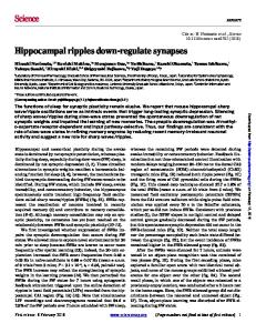
Figure 1. Response of a typical layer II SC to long hyperpolarizing and depolarizing current steps (panel A). In response to presynaptic stimulation within layer II, SCs show a compound response (panel B, left plot), which can be pharmacologically separated into an excitatory component (panel B, middle plot), and an inhibitory component (panel B, right plot).
Spike-timing-dependent plasticity of inhibitory synapses
∆t = +8 ms
A
JN-00551-2006.R1
∆t = −9 ms
B
14
↓
IPSP slope [V/ms]
12 10 8 6
↓
4 2
MΩ
−10
0
100 80 60
10 20 Time [min]
0.2 mV 20 ms 5 min
30
40
−10
0
10 20 Time [min]
30
0.2 mV 20 ms 5 min
Figure 2. After pairing presynaptic stimulations with evoked postsynaptic spikes, we monitored the evolution of changes in postsynaptic IPSPs. We observed long-lasting potentiation of IPSPs for pairings with ∆t = t post − t pre > 0 (A, upper panel: individual and mean initial slopes for one cell in elapsed time; middle panel: input resistance; lower panel: each trace is the mean of 30 IPSPs; traces are shown over elapsed time). We observed long-lasting depression of IPSPs for pairings with ∆t = t post − t pre < 0 (B, upper panel: individual and mean initial slopes for one cell in elapsed time; middle panel: input resistance; lower panel: each trace is the mean of 30 IPSPs; traces are shown over elapsed time).
Spike-timing-dependent plasticity of inhibitory synapses
JN-00551-2006.R1
A
∆IPSP [norm]
2
1.5
1
0.5
0 −20
−10
0
10
20
∆ t [ms] B
↓
1.4 1 −10
−5
0
5
10
15
20
25
30
Time [min]
Figure 3. Panel A: Summary results of change in postsynaptic IPSP initial slope (∆IPSP), expressed as a function of ∆t = t post − t pre . No change corresponds to normalized IPSP slope equal to unity (equal to the baseline value before pairing). Each point represents data from one cell. Change in slope is evaluated as the mean IPSP slope between 20 and 30 min. following pairings, normalized to the mean of the slopes for 15 minutes preceding pairings. Data for which the change in IPSP slope was significant (p<.01, Anova) are plotted in open symbols. The dashed line is a least-square error fit, used in modeling, and is described in the text. Panel B: Pooled time series for potentiating synapses (circles) and depressing synapses (squares), evaluated for IPSP slopes showing significant (p<.01) potentiation following pairings between +5 to + 15 ms, and significant (p<.01) depression following pairings between -15 to -5 ms. Potentiation evolved more slowly than depression.
Spike-timing-dependent plasticity of inhibitory synapses
JN-00551-2006.R1
A
∆ t = +10 ms
↓
IPSP Slope [V/ms]
30
10 ∆ t = −10 ms
↓
30
10 −10
−5
0
5
10 15 Time [min]
B 2
∆ t = +10 ms
C
25
30
35
Summary
∆ t = −10 ms 0.5 mV 20 ms
∆IPSP [norm]
0.5 mV 20 ms
20
1
0 −20
−10
0 ∆ t [ms]
10
20
Figure 4. Introducing BAPTA into the intracellular solution revealed a calcium dependence of inhibitory STDP. We observed no significant potentiation in IPSP slope after pairing for ∆ t> 0 (A, top) or depression for ∆t < 0 (A, bottom). Representative sets of IPSPs (mean of 30 traces) before and after pairings for each temporal order of pairings are shown in panel B. Summary data for n=10 neurons (some data overlap completely) is shown in panel C.
Spike-timing-dependent plasticity of inhibitory synapses
JN-00551-2006.R1
A
8 ∆ t = +10 ms
↓
IPSP Slope [V/ms]
6 4 2 30
∆ t = −10 ms
↓
20 10 −10
−5
0
5
10
15
Time [min]
B
C
0.1 mV 20 ms
∆ t = −10 ms 0.5 mV 20 ms
∆IPSP [norm]
∆ t = +10 ms
Summary
2
1
0 −20
−10
0 ∆ t [ms]
10
20
Figure 5. With nimodipine in the bath solution, we show that calcium enters the postsynaptic cell through L-type voltage-gated calcium channels to participate in the induction of inhibitory STDP. We observed no significant change in IPSP slope after pairing for ∆ t> 0 (A, top) or for ∆t < 0 (A, bottom) for this condition. Representative sets of IPSPs (mean of 30 traces) before and after pairings for each temporal order of pairings are shown in panel B. Summary data for n=9 cells (some data overlap completely) is shown in panel C.
Spike-timing-dependent plasticity of inhibitory synapses
JN-00551-2006.R1
A Paired−Pulse Ratio
1 0.8 0.6 0.4 0.2 0 45
50
55
60
65 70 75 Pulse Interval [ms]
B
80
85
90
95
C
0.25 mV 25 ms
Figure 6. Paired-pulse ratios are unaffected by plasticity induction. SCs exhibit pairedpulse depression (A, summary of n=10 cells) for pairs of presynaptic stimuli separated by less than 100 ms, both before IPSP-spike pairings (A, dotted line and squares, mean ± SEM) and after pairings (B, solid lines and open circles, mean ± SEM). Spike-IPSP pairings did not affect paired-pulse ratios for any of the delays tested (p>.4, Anova). Sample traces of paired-pulse IPSPs are shown in B (before pairings, mean of 22 traces) and C (after pairings, mean of 17 traces).
Spike-timing-dependent plasticity of inhibitory synapses
Activity
B 60
SC2
SC3
SC4
SC5
SC 6
SC14
SC7
SC15
50
g [nS]
SC1
40
1
SC10
SC9
SC8
30 20 10 0
I
excitatory synapse
20
40
inhibitory synapse
C
D
Inhibitory synaptic conductance [nS]
90
g
EE
= 15 nS
g
EE
= 18 nS
g
EE
< 21 nS
g
EE
g EE = 18 nS
g EE = 21 nS
g EE = 24 nS
g EE = 27 nS
g EE = 30 nS
g EE = 33 nS
g EE = 36 nS
g
= 39 nS
g
= 42 nS
g
= 45 nS
g
= 51 nS
g
= 54 nS
g
= 57 nS
g
0
90
90
60
60
g EE= 27 nS
g EE= 30 nS
g EE= 33 nS
30
g EE= 36 nS
0
STDP
90
90
EE
EE
EE
EE
= 48 nS
60
60
g EE= 39 nS
g EE= 42 nS
g EE= 45 nS
30
g EE= 48 nS
0
STDP
90
90
g
EE
EE
EE
EE
= 60 nS
60
60 30
g EE = 15 nS
30
= 24 nS
STDP
30
90 60
60
30
0 100
80
Time [s]
STDP
30
60
SDF [norm.]
A
JN-00551-2006.R1
g
EE
SC1
= 51 nS
g
EE
= 54 nS
SC15 SC1
g
EE
= 57 nS
SC15 SC1
SC15
g
EE
30
= 60 nS
SC1
SC15
Neuron number 0.25
SC1
SC15
SC1
SC15
SC1
Neuron number
normalized spike rate 0
0
0.5
0.75
1
Figure 7. A simple model of an excitatory chain illustrates the effects of inhibitory plasticity on propagation of activity. A) Geometry of the network. SCs are connected in a unidirectional excitatory chain, and each SC is reciprocally connected to an interneuron. Input delivered to SC1 initiates propagation of activity from left to right in the SCs. B) Conductances (solid lines) and normalized spike density functions (dotted lines) for three SCs over the course of one run. As inhibition increases in the postsynaptic SC, firing rate decreases. With enough inhibition, postsynaptic SCs no longer spike, thus removing one of the requirements for STDP. C) Average firing rate of SCs in a color code for the chain with different excitatory and fixed inhibitory synaptic strengths. Each panel corresponds to simulations with a given excitatory synaptic strength. Inhibitory strength is varied along the y-axis and the SCs of the chain are represented on the xaxis, such that each horizontal dataset corresponds to one simulated experiment with a constant level of inhibition. Rate is normalized to the rate of the most active SC (typically
SC15
SC1
SC15
Spike-timing-dependent plasticity of inhibitory synapses
JN-00551-2006.R1
SC1). Each panel exhibits a clear transition from uninterrupted activity propagation (low values of inhibitory synaptic strength) to interrupted propagation (high inhibitory synaptic strength). Yellow lines mark the minimal amount of inhibitory synaptic strength at which activity propagation was terminated. The additional color bars on top of the panels show the activity of SCs for each system with the same excitatory strengths, but plastic inhibitory synapses, after 150 s simulated activity. Propagation of activity is reliably terminated in each of these cases. D) Final synaptic conductance of plastic synapses from the local interneuron to each of the SCs (x axis) after 150 s simulated time. Panels correspond to excitatory coupling strength, as in (C). Red lines mark the minimal amount of inhibitory synaptic strength for which activity propagation was terminated for each panel, as in (C). Black lines show the final, stable conductances of the plastic inhibitory synapses from the interneuron to each SC. For plastic synapses, only a few synapses are potentiated, and the strongest synapse approximately matches the minimal necessary strength.
Spike-timing-dependent plasticity of inhibitory synapses
A
excitatory connection inhibitory connection
JN-00551-2006.R1
Input IN 100
B
IN 401 − 500 IN 1
Input
SC400
SC 1 − 400
C
SC1 5500
5600
5700
5800
5900
6000
g
inh
0.0005
0.002
0.005
0.02
0.05
time nS
5533
5553
5573
5593
5613
66800
66550
66300
time [ms]
time [ms]
5633
5653 ms
66381
66401
66421
66441
Figure 8. A two-dimensional layer model extends the results on control of activity propagation. A) Network geometry. A layer of locally, randomly connected SCs is locally and reciprocally connected to a layer of inhibitory interneurons, through synapses obeying the experimentally observed plasticity rule. B) Spike rasters of all neurons, shown in different colors for different neurons to distinguish multiple spikes from spikes in neighboring neurons. Initially, with weak inhibition, inputs to 3 cells in the middle of the SC layer propagate across the whole layer, involving every SC in that event (left panel). After plasticity took effect, the same inputs activate only a well-defined patch of SCs (right panel). Color maps (below) show each SC’s first spike time within the gray shaded areas of the spike rasters. Color corresponds to spike time, from early (blue) to late (red) in the shown event. The SCs in which activity originated are outlined in white.
66461
66481
66501 ms
Spike-timing-dependent plasticity of inhibitory synapses
JN-00551-2006.R1
C) Final synaptic strength of inhibitory synapses, in a logarithmic color code. Panels correspond to interneurons (as in A), and each panel shows the strength of the synapses from one interneuron to all the SCs. Individual pixels within each panel correspond to synapses onto the SCs, and color codes the conductance of each synapse. Remarkably, only a few synapses are potentiated to control the initial event in (B). These potentiated synapses tend to form ring-like structures that efficiently control activity propagation from an active focus to the rest of the network.
Appendix – Numerical methods Modeling: We developed a one compartment Hodgkin-Huxley model for SCs (excitatory population) and used a standard model for the inhibitory interneurons. SCs had seven currents I Na , I Kd , I h1 , I h2 , I Ks , and leak currents I leak and I leak,K ; i.e. membrane voltage V (t ) was described by
C
dV = − I Na − I Kd − I h1 − I h2 − I Ks − I leak − I leak,K − I syn . dt
where C denotes membrane capacitance. Currents are described in more detail in Table 1. Each model neuron was excited by 25 stochastically firing background neurons, whose spike trains followed Poisson statistics with firing frequency f (t ) , which was sinusoidally modulated at theta frequency (6 Hz). The maximum spike rate of the background neurons was 4 Hz . The first SC received additional input from a Poisson neuron that was firing at 30 Hz during the peak 90% of each Theta cycle resulting in zero to two spikes per Theta cycle. Synapses were modeled with a generic two parameter, first order synapse model, with synaptic current I syn related to synaptic conductance g syn and pre- and post-synaptic membrane voltages Vpre and Vpost by
I syn = g syn S (t )(Vrev − Vpost ) . The fraction S ( t ) of active neurotransmitter obeys an equation of the form
dS (t ) = α (1 − S (t ))R(t ) − βS (t ) , dt where R (t ) is 1 for a given time t release and 0 otherwise. For 1 ms after the release has ended, the synapse is refractory and no further release can occur. Rate constants were α E = 1 kHz and β E = 0.03 kHz for excitatory synapses between SCs, from SCs to interneurons, and from background and input Poisson neurons. For all inhibitory synapses, α = 0.5 kHz and β = 0.02 kHz . A presynaptic spike was detected by crossing of the presynaptic membrane potential Vpre across the release potential Vth = −20mV from below. Reversal potentials were Vrev = 0mV for all
excitatory and Vrev = −80mV for all inhibitory synapses. The release times were
t release,E = 0.5 ms for excitatory synapses and t release,I = 15 ms for inhibitory synapses. All synapse parameters were fitted to match the model to the observed compound EPSPs and IPSPs in SCs, see figures 1 and 9. In the one-dimensional model, the conductance of excitatory synapses between SCs, g syn,EE , was varied in the range given in the main text to test for its influence on the observed activity control and the excitation from SCs to the inhibitory neuron had conductance g syn,EI = 2 nS + σ , where σ is a Gaussian random variable with standard deviation 0.1 nS . The excitation from background Poisson neurons was through synapses with g syn,E = 1 nS for SCs and g syn,E = 0.5 nS for the inhibitory neuron. The inhibitory neuron forms an inhibitory self-synapse with g syn,II = 50 nS . The initial conductance of the plastic inhibitory synapses was g syn,IE,initial = 1 nS . The synaptic conductances in the two-dimensional model were essentially the same, however the excitation between SCs was fixed to g syn,EE = 15 nS and the initial strength of the plastic inhibitory synapses was again g syn,IE,initial = 1 nS . Plasticity of inhibitory synapses was realized through an additive STDP learning rule,
∆g = rF (t post − t pre ) , where tpost and tpre denote the spike times of presynaptic and
postsynaptic neurons, the learning amplitude r was set to 5 nS , and F is the fitted learning function, see main text, figure 3. To account for the fact that potentiation of synapses is only observed some time after induction, the synaptic changes due to this learning rule take effect 10 s after the occurrence of pre-and postsynaptic spike pairing. The model was implemented in C++ and integrated using a 5/6 order variable time step Runge Kutta algorithm. Simulations were compiled with gcc 3.2 and executed on an AMD64 3400+ running RedHat Fedora Core 2. A simulation of 100 simulated seconds took 26 minutes in the one-dimensional model and about 4 days for the twodimensional model.
The network geometry of the two-dimensional model is illustrated in figure 8. In addition to the neurons shown in the figure, a pool of 500 Poisson neurons, which again had modulated firing rates with maximal rate 4 Hz , was simulated and each SC and IN received 10 randomly chosen inputs from this pool. The size of the SC grid was 20x20, the IN grid 10x10. Each SC connected to neighboring SCs with probability pEE = 0.3 , if the distance in 1-norm was less than rEE = 4 , i.e., x1 − x2 + y1 − y2 < 4 where x and y denote the position in x and y direction in full integers. For connections between SCs and interneurons the connection probability was pEI = 0.8 , and the radius was rEI = 3 (with the same metric as above). For connections from inhibitory neurons to SCs,
pIE = 0.8 and rIE = 9 . The input to the two-dimensional layer was delivered through a single Poisson neuron as in the one-dimensional case. This input neuron synapsed onto three neighboring cells in the middle of the layer.
Ix = gx A(y)(V − Ex ),
dy = αy (V )(1 − y) − βy (V )y, dt
I
A(y)
INa
m3 h
where
x = {Na, Kd, h1, h2, Ks}
where y = {m, h, n, mh1, mh2 , mKs , hKs }
activation and inactivation functions
αm = 0.32
−52 − V e(−52−V )/4
αh = 0.128 e
−1
,
βm = 0.28
(−48−V )/18
−50 − V
,
βh =
IKd
n
Ih1
mh1
αh1 =
−0.00289 V − 0.445 , 1 − e(V +0.445/0.00289)/24.02
βh1 =
Ih2
mh2
αh2 =
−0.00318 V − 0.695 , 1 − e(V +0.695/0.00318)/26.72
βh2 =
IKs
mKs h3Ks
αn = 0.032
αmKs = αhKs =
e(−50−V )/5
−1
1 , 1 + e(20−V )/10 1 1+
e(60+V )/15
,
,
25 + V e(25+V )/5
−1
4 e(−25−V )/5 + 1
βn = 0.5 e(−55−V )/40
βmKs = βhKs =
0.0271 V − 1.024 1 − e(V −1.024/0.0271)/(−17.4) 0.0216 V − 1.065 1−
e(V −1.065/0.0216)/(−14.25)
1 1 + e(20−V )/25 1 1+
e(20−V )/10
Table 1. Overview of the membrane currents. The leak currents were
I leak ( t ) = g leak (V ( t ) − Eleak ) and I leak,K ( t ) = g leak,K (V ( t ) − EK ) and the remaining parameters C = 0.286 nF , g leak = 0.021 µ S , g leak,K = 0.055 µS , Eleak = −55 mV ,
g Na = 7.15 µ S , ENa = 50 mV , g Kd = 1.43 µ S , EK = −95 mV , g h1 = 0.074 µS , g h2 = 0.04 µS , Vh = −20 mV , and g Ks = 0.1 µS . The inhibitory interneuron(s) had the same I Na , I Kd , and leak currents except a smaller potassium leak conductance,
g leak,K = 5.72 nS , and no h or slow potassium currents. I Na ( t ) and I K ( t ) were taken
from Miles et al. (Neuronal Networks of the Hippocampus, Cambridge Univ. Press 1991), the h-currents from Dickson et al. (J. Neurophys., 2000)
Figure 9. Response of the model SC to long hyperpolarizing and depolarizing current steps (panel A). In response to presynaptic input, SCs show a summed compound response (panel B, left plot), which can be separated into an excitatory component (panel B, middle plot), and an inhibitory component (panel B, right plot).