APPLIED AND ENVIRONMENTAL MICROBIOLOGY, Dec. 2006, p. 7718–7722 0099-2240/06/$08.00⫹0 doi:10.1128/AEM.01578-06 Copyright © 2006, American Society for Microbiology. All Rights Reserved.
Vol. 72, No. 12
Can Anopheles gambiae Be Infected with Wolbachia pipientis? Insights from an In Vitro System䌤 Jason L. Rasgon,* Xiaoxia Ren,† and Michael Petridis† The W. Harry Feinstone Department of Molecular Microbiology and Immunology, Bloomberg School of Public Health, Johns Hopkins University, Baltimore, Maryland 21205, and Johns Hopkins Malaria Research Institute, Baltimore, Maryland 21205 Received 7 July 2006/Accepted 2 October 2006
Wolbachia pipientis are maternally inherited endosymbionts associated with cytoplasmic incompatibility, a potential mechanism to drive transgenic traits into Anopheles populations for malaria control. W. pipientis infections are common in many mosquito genera but have never been observed in any Anopheles species, leading to the hypothesis that Anopheles mosquitoes are incapable of harboring infection. We used an in vitro system to evaluate the ability of Anopheles gambiae cells to harbor diverse W. pipientis infections. We successfully established W. pipientis infections (strains wRi and wAlbB) in the immunocompetent Anopheles gambiae cell line Sua5B. Infection was confirmed by PCR, antibiotic curing, DNA sequencing, and direct observation using fluorescence in situ hybridization. The infections were maintained at high passage rates for >30 passages. Our results indicate that there is no intrinsic genetic block to W. pipientis infection in A. gambiae cells, suggesting that establishment of in vivo W. pipientis infections in Anopheles mosquitoes may be feasible. include (i) identification and engineering of effector genes that block pathogen uptake, development, and/or transmission in the mosquito vector, (ii) integration and successful expression of these effector genes in the mosquito, and (iii) spread of the transgene into natural mosquito populations to a high enough frequency to interrupt pathogen transmission cycles (i.e., development of a drive mechanism) (14, 17, 27). While there has been significant progress toward items one and two (13), there is as yet no effective drive mechanism available to spread transgenes into natural Anopheles populations. There are, however, several potential options under theoretical consideration. One such potential transgene driver is the obligate endosymbiont Wolbachia pipientis (17, 23, 27). In mosquitoes, the maternally inherited bacterial symbiont W. pipientis induces crossing sterilities known as cytoplasmic incompatibility, i.e., reduced egg hatch when uninfected females mate with infected males. Matings between infected females and infected or uninfected males are fertile. Consequently, infected females have a reproductive advantage, allowing W. pipientis infection to spread rapidly through host populations. If a transgene of interest is inserted into the W. pipientis genome or placed on a separate, maternally inherited construct, the transgene will “hitchhike” with the symbiont into the population (23, 27). While W. pipientis symbionts have been identified in many mosquito species, no infections have ever been identified in any species of Anopheles (16, 18, 19). Since preexisting natural infections can interact with and alter the behavior of introduced infections (27), the naive infection status of natural Anopheles gambiae populations offers a clean slate for W. pipientis-based malaria control strategies. Thus far, it is unclear whether the absence of infection in anopheline mosquitoes reflects the evolutionary history of W. pipientis horizontaltransfer events or a physiological incapability of anopheline mosquitoes to harbor W. pipientis infections (23). If the latter
Human malaria, caused by four protozoan parasites of the genus Plasmodium, is a disease responsible for inordinate mortality, morbidity, and economic loss worldwide. Malaria infects up to 500 million people per year and is responsible for almost 3 million deaths annually (10, 24). Plasmodium parasites responsible for human malaria are obligately dependent on mosquitoes of the genus Anopheles for transmission from one human host to the next, with Anopheles gambiae being the primary vector in Africa (8). There is no vaccine currently available for malaria treatment, and therefore, control of the disease is limited to antiparasitic drugs and control of the mosquito vectors (2, 22). Control of the disease has been hampered by evolution of drug resistance in Plasmodium species (25), evolution of insecticide resistance in the mosquito vectors (12), and lack of basic public health infrastructure to sustain vector control efforts in problem areas (6, 26). These concerns have stimulated interest in new methods for malaria control (13). One innovative method for malaria control is replacement of wild Plasmodium-susceptible Anopheles strains with genotypes that are refractory to Plasmodium transmission (population replacement) (14, 27). While the ecological impacts of such a replacement are unknown (21), there has been considerable laboratory progress toward developing genetically-modified Anopheles mosquitoes with impaired capacity to transmit Plasmodium (13). Before successful population replacement strategies for malaria control can be implemented, there are, at minimum, three critical milestones that must be met. These
* Corresponding author. Mailing address: The W. Harry Feinstone Department of Molecular Microbiology and Immunology, Johns Hopkins Malaria Research Institute, Bloomberg School of Public Health, Johns Hopkins University, E4626, 615 N. Wolfe Street, Baltimore, MD 21205. Phone: (410) 502-2584. Fax: (410) 955-0105. E-mail: jrasgon @jhsph.edu. † X.R. and M.P. contributed equally to this work. 䌤 Published ahead of print on 6 October 2006. 7718
INFECTION OF A. GAMBIAE CELLS WITH W. PIPIENTIS
VOL. 72, 2006
7719
case is true, then the potential for W. pipientis gene drive as part of a malaria control strategy needs to be reevaluated. Establishing artificial W. pipientis infections in Anopheles mosquitoes would address this critical question. Although artificial W. pipientis transfections are routine in Drosophila species, they have only recently succeeded in Aedes mosquitoes (30) and have never been accomplished in Anopheles mosquitoes (23). One potential method to evaluate the ability of Anopheles to harbor W. pipientis infections in the absence of artificial mosquito transfection protocols is to use a cell culture system that mimics potential immune responses of Anopheles to W. pipientis symbionts. While the reductionist approach of an in vitro system cannot match the biological complexity of an intact organism, cell line experiments have proven invaluable for providing insight into numerous aspects of mosquito biology, including host-pathogen interactions, endosymbiont host range expansion, and vector immunology (4, 5, 7, 15). Here, we show that immunocompetent, cultured Anopheles gambiae cells are able to be infected with two different strains of W. pipientis, and thus there is no a priori reason to suggest that Anopheles are refractory to W. pipientis infection.
MATERIALS AND METHODS Cell maintenance. We chose to perform experiments with the immunocompetent, hemocyte-like cell line Sua5B (split from line Sua1B) because this cell line has immune responses to microbial challenge similar to those of an intact mosquito (4; G. Dimopoulos, personal communication). While we do not suggest that the behavior of W. pipientis in this cell line will be indicative of how the symbiont will behave in transfected mosquitoes, it is reasonable to assume that, if immunocompetent cells can be infected, other cell types (germ line and somatic) can potentially be infected as well. The cells were grown without antibiotics in 25-cm2 plastic Corning culture flasks (Fisher Scientific, Pittsburg, PA) at room temperature in Schneider medium (20) (Sigma-Aldrich, St. Louis, MO) supplemented with 10% heat-inactivated fetal bovine serum (FBS) (SigmaAldrich) and were passaged once per week. Experimental infections. Two different W. pipientis strains were used for experimental infections: (i) strain wAlbB, purified from the Aedes albopictus cell line Aa23, and (ii) strain wRi from Drosophila simulans embryos. Infection with strain wAlbB. Aa23 cells were cultured as described for Sua5B cells in 75-cm2 culture flasks to ⬃90% confluence. W. pipientis symbionts were purified from Aa23 cells as described for Rickettsia (1), with modifications. Briefly, the cells were dislodged by shaking, pelleted by low-speed centrifugation (2,500 ⫻ g), and suspended in 10 ml Schneider’s medium plus FBS in a 50-ml conical tube. The cells were lysed by vortexing for 5 min with 50 sterile 3-mm borosilicate glass beads, and the lysate was centrifuged at 2,500 ⫻ g for 10 min to pellet cellular debris. The supernatant was passed through a 5-m Millex syringe filter (Millipore, Billerica, MA) to remove debris and centrifuged at 18,400 ⫻ g for 5 min on a 250-mM sucrose cushion to pellet the W. pipientis bacteria. The W. pipientis pellet was resuspended in 1 ml Schneider’s medium plus FBS and passed through a 2.7-m syringe filter (Whatman, Florham Park, NJ) to remove any remaining cellular material. Before being infected, Sua5B cells were grown in the wells of 48-well plates to 80% confluence. Five-hundred microliters of the W. pipientis suspension was layered onto cells. The plates were centrifuged at 2,500 rpm at 15°C for 1 h and then incubated at room temperature overnight. The cells were transferred to 6-well plates containing 1 ml of growth medium/well. The medium was changed every 2 to 3 days until cell growth was observed. The cells were then transferred to 25-cm2 flasks and cultured as previously described. Infection with strain wRi. Seven-day-old Drosophila simulans flies (DSR strain) were allowed to oviposit on grape juice agar plates smeared with autoclaved Saccharomyces cerevisiae and propionic acid. The plates were checked at 30-min intervals. One thousand to two thousand embryos were collected, dechlorinated in 50% bleach, rinsed in distilled water, surface sterilized in 70% ethanol, and rinsed in Schneider’s medium. The embryos were homogenized in 150 l Schneider’s medium plus FBS. Additional medium plus 10% FBS was added to the homogenate so that the final volume equaled 500 l. Sua5B
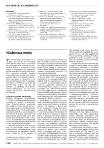
FIG. 1. W. pipientis bacteria visualized in Anopheles gambiae Sua5B cells by FISH, using rhodamine-labeled W. pipientis-specific oligonucleodide probes. (A) Strain wAlbB; (B) strain wRi; (C) strain wRi dividing synchronously in an Sua5B cell. Red, W. pipientis bacteria; blue, cell nuclei (stained with DAPI).
monolayers were grown in 48-well plates to 80% confluence. The medium was removed, the embryo homogenate was layered onto cells, and the cells were infected as described above. This procedure was performed four times with the same cell line to increase the proportion of infected cells. After the cells were
7720
RASGON ET AL.
APPL. ENVIRON. MICROBIOL.
FIG. 2. Confirmation of W. pipientis infection, antibiotic susceptibility, and lack of contaminating Drosophila DNA. (A) Amplification of W. pipientis-specific 16S rDNA sequences from wAlbB-infected cells. Sua5B, uninfected A. gambiae cells; SBA, Sua5B cells infected with W. pipientis strain wAlbB; SBAT, SBA cells cured by tetracycline treatment; Aa23, wAlbB-infected A. albopictus cells (positive control); (-), no-template negative control. (B) Amplification of W. pipientis-specific 16S rDNA sequences from wRi-infected cells. SBR, Sua5B cells infected with W. pipientis strain wRi; SBRT, SBR cells cured by tetracycline treatment; DSR, wRi-infected Drosophila simulans (positive control); (-), no-template negative control. (C) Drosophila DNA (SUF) is undetectable in wRi-infected Sua5B cells and is detectable only in the Drosophila control.
infected four times, W. pipientis was purified out of the cell line, concentrated, and reinoculated into the cells as described above to further increase the infection level of the cells. This procedure was performed three times. W. pipientis PCR. DNA was extracted from the cells, using DNeasy kits (QIAGEN, Valencia, CA) according to the manufacturer’s suggested protocol. W. pipientis infection was confirmed by diagnostic PCR amplification of a 440base pair (bp) fragment of the W. pipientis 16srRNA gene, using primers WspecF and WspecR as previously described (29). An approximately 600-bp fragment from the W. pipientis surface protein gene was amplified, using primers 81F and 691R as previously described (31), purified using Qiaquick spin columns (QIAGEN), and directly sequenced in both directions. ND4 sequencing. Primers ND4⫹ and ND4⫺ were used to amplify an approximately 400-bp fragment from the NADH dehydrogenase subunit 4 (ND4) gene from the cultures. Primer sequences and PCR conditions were as previously stated (9). Amplified fragments were purified and sequenced as described above. Drosophila-specific PCR. An approximately 450-bp fragment of the single-copy nuclear gene suppressor-of-forked (SUF) was amplified from wRi-infected cultures using primers su(f) (forward) and su(f) (reverse). Primer sequences and PCR conditions were as previously stated (28). Visualization of W. pipientis cells. W. pipientis bacteria were visualized with two oligonucleotide probes (W1 and W2) 5⬘-end labeled with rhodamine (11). Cell monolayers were grown on 8-well chamber slides to ⬃50% confluence and then fixed for 10 min with 4% formalin. One hundred nanograms of each probe was added to 20 ml hybridization buffer (50% formamide, 5⫻ SSC, 200 g/liter dextran sulfate, 250 mg/liter poly(A), 250 mg/liter salmon sperm DNA, 250 mg/liter tRNA, 0.1 M dithiothreitol (DTT), 0.5⫻ Denhardt’s solution), and the slides were incubated at 37°C overnight (approximately 18 h). The next day, the slides were washed twice in 1⫻ SSC–10 mM DTT and twice in 0.5⫻ SSC–10 mM DTT at 55°C. The slides were rinsed in deionized water, mounted with glycerol containing 1:1,000,000 DAPI (4⬘,6⬘-diamidino-2-phenylindole), and viewed on an Olympus BX-41 compound microscope fitted with epifluorescent optics. Antibiotic curing. wRi- and wAlbB-infected Sua5B cells were maintained as previously described with the addition of tetracycline (10 g/ml) for four passages.
RESULTS AND DISCUSSION Infection with strain wAlbB. W. pipientis strain wAlbB was purified from infected Aa23 cells using a protocol that results in very pure W. pipientis preparations without contaminating the host material. Sua5B cells were infected at a very high level (⬎90% of cells infected, as determined by fluorescence in situ hybridization [FISH]), likely due to the large amount of W. pipientis bacteria used for experimental infections. The level of infection subsequently increased to almost 100% and had been stable at this level for ⬎30 passages at the time the manuscript was submitted (Fig. 1A). Infection was further confirmed by specific PCR amplification of a portion of the W. pipientis 16SrDNA gene. Elimination of detectable bacteria by tetracycline treatment confirmed that results were due to true establishment of infection rather than by contaminating W. pipientis DNA (Fig. 2A). Sequencing a portion of the W. pipientis surface protein (wsp) gene confirmed that the cells were infected with wAlbB (100% match to GenBank accession number AF020059). Sequence analysis of a 349-bp fragment of the mitochondrial ND4 gene from infected Sua5B cells indicated a 100% match with a corresponding fragment of A. gambiae (GenBank accession number L20934) 100%. Infection with strain wRi. Initial attempts to infect Sua5B cells succeeded with low infection rates (approximately 1 infected cell per 10,000 cells, as determined by FISH). After four sequential attempts to introduce infection into the same cell line, the infection rate increased 1,000-fold (approximately 10% of cells infected). By purifying W. pipientis bacteria out of
INFECTION OF A. GAMBIAE CELLS WITH W. PIPIENTIS
VOL. 72, 2006
the cell line, concentrating them, and reinoculating them back into the cells three times, as described above, we further increased the rate to approximately 30% of cells infected. After 15 passages, infection levels returned to approximately 10% and had been stable at this level for ⬎20 passages at the time the manuscript was submitted (Fig. 1B). We were able to observe W. pipientis replication by FISH staining, and interestingly, all symbionts in the cell were replicating synchronously (Fig. 1C). W. pipientis bacteria were detected by PCR and were eliminated by antibiotic treatment (Fig. 2B). Sequencing a portion of the wsp gene confirmed that the infection was of strain wRi (100% match to GenBank accession number AF020070). Because in this experiment we transferred W. pipientis bacteria from infected embryos, we were concerned that our results could be explained by heterologous culturing of infected Drosophila cells rather than by infection of Anopheles cells. To confirm that our results were due to infection of Anopheles cells and not by inadvertent culturing of Drosophila cells, we amplified a portion of the SUF gene, using Drosophila-specific primers. Amplification succeeded only from the Drosophila control (Fig. 2C). Sequence analysis of a 349-bp fragment of the mitochondrial ND4 gene from infected Sua5B cells indicated a 100% match with a corresponding fragment of A. gambiae (GenBank accession number L20934). Our results demonstrate that two distinct W. pipientis strains can infect immunocompetent A. gambiae cells in vitro. wAlbB infection was maintained at a higher rate than wRi infection (⬃100% versus ⬃10% of cells infected). The wRi and wAlbB strains belong to two phylogenetically distinct W. pipientis clades (wRi, “A” supergroup; wAlbB, “B” supergroup) that diverged approximately 32 million years ago (3). It is possible that observed differences in cell infection levels may reflect genetic divergence between these two W. pipientis strains. Alternatively or in concert with genetic divergence, differences in infectious phenotypes may reflect adaptation of the wAlbB strain to cell culture conditions, as this strain has been in the cell line for ⬎10 years. Further experiments using W. pipientis bacteria from a variety of phylogenetic clades, hosts, and cell lines may help to clarify this issue. Although care must be taken in extrapolating from in vitro results to in vivo systems, our data indicate that there is no intrinsic genetic block to W. pipientis infection of Anopheles gambiae cells and thus that there is no a priori reason to suggest that Anopheles mosquitoes are refractory to W. pipientis infection. Therefore, with proper technique, establishment of in vivo Anopheles infections may well be feasible. W. pipientis-infected Sua5B cells may provide a source of Anophelesadapted W. pipientis bacteria which could increase the probability of establishing in vivo infections. We suspect that infected Sua5B cells will also be useful in a system to investigate genomic and physiological factors influencing W. pipientis host range expansion. ACKNOWLEDGMENTS This work was supported by a Johns Hopkins Malaria Research Institute Pilot Grant to J.L.R. We thank G. Dimopoulos for providing Sua5B cells, S. Dobson and C. Khoo for providing Aa23 cells, and T. Scott, A. Scott, G. Glass, F. Gould, and M. Jacobs-Lorena for comments on a draft of the manuscript.
7721
REFERENCES 1. Baldridge, G. D., N. Burkhardt, M. J. Herron, T. J. Kurtti, and U. G. Munderloh. 2005. Analysis of fluorescent protein expression in transformants of Rickettsia monacensis, an obligate intracellular tick symbiont. Appl. Environ. Microbiol. 71:2095–2105. 2. Beaty, B. J. 2000. Genetic manipulation of vectors: a potential novel approach for control of vector-borne diseases. Proc. Natl. Acad. Sci. USA 97:10295–10297. 3. Clark, M. A., N. A. Moran, and P. Baumann. 1999. Sequence evolution in bacterial endosymbionts having extreme base compositions. Mol. Biol. Evol. 16:1586–1598. 4. Dimopoulos, G., A. Richman, H. M. Muller, and F. C. Kafatos. 1997. Molecular immune responses of the mosquito Anopheles gambiae to bacteria and malaria parasites. Proc. Natl. Acad. Sci. USA 94:11508–11513. 5. Dobson, S. L., E. J. Marsland, Z. Veneti, K. Bourtzis, and S. L. O’Neill. 2002. Characterization of Wolbachia host cell range via the in vitro establishment of infections. Appl. Environ. Microbiol. 68:656–660. 6. Epstein, D. 1999. Malaria: failure, puzzle, challenge. Perspect. Health 4:2–7. 7. Fallon, A. M., and D. Sun. 2001. Exploration of mosquito immunity using cells in culture. Insect Biochem. Mol. Biol. 31:263–278. 8. Fontenille, D., and F. Simard. 2004. Unravelling complexities in human malaria transmission dynamics in Africa through a comprehensive knowledge of vector populations. Comp. Immunol. Microbiol. Infect. Dis. 27:357– 375. 9. Gorrochotegui-Escalante, N., C. Gomez-Machorro, S. Lozano-Fuentes, L. Fernandez-Salas, M. De Lourdes Munoz, J. A. Farfan-Ale, J. GarciaRejon, B. J. Beaty, and W. C. Black IV. 2002. Breeding structure of Aedes aegypti populations in Mexico varies by region. Am. J. Trop. Med. Hyg. 66:213–222. 10. Hay, S. I., C. A. Guerra, A. J. Tatem, A. M. Noor, and R. W. Snow. 2004. The global distribution and population at risk of malaria: past, present, and future. Lancet Infect. Dis. 4:327–336. 11. Heddi, A., A. M. Grenier, C. Khatchadourian, H. Charles, and P. Nardon. 1999. Four intracellular genomes direct weevil biology: nuclear, mitochondrial, principal endosymbiont, and Wolbachia. Proc. Natl. Acad. Sci. USA 96:6814–6819. 12. Hemingway, J., and H. Ranson. 2000. Insecticide resistance in insect vectors of human disease. Annu. Rev. Entomol. 45:371–391. 13. Ito, J., A. Ghosh, L. A. Moreira, E. A. Wimmer, and M. Jacobs-Lorena. 2002. Transgenic anopheline mosquitoes impaired in transmission of a malaria parasite. Nature 417:452–455. 14. James, A. A. 2005. Gene drive systems in mosquitoes: rules of the road. Trends Parasitol. 21:64–67. 15. Kempf, B. J., C. D. Blair, and B. J. Beaty. 2006. Quantitative analysis of La Crosse virus transcription and replication in cell cultures and mosquitoes. Am. J. Trop. Med. Hyg. 74:224–232. 16. Kittayapong, P., K. J. Baisley, V. Baimai, and S. L. O’Neill. 2000. Distribution and diversity of Wolbachia infections in Southeast Asian mosquitoes (Diptera: Culicidae). J. Med. Entomol. 37:340–345. 17. Rasgon, J. L., and T. W. Scott. 2003. Wolbachia and cytoplasmic incompatibility in the California Culex pipiens mosquito species complex: parameter estimates and infection dynamics in natural populations. Genetics 165:2029– 2038. 18. Rasgon, J. L., and T. W. Scott. 2004. An initial survey for Wolbachia (Rickettsiales: Rickettsiaceae) infections in selected California mosquitoes (Diptera: Culicidae). J. Med. Entomol. 41:255–257. 19. Ricci, I., G. Cancrini, S. Gabrielli, S. D’Amelio, and G. Favia. 2002. Searching for Wolbachia (Rickettsiales: Rickettsiaceae) in mosquitoes (Diptera: Culicidae): large polymerase chain reaction survey and new identifications. J. Med. Entomol. 39:562–567. 20. Schneider, I. 1972. Cell lines derived from late embryonic stages of Drosophila melanogaster. J. Embryol. Exp. Morph. 27:353–365. 21. Scott, T. W., W. Takken, B. D. J. Knols, and C. Boete. 2002. The ecology of genetically modified mosquitoes. Science 298:117–119. 22. Shiff, C. 2002. Integrated approach to malaria control. Clin. Microbiol. Rev. 15:278–293. 23. Sinkins, S. P. 2004. Wolbachia and cytoplasmic incompatibility in mosquitoes. Insect Biochem. Mol. Biol. 34:723–729. 24. Snow, R. W., C. A. Guerra, A. M. Noor, H. Y. Myint, and S. I. Hay. 2005. The global distribution of clinical episodes of Plasmodium falciparum malaria. Nature 434:214–217. 25. Talisuna, A. O., P. Bloland, and U. D’Alessandro. 2004. History, dynamics, and public health importance of malaria parasite resistance. Clin. Microbiol. 17:235–254. 26. Townson, H., M. B. Nathan, M. Zaim, P. Guillet, L. Manga, R. Bos, and M. Kindhauser. 2005. Exploiting the potential of vector control for disease prevention. Bull. W. H. O. 83:942–947.
7722
RASGON ET AL.
27. Turelli, M., and A. A. Hoffmann. 1999. Microbe-induced cytoplasmic incompatibility as a mechanism for introducing transgenes into arthropod populations. Insect Mol. Biol. 8:243–255. 28. Turelli, M., and A. A. Hoffmann. 1995. Cytoplasmic incompatibility in Drosophila simulans: dynamics and parameter estimates from natural populations. Genetics 140:1319–1338. 29. Werren, J. H., and D. M. Windsor. 2000. Wolbachia infection frequencies in
APPL. ENVIRON. MICROBIOL. insects: evidence of a global equilibrium? Proc. Biol. Sci. London Ser. B 267:1277–1285. 30. Xi, Z., C. C. Khoo, and S. L. Dobson. 2005. Wolbachia establishment and invasion in an Aedes aegypti laboratory population. Science 310:326–328. 31. Zhou, W., F. Rousett, and S. L. O’Neill. 1998. Phylogeny and PCR-based classification of Wolbachia strains using wsp gene sequences Proc. Biol. Sci. Lond. B 265:509–515.