Volume 1 (1) 2010
CASE REPORT
PROTEUS SYNDROME Vipin George Kuriakose*, Anu Mary Abraham** *Intern,TD Medical College,Alapuzha,Kerala
** Intern,MOSC Medical College,Kolencherry,Kerala
ABSTRACT: A 26 year old lady, born of a nonconsanguineous marriage, presented to the Department of Physical Medicine and Rehabilitation of T.D.M.C, Alappuzha in September 2006, being referred from the Department of Neurosurgery with acute weakness of both lower limbs and urinary incontinence, for the purpose of rehabilitation. Clinical examination revealed multiple developmental deformities and soft tissue swellings, fulfilling the diagnostic criteria of Proteus Syndrome. She also had compressive myelopathy at the level of D 10. Summary Proteus syndrome is a complex hamartomatous disorder first described by Cohen and Hayden in 1979. It was so named by Wiedemann et al in 1983 after the giant Greek god of the sea, who could change the shape of his body to avoid capture. The name Proteus thus refers to polymorphism. Proteus syndrome is a very rare, sporadic, congenital condition comprising malformations and overgrowth of multiple tissues. The disease causes tissue overgrowth in a mosaic pattern and may affect tissues derived from any germinal layer. The syndrome has multiple, diverse, somatic manifestations that evolve over time and involve the skeletal system, soft tissues, skin, and vascular system. These signs include partial gigantism of the hands and/or feet, asymmetry of the limbs, plantar hyperplasia, macrodactyly, bony exostoses, soft-tissue tumors (hemangioma, lymphangioma, lipoma), varicosities, verrucous epidermal nevi, and long-bone overgrowth , vertebral abnormalities, asymmetric limb overgrowth and length discrepancy, hyperostosis, abnormal and asymmetric fat distribution, asymmetric muscle development, connective-tissue nevi, and vascular malformations. Case report
Vipin GK
A 26 year old lady was referred from the Neurosurgery department of T.D.Medical College, Alappuzha, Kerala to the department of Physical Medicine and Rehabilitation, for the purpose of rehabilaitation as she was diagnosed with spastic paraplegia at the D10 spinal level, also associated with urinary incontinence. She was the second child born of a nonconsanguineous marriage via normal vaginal delivery weighing 2.6 kg. Antenatal and Postnatal periods were uneventful. At the age of 6, she presented to the Pediatric opd for evaluation of asymmetric growth of her body with hemihypertrophy of the right side of the body, also left side of the neck. She was diagnosed as a case of Proteus syndrome. Her cognitive and behavioural developmental was normal. Inspite of her condition she was able to her daily activities. She had also achieved a pass in tenth grade. Her syndrome was characterised by the following physical features: Disproportionate overgrowth of body mostly asymmetric in nature Hyperostosis of skull Hemifacial hypertrophy Hemihypertrophy of the tongue Large long neck with asymmetrical overgrowth of the left side Multiple lipomatosis mostly over the trunk Kyphoscoliosis of the thoracolumbar spine Deformed toes Cerebriform nevus on the sole of the left foot In August, 2000 she underwent surgical excision of the cerebriform nevus followed by skin grafting over the area. She was brought to the orthopaedician requiring consultation for history of recurrent falls and a diagnosis of habitual dislocation of the left patella., for which Quadriceps plasty was done. Incidentally she developed spastic paraplegia, with bowel and bladder
Volume 1 (1) 2010
CASE REPORT
involvement following the surgical procedure.
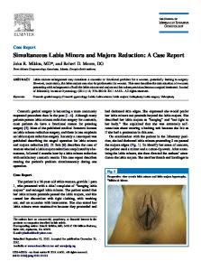
ASYMMETRIC GROWTH OF THE LEFT SIDE OF NECK
HEMIHYPERTROPHY OF THE TONGUE
HYPEROSTOSIS OF THE SKULL RECURRENCE OF CEREBRIFORM NEVUS OVER THE DISTAL END OF THE PREVIOUSLY EXCISED AREA
DEFORMED TOES OF THE LEFT FOOT ROENTGENOGRAM SHOWING KYPHOSCOLIOTIC THORACOLUMBAR SPINE
Vipin GK
Volume 1 (1) 2010
CASE REPORT
MRI spine revealed : Severe degenerative changes of dorsal spine associated with ligamentum hypertrophy , disc herniation indenting the cord with spinal compression at D8 & D9 level with consequent myelomalacia at this level. A spinal cord stenosis is noted in the dorsal spine. Congenital hypertrophy of posterior elements at all levels in the dorso lumbar spine A cystic lesion at D 12 & L1 compressing the left exiting D12 nerve root (likely a Tarlov’s / perineural cyst through a small cystic nerve tumour is another possibility) USG abdomen was within normal limits Surgical decompression of the spinal cord with laminectomy at D9 and D 10 spinal levels was done. Post operatively she was referred to the PMR department of the hospital undergoing rehabilitation for a period of six months as she had persistence of her symptoms. She had pain of her left lower limb and a Doppler study revealed deep vein thrombosis of the external iliac veins,common femoral, superficial, proximal deep femoral and popliteal veins. She was initiated on heparin therapy for the same. Discussion Proteus syndrome can present a range of symptoms from birth. The disease is polymorphous with variable expression. The disease process is not usually apparent at birth but develops rapidly in childhood. The condition is the result of one of several sporadic somatic gene mutations, including the phosphatase tensin homolog (10q23.3) mutation. These mutations result in a varied phenotypic presentation resulting from postnatal dysregulated tissue growth occurring in a mosaic distribution (mosaicism resulting from a somatic alteration of a dominant lethal gene). Because of the high variability in clinical presentation, and the potential to misdiagnose patients with other tissue overgrowth syndromes, diagnostic criteria for Proteus syndrome were established in 1998 by Biesecker and colleagues. The condition has to occur sporadically, the lesions have to show a mosaic distribution, and the course of tissue overgrowth has to be progressive. If these general criteria are met, a number of specific criteria have to be present to finalize diagnosis. The presence of a connective tissue nevus is almost pathognomic for Proteus syndrome and is sufficient to confirm the diagnosis. These nevi occur most commonly on the plantar surface of the feet, but may also be on the abdomen, the hands, and the nose.
Vipin GK
The recent recommendations on diagnostic criteria for Proteus syndrome have greatly aided in the diagnostic process. The difficulty in diagnosing these patients is highlighted by our patient,who had a long delay before a correct diagnosis was made. Before 1983, Proteus syndrome was in most cases, presumed to be Klippel–Trenaunay–Weber syndrome, which consists of a combination of slow-flow vascular malformations with limb hypertrophy. Subcutaneous lesions on the upper or lower limbs and/or trunk are always combined capillary ,lymphatic, and venous malformations and not lipoid tissue, as the lesions in Proteus syndrome can be. Overgrowth is present at birth and is commonly severe, in contrast to Proteus syndrome in which overgrowth is usually mild or absent at birth. Hemihyperplasia/lipomatosis syndrome, which is a subset of hemihyperplasia, can present with cutaneous capillary malformation in combination with multiple lipomas and asymmetrical growth development. Although mild to moderate signs may be present at birth, progressive overgrowth does not occur. The gene mutation responsible for Type I
Volume 1 (1) 2010
Neurofibromatosis can present with hemihypertrophy and fibromatous tumors of the skin. This aids in diagnosing the condition. The neurofibromas evolve from the neural crest. Neurofibromas have not been reported in Proteus syndrome. The gene mutation responsible for the Bannayan–Riley–Ruvalcaba syndrome is also well understood. This syndrome shares macrodactyly, subcutaneous lipomas, capillary malformations, and macrocephaly with Proteus syndrome, but is lacking asymmetrical growth, hyperostosis of the skull, epidermal nevi, and skeletal deformities. Parkes–Weber syndrome shows fast-flow vascular malformations with cutaneous warmth, bruit, and thrills involving the upper and/or lower limbs. Maffucci syndrome, which can present with venous anomalies in different locations and can be differentiated from Proteus syndrome by enchondromas, which have not been reported in Proteus syndrome. Deformities in Proteus syndrome become apparentat various ages. Most anomalies, however, are expressed at an early age. Progression of excessive growth of fingers and toes, and softtissue hypertrophy is possible. New soft-tissue swellings may develop, but regression of, for example, soft-tissue tumors is also possible. Adenoma of the parotid gland, testicular tumors, ovarian cystadenoma, and central nervous system tumors have been reported in association with Proteus syndrome. Soft-Tissue Abnormalities Proteus syndrome is characterized by overgrowth of specific types of tissue. Adipose tissue overgrowth is most commonly manifested as asymmetry in the anterior or posterior body wall or in the subcutaneous fat of the extremities. Overgrowth of fat and muscle may coexist and result in abnormally large muscle groups that contain interspersed fat . Adipose tissue overgrowth can be seen also in more complex soft-tissue enlargements that contain lymphatic, capillary, or venous vascular malformations combined with fat. Muscularcalcifications are infrequent in Proteus syndrome, but their presence should bring this entity into the differential diagnosis. Connective- tissue nevi that cause a corrugated appearance of the skin, particularly in the plantar aspect of the foot are a highly specific finding but are not universal among patients with Proteus syndrome. Commonly referred to as moccasin sole or cerebriform nevus. Skeletal abnormalities Skeletal abnormalities are the most frequent findings in Proteus syndrome with macrodactyly being the most striking manifestation. It may occur in association with clinodactyly( permanent medial or lateral deflection of one or more fingers). Syndactyly (fusion of the bones in fingers or toes, or webbing of soft tissue
Vipin GK
CASE REPORT
between the digits) and polydactyly also are occasionally present. Conclusion Proteus syndrome is a rare polymorphous disease with deformities. The occurrence of mosaicism of the syndrome results in various ways of expression at a young age. The long-term prognosis is not clear. It is important to diagnose Proteus syndrome at an early age so that optimal treatment at the youngest age possible can be established and performed.
REFERENCES: 1. CohenMMJr. Proteus syndrome: clinical evidence for somatic mosaicism and selective review. Am J Med Genet 1993; 47:645–652. 2. Wiedemann H-R, Burgio GR, Aldenhoff P, Kunze J, KaufmannHJ, Schirg E. 1983. The proteus syndrome, partial gigantism of the hands and/or feet, nevi, hemihypertrophy, subcutaneous tumours, macrocephaly, skull anomalies and possible accelerated growth and visceral affections. Eur J. Pediatr 140:5- 12. 3. McGaughran JM, Harris DI, Donnai D, et al. A clinical study of type I neurofibromatosis in north west England. J Med Genet 1999;36:197–203 4.Sorensen SA, Mulvihill JJ, Nielsen A. Long-term follow-up of von Recklinghausen neurofibromatosis: survival and malignant neoplasms. N Engl J Med 1986;314: 1010–1015 5.Cohen Jr MM. Klippel–Trenaunay syndrome. Am J Med Genet 2000;93:171–175 [Editorial] 6.Servelle M. Klippel and Trenaunay’s syndrome: 768 operated cases. Ann Surg 1985;201:365–373 Vaughn RY, Selinger AD, Howell CG, et al. Proteus syndrome: diagnosis and surgical management. J Pediatr Surg 1993;28:5–10 7. Chalk CH, Mills KR, Jacobs JM, et al. Familial multiple symmetric lipomatosis with peripheral neuropathy. Neurology1990;40:1246–1250 8.Gordon PL, Wilroy RS, Lasater OE, et al. Neoplasms in Proteus syndrome. Am J Med Genet 1995;57:74–78 9. Anderson IF. Maffucci’s syndrome: report of a case with a review of the literature. S Afr Med J 1965;39:1066–1070 3
Volume 1 (1) 2010
10. Burgio GR, Wiedemann HR. Further and new details on the Proteus syndrome. Eur J Pediatr 1984;143:71–7
Vipin GK
CASE REPORT
Vipin GK