ijhns IJHNS
CASE REPORT
Congenital Midline Cervical Cleft: A 10.5005/jp-journals-10001-1301 Case Report with Review of Literature
Congenital Midline Cervical Cleft: A Case Report with Review of Literature 1
Ashwin A Jaiswal, 2Bikram K Behera, 3Ravindranath Membally, 4Manoj K Mohanty
ABSTRACT
introduction
Aim: To highlight a rare case of a congenital midline cervical cleft (CMCC) in context with embryological theories/hypothesis, presentation, and management along with review of literature.
Congenital midline cervical cleft (CMCC) is an uncommon malformation of the anterior neck with less than 100 cases reported.1 The first recorded case of CMCC was in 1848 by Luschka,2 while Bailey3 documented the first description of this abnormality in 1924; however, it was completely described by Ombreadanne in 1946.4 It represents a variant of the cleft category number 30 of the Tessier classification system of craniofacial defects.5 Embryologically, impaired fusion of branchial arches is considered as the most accepted theory explaining the etiology of CMCC. Inadequate treatment may cause secondary complications, such as impaired neck extension, microgenia, exostosis, torticollis, or infection.1 Surgical excision with Z-plasty is the recommended treatment of choice for CMCC.
Introduction: Congenital midline cervical cleft is a rare but interesting anterior neck anomaly with controversial theories/ hypothesis regarding its embryogenesis. Case report: We describe here a classical case of midline cervical cleft that presented at birth with a cephalocaudal orientation, extending from the level below the hyoid bone to the suprasternal notch with a length of 3 cm and width of 0.5 cm. At 6 months of age, the lesion was excised and closure was done by multiple Z-plasty, with satisfactory results. Discussion: Although the diagnosis is clinical, it is frequently misdiagnosed. The associated clinical features could include thyroglossal duct cysts, cleft lip/mandible/sternum, cervical contractures, mandibular spurs, microgenia, and/or bronchogenic cysts. If it is not treated at an early age, it can result in complications like webbing of the neck, dental malocclusion, and restricted neck movements. Conclusion: Earliest recognition of CMCC and proper intervention can provide better esthetic and functional prognosis. Clinical significance: A correct earlier recognition of the lesion and appropriate surgical management are key to avoid longterm complications. Keywords: Branchial arches, Congenital midline cervical cleft, Thyroglossal duct cysts, Z-plasty. How to cite this article: Jaiswal AA, Behera BK, Membally R, Mohanty MK. Congenital Midline Cervical Cleft: A Case Report with Review of Literature. Int J Head Neck Surg 2017;8(1):25-30. Source of support: Nil Conflict of interest: None
1
Specialist, 2Consultant, 3Joint Director, 4Head
1
Department of ENT and Head-Neck Surgery, JLN Hospital & Research Centre, Bhilai, Chhattisgarh, India
2
Department of Anaesthesia, JLN Hospital & Research Centre Bhilai, Chhattisgarh, India
3
Department of Pathology, JLN Hospital & Research Centre Bhilai, Chhattisgarh, India
4
Department of Paediatric Surgery, JLN Hospital & Research Centre, Bhilai, Chhattisgarh, India Corresponding Author: Ashwin A Jaiswal, Specialist Department of ENT and Head-Neck Surgery, JLN Hospital & Research Centre, Bhilai, Chhattisgarh, India, Phone: +919407983605, e-mail:
[email protected]
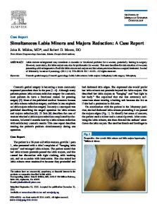
CASE report A full-term baby weighing 2,800 gm born by normal spontaneous vaginal delivery after an uneventful pregnancy was referred to us with a congenital neck abnormality noted at birth. Pregnancy scans at 12 and 20 weeks were reported normal. Her parents were healthy and nonconsanguineous. The patient had no airway or feeding difficulties, and her cry was normal. On examination, the cervical lesion had the configuration of a linear cleft with a cephalocaudal orientation, extending from the level below the hyoid bone to the suprasternal notch with a length of 3 cm and width of 0.5 cm. It was composed of three components, a notch structure with nipple-like skin tag at cephalic end, a blind sinus of 0.3 cm depth at the caudal end, and a midline longitudinal mucosal surface between them with underlying fibrotic tissue (Fig. 1A). On extension of the neck, a skin web was formed between the cleft and the mandible (Fig. 1B). The lesion did not move on deglutition or protrusion of the tongue and had no fixity to the underlying structures. There was a seromucinous transparent discharge from its moist pink colored mucosal surface that was gradually reduced during the first weeks of infancy. The sinus was examined by a small lacrimal probe, and a narrow tract coursing toward the suprasternal notch was observed. Examination of the rest of the face, neck, and oral cavity was normal; there was no other sinus or
International Journal of Head and Neck Surgery, January-March 2017;8(1):25-30
25
Ashwin A Jaiswal et al
cleft or features suggestive of any syndromic association. A clinical diagnosis of CMCC was made and the patient was planned for surgery at a later date. At 6 months of age, she revisited our outpatient department with no history of discharge, swelling, or change in the appearance of the lesion. A fistulogram performed showed the sinus to be a narrow channel, 0.3 cm in length, coursing toward the suprasternal notch and ending abruptly. There was no connection with other structures. A computed tomography scan of neck with thorax was done, which showed a small delineated track in the midline submental location with small area of nodularity and a blind end almost at the thoracic inlet. No communication or cyst or collection was seen and any cervical or vertebral anomalies were ruled out. At laryngobronchoscopy, pharynx, larynx, and the tracheobronchial tree were normal. Any other systemic/physical deformity was not seen on examination of the patient. The patient was treated surgically. A vertical elliptical incision was given and the cleft with nipple-like skin tag at cephalic end, underlying fibrotic tissue, and the sinus at the caudal end were excised. The fibrous cord extending up to the manubrium was removed completely. The skin defect was closed by serial Z-plasties. The resultant flaps were sutured first by vicryl 4-0 for dermal suture and by
A
C
prolene 5-0 for skin closure (Figs 2A to D). Postoperative course was uneventful, and there was no wound infection. In follow-up examinations at 1 month, 3 months, and 1 year after operation, there was excellent wound healing of the Z-plasty with no wound contracture in the neck and a wide range of neck movements (Figs 1C to E). Histological examination of the excised tissue showed (1) cleft lined by stratified squamous epithelium with surface parakeratosis, (2) scarcity of adnexal structures in underlying dermis, (3) striated muscle bundles present in the deeper dermis, and (4) presence of inflammatory infiltrate with more lymphocytes and neutrophils in the dermis (Figs 3A to D).
DISCUSSION Congenital midline cervical cleft constitutes 2% of all congenital cervical malformations.4 The prevalence of CMCC in all cases of thyroglossal cyst and brachial cleft sinuses is 1.7%.6 It predominantly affects white female patients. A female to male ratio of 2:1 is reported, with a sporadic presentation.1 The age of presentation ranges from birth to 23 years.7 The lesion is located in the midline of the anterior neck at any point between the mandible and the sternum.1 On its typical presentation, CMCC consists of three anatomic parts: An superior nipple-like skin tag
B
D
E
Figs 1A to E: Clinical photograph of the patient: (A) Front view showing classical three components, a notch structure with nipple-like skin tag at cephalic end, a blind sinus of 0.3 cm depth at the caudal end, and a midline longitudinal mucosal surface between them with underlying fibrotic tissue; (B) Front view with extension of neck showing formation of skin web between the cleft and the mandible. Postoperative follow-up photographs; (C) Front view after 1 month of follow-up showing excellent wound healing of the Z-plasty and no wound contracture in the neck; (D) Front view after 3 months of follow-up showing no wound contracture in the neck and full neck movements; and (E) Front view after 1 year of follow-up showing wide range of neck movement postoperatively
26
ijhns Congenital Midline Cervical Cleft: A Case Report with Review of Literature
A
B
C
D
Figs 2A to D: Intraoperative photographs: (A) Elliptical cervical incision with serial Z-plasties; (B) excision carried at the upper end in full thickness; (C) excision carried out at the lower end with the fibrotic cord extending up to the manubrium; and (D) resultant flaps after Z-plasties sutured in two layers
which hoods a linear area of a red or pink moist surface of atrophic epidermis without adnexal structures, to end to a posterior duct, usually shallow and blind but occasionally going all the way down to the area of the manubrium or the sternum, or toward the hyoid bone. Mucous drain may exit from the inferior duct.7 The seromucinous discharge resolves gradually during the first months of infancy. With time, the cleft heals and a longitudinal scar is formed, resulting in the formation of web, which causes contracture of the neck, limits neck mobility, particularly extension, or torticollis.7 Three clinical outcomes emerge from this evolution. The first is neck contracture and functional compromise, the second is secondary anatomical disarrangement, such as formation of micrognathia, or bony spur (exostosis) of the mandible or sternum, and the third is misdiagnosis later in life, when the cleft achieves the form of a midline linear spot-like scar, rather
than the typical presentation after birth.7 Patients with the lesion were sometimes referred to dermatologists by primary care physicians with the possible diagnosis of a thyroglossal duct cyst or an “unusual birthmark.”8 The spectrum of severity ranges from ventral cervical webs to mentosternal clefts, leading to pterygium colli medianum with severe regional hypoplasia.9 Although most patients are asymptomatic at diagnosis with an apparent cosmetic concern due to ugly appearance of the CMCC, neck contractures and mandibular or sternal growth abnormalities may develop in untreated patients. An exostosis from the midpoint of the mandible can form, resulting from persistent traction from the contracting fibrous cord underneath the cleft. Congenital midline cervical cleft can prevent full extension of the neck, resulting in micrognathia and torticollis, predispose patients to infection, and can coexist with other clefting
International Journal of Head and Neck Surgery, January-March 2017;8(1):25-30
27
Ashwin A Jaiswal et al
A
B
C
D
Figs 3A to D: Histopathological examination: Operative specimen sections showing: (A) cleft lined by stratified squamous epithelium with surface parakeratosis (hematoxylin and eosin [H&E], 40×); (B) scarcity of adnexal structures in underlying dermis (H&E, 10×); (C) striated muscle bundles present in the deeper dermis (H&E, 10×); and (D) inflammatory infiltrate with more lymphocytes and neutrophils in the dermis (H&E, 40×)
defects or cysts.10 The midline cervical cleft may be a solitary deformity, but there are cases where it is combined with thyroglossal duct cyst, ectopic bronchogenic cyst, branchial cyst, midline hemangioma, ectopia cordis, cleft lip, mandible or tongue, cleft sternum, absence of hyoid bone or thyroid cartilage, or congenital heart disease.7,11,12 This eventually results in a fourth clinical issue, failure of diagnosis of any of these disorders.1 Different theories have been proposed on the embryological origin of the CMCC. Most investigators believe that the defect is the result of fusion failure of the first and second branchial arches in the midline.1 Mechanisms proposed to be implicated with incomplete branchial fusion are vascular anomalies (ischemia, necrosis, and scarring), persistence of remnants of the thyroglossal duct and sinus cysts, increased pressure on the cervical area from the pericardial roof in early stages of developing embryo, rupture of a pathologic adhesion between the epithelium of the cardiohepatic fold with that of the ventral part of the first branchial arch, and absence of mesenchymal tissue in the cervical midline.1,7 Congenital
28
midline cervical cleft was found to be associated with chromosomes 13/14 de novo Robertsonian translocations as well as midline deformities including a sacral tuft and a minor tongue-tie.13 The proper description of the pathology includes three different anatomic areas. The superior skin tag part may present normal skin, or stratified squamous epithelium with parakeratosis. Presence of cartilage or skeletal striated muscle has been reported. Stratified squamous epithelium with surface parakeratosis continues all the way down the main part of the lesion; combined with the absence of adnexal structures in the underlying dermis is the hallmark of histological presentation of the major part of the malformation.1,7 The inferior sinus tract consists of pseudostratified ciliated columnar epithelium with seromucinous glands.1,7 In some cases, this tract may contain skin epithelium, muscle, or cartilage.7 Management includes excision of cleft with reconstruction of the defects. The severity of regional hypoplasia decides the reconstructive armamentarium ranging from Z- and V-Y plasty for simple webs to tissue expansion
ijhns Congenital Midline Cervical Cleft: A Case Report with Review of Literature
or myocutaneous flaps for severe regional hypoplasia and geniosternoplasty for mentosternal clefts.14 Surgical intervention is necessary to avoid potential long-term complications, such as scarring, contractures, and limitation of neck mobility. Complete surgical excision of the cleft including the underlying fibrous cord is the recommended procedure of choice preferably in infancy. The reconstruction involves multiple Z-plasty procedures to avoid the formation of hypertrophic scars and provide enough length to avoid contracture at a later date. This is of cosmetic and functional importance to avoid the inevitable scarring and contractures that follow later in life. The most frequently used techniques are variations of Z-type plasty, in order to achieve uncompromised neck extension.1,7,8,10,11 Simple short sinuses less than 2 cm length may be excised through stair step incisions, with a technique similar to that used for some second branchial clefts. More complicated clefts are excised with a series of Z-plasty incisions that improve the functional and cosmetic results. Spencer Cochran et al15 recommended single Z-plasty to be appropriate for lesions less than 2 cm, and serial Z-plasties for longer lesions.4 We used serial Z-plasties because the resultant defect after the excision of the lesion was more than 2 cm in length. Z-plasty is a common technique and a versatile surgical maneuver. Z-plasty allows the surgeon to (1) lengthen a contracted scar; (2) reorient the direction of a scar or defect; (3) break up a straight line; and (4) shift soft tissue contour.16,17 The Z-plasty technique involves creating two opposing triangular transposition flaps that are rotated synchronously to close a central defect by redistribution and rearrangement of tissue.18 The Z-plasty is symmetrically designed so that the lateral limbs are equal in length to the tissue defect (central limb) and that the angles between the lateral limbs and the central limb are 60°. Reapproximation of the central defect at the skin level prior to designing the Z-plasty flaps has been noted to aid in constructing a more precise Z-plasty.19,20 The angle of the Z-plasty flaps may vary, and the gain in length varies directly with the angle of the Z-plasty. The optimal angle has been determined to be 60°, which has a theoretical gain in length of 75%. The angle may be greater than 60°, but the tension required to transpose the skin flaps increases as the angle of the Z-plasty increases, such that angles greater than 75° cause tissue distortion and dog-ear deformities, but angles less than 20° present problems with flap viability secondary to compromised blood flow at the flap tips.21 Many authors suggest as proper for surgery the age before the second year of life, with earlier repair indicated in more severe cases.7 Early repair prevents contracture and cosmetic deformities. Ercocen et al16 suggested that early intervention avoids the disfiguring appearance of
the malformation and also prevents subsequent limitation of neck motion. Derbez et al4 reported five cases of CMCC. All were treated in early life at age ranging from 1 month to 2 years. They concluded that surgical repair should be done as soon as possible to reduce the risk of recurrence and avoid limitation of neck extension. We believe that the age of surgical intervention should be early infancy during the first 6 months of life. More advanced cases having hypoplasia of mandible, absent hyoid and/or thyroid cartilage, or other supporting structures of the neck warrant extensive teamwork between plastic surgeon, head and neck surgeon, faciomaxillary surgeon supported by psychologists, speech therapists, pediatrician, and very dedicated nursing care. These cases require multiple surgical procedures to be able to lead a meaningful life.22
CONCLUSION Congenital midline cervical cleft is a rare congenital anomaly of the neck, with much controversy on its etiology. Complete excision of cleft with reconstruction of the defect with multiple Z-plasty technique is the treatment of choice. Earliest recognition of CMCC by neonatologists, ear, nose, and throat surgeons, pediatric and plastic surgeons and proper intervention can provide better esthetic and functional prognosis. More advanced cases warrant multidisciplinary team approach along with multiple surgical procedures.
CLINICAL SIGNIFICANCE A correct earlier recognition of the lesion and appropriate surgical management are key to avoid long-term complications.
REFERENCES 1. Eastlack JP, Howard RM, Frieden IJ. Congenital midline cervical cleft: case report and review of the English language literature. Pediat Dermatol 2000 Mar-Apr;17(2):118-122. 2. Luschka H. Veber fistula colli congenital. Arch Physiol 1848;7:25. 3. Bailey H. Thyroglossal cysts and fistulae. Br J Surg 1924;12: 579-589. 4. Derbez R, Nicollas R, Roman S, Estève A, Triglia JM. Congenital midline cervical cleft of the neck: a series of five cases. Inter J Pediatr Otorhinolaryngol 2004 Sep;68(9):1215-1219. 5. Saha S, Misra S, Saha VP, Mondal AR. Midline cervical cleft: a report of two cases. Indian J Otolaryngol Head Neck Surg 2005 Jan;57(1):78-79. 6. Sinopidis X, Kourea HP, Panagidis A, Alexopoulos V, Tzifas S, Dimitriou G, Georgiou G. Congenital midline cervical cleft: diagnosis, pathologic findings, and early stage treatment. Case Rep Pediatr 2012 Sep;2012(1):951040.
International Journal of Head and Neck Surgery, January-March 2017;8(1):25-30
29
Ashwin A Jaiswal et al 7. Mlynarek A, Hagr A, Tewfik TL, Nguyen VH. Congenital midline cervical cleft: a case report and review of the literature. Int J Pediatr Otorhinolaryngol 2003 Nov;67(11):1243-1249. 8. Warden C, Millar AJW. A rare congenital midline cervical cleft. S Afr J Surg 2010 Jul;48(3):98-99. 9. Godbersen S, Heckel V, Wiedemann HR. Pterygium colli medianum and midline cervical cleft: midline anomalies in the sense of a developmental field defect. Am J Med Genet 1987 Jul;27(3):719-723. 10. McInnes CW, Benson AD, Verchere CG, Ludemann JP, Arneja JS. Management of congenital midline cervical cleft. J Craniofac Surg 2012 Jan;23(1):36-38. 11. Hirokawa S, Uotani H, Okami H, Tsukada K, Futatani T, Hashimoto I. A case of congenital midline cervical cleft with congenital heart disease. J Pediatr Surg 2003 Jul;38(7):1099-1101. 12. Vure S, Pang K, Hallam L, Lui M, Croaker D. Congenital midline cervical cleft with an underlying bronchogenic like cyst. Pediatr Surg Int 2009 Sep;25(9):811-813. 13. Agag R, Sacks J, Silver L. Congenital midline cervical cleft. Cleft Palate Craniofac J 2007 Jan;44(1):98-101.
30
14. Nicklaus PJ, Forte V, Friedberg J. Congenital mid-line cervical cleft. J Otolaryngol 1992 Aug;21(4):241-243. 15. Cochran CS, DeFatta RJ, Brenski AC. Congenital midline cervical cleft: a practical approach to Z-plasty closure Int J Pediatr Otorhinolaryngol. 2006 Mar;70(3):553-559. 16. Ercocen AR, Yilmaz S, Aker H. Congenital midline cervical cleft: a case report and review of the literature. J Oral Maxillofac Surg 2002;60(5):580-585. 17. Hudson DA. Some thoughts on choosing a Z-plasty: the Z made simple. Plast Reconstr Surg 2000 Sep;106(3):665-671. 18. Rohrich RJ, Zbar RI. A simplified algorithm for the use of Z-plasty. Plast Reconstr Surg 1999 Apr;103(5):1513-1517. 19. Bajaj Y, Dunaway D, Hartley BEJ. Surgical approach for congenital midline cervical cleft. J Laryngol Otol 2004 Jul;118(7):566-569. 20. Daw JL Jr, Patel PK. Double-opposing Z-plasty for correction of midline cervical web. J Craniofac Surg 2003 Sep;14(5):774-778. 21. Davis WE, Schrick RE, Templer J. An introduction to Z-plasty. Ear Nose Throat J 1981 Jan;60(1):29-34. 22. Kara CO, Kara IG. Congenital midline cervical cleft. Otolaryngol Head Neck Surg 2006 Dec;135(6):953-954.