Experimental Oncology 24, 173-179, 2002 (September)
173
EVALUATION OF NAPROMUSTINE, A NITROGEN MUSTARD DERIVATIVE OF NAPHTHALIMIDE, AS A RATIONALLY DESIGNED MIXED-FUNCTION ANTICANCER AGENT A. Pain1, S. Samanta1, S. Dutta1, A.K. Saxena2, M. Shanmugavel2, H. Kampasi2, G.N. Qazi2, U. Sanyal1,* 1 Chittaranjan National Cancer Institute, Calcutta 700026, India 2 Regional Research Laboratory, Jammu-Tawi 180001, India
ÈÇÓ×ÅÍÈÅ ÏÐÎÒÈÂÎÎÏÓÕÎËÅÂÛÕ ÑÂÎÉÑÒ ÍÀÏÐÎÌÓÑÒÈÍÀ, ÑÈÍÒÅÇÈÐÎÂÀÍÍÎÃÎ ÍÀ ÎÑÍÎÂÅ ÍÀÔÒÀËÈÌÈÄÀ À. Ïåéí1, Ñ. Ñàìàíòà1, Ñ. Äàòòà1, À.Ê. Ñàêñåíà2, Ì. Øàíìàãàâåë2, Ã. Êàìïàñè2, Ã.Í. Êâàçè2, Ó. Ñàíüÿë1,* 1 Íàöèîíàëüíûé ðàêîâûé èíñòèòóò ×èòòàðàíüÿí, Êàëüêóòòà, Èíäèÿ 2 Îáëàñòíàÿ Èññëåäîâàòåëüñêàÿ ëàáîðàòîðèÿ, Äæàììè-Òàâè, Èíäèÿ Napromustine, a new nitrogen mustard of substituted naphthalimide, has been synthesized as a new mixed-function anticancer agent from N-(3-bromopropyl) naphthalimide. Chemical alkylating activity of Napromustine exceeded that of N-di(2-chloroethyl)amine as a standard alkylating compound. Napromustine has displayed an excellent and reproducible antitumor activity in vivo against Sarcoma-180 and Ehrlich ascites carcinoma comparable to that of fluorouracil judging by the increase in median survival times of treated animals. Napromustine also significantly increased the life span of mice bearing highly advanced tumors for 10 days before the drug challenge. The compound under study did not affect hematopoiesis or induce hepatotoxicity and nephrotoxicity. This compound inhibits the synthesis of DNA and RNA in S-180 tumor cells. Meanwhile in vitro screening in 3 different human tumor cell lines did not reveal any significant cytotoxic activity. Key Words: anticancer agent, screening, alkylating activity, mustine derivative. Íà îñíîâå N-(3-áðîìïðîïèë)íàôòàëèìèäà êàê èñõîäíîãî ïðîäóêòà áûë ñèíòåçèðîâàí è èñïûòàí ïðåïàðàò íàïðîìóñòèí, îáëàäàþùèé áîëåå âûñîêîé àëêèëèðóþùåé àêòèâíîñòüþ, ÷åì ñòàíäàðòíûé àëêèëèðóþùèé ïðåïàðàò N-äè(2-õëîðýòèë)àìèí. Ïðîòèâîîïóõîëåâûé ýôôåêò ïðåïàðàòà ïðè èñïîëüçîâàíèè øòàììîâ ñàðêîìû-180 è àñöèòíîãî ðàêà Ýðëèõà îêàçàëñÿ áîëåå âûñîêèì, ÷åì ó ôëóîðîóðàöèëà. Ââåäåíèå íàïðîìóñòèíà äàæå íà ïîçäíèõ ñðîêàõ ïåðåâèâêè îïóõîëè ïðèâîäèëî ê ñóùåñòâåííîìó óâåëè÷åíèþ ìåäèàíû âûæèâàåìîñòè æèâîòíûõîïóõîëåíîñèòåëåé. Ïðåïàðàò íå óãíåòàë ôóíêöèþ êðîâåòâîðåíèÿ, íå îáëàäàë ãåïàòî- è íåôðîòîêñè÷íîñòüþ è èíãèáèðîâàë ñèíòåç ÄÍÊ è ÐÍÊ â êëåòêàõ S-180.  òî æå âðåìÿ ïðè èñïûòàíèÿõ in vitro íà 3 ðàçëè÷íûõ ïåðåâèâàåìûõ ëèíèÿõ êëåòîê îïóõîëåé ÷åëîâåêà ïðåïàðàò íå ïðîÿâëÿë öèòîòîêñè÷åñêîãî ýôôåêòà. Êëþ÷åâûå ñëîâà: ïðîòèâîîïóõîëåâîå ñîåäèíåíèå, ñêðèíèíã, àëêèëèðóþùàÿ àêòèâíîñòü, ïðîèçâîäíîå íàôòàëèìèäà.
Nitrogen mustards (mustines) containing the established anticancer functionality N, N/-bis(2-chloroethyl)amino group, best represented by mechlorethamine (HN2), cyclophosphamide (Fig. 1, Structure A), ifosphamide, chlorambucil, melphalan etc [1] are important alkylating agents. However in spite of their wide application in the management of several human tumors they have undesirable side effects. Hence efforts are ongoing to develop new mustine derivatives possessing better therapeutic efficacy and lower toxicity [2]. Although various other structural patterns have been used as the carrier molecules for mustine group, naphthalimide ring has not yet been explored, to our knowReceived: August 16, 2002. *Correspondence. Fax : +91-33-475-7606; E-mail :
[email protected] Abbreviations used: FU — fluorouracil; BUN — blood urea nitrogen; MST — median survival time; Napromustine — 2-[3-{bis(2chloroethyl)amino}propyl]-1H-benz[de]isoquinoline-1,3-dione; SAKP — serum alkaline phosphatase; SGOT — glutamic oxaloacetic transaminase; SGPT — glutamic pyruvic transaminase.
ledge. This structural pattern is particularly interesting since some substituted naphthalimides containing N-(2,2-dimethylaminoethyl) chain have demonstrated substantial antineoplastic activity in various tumors [3]. To that group belongs Amonafide and Mitonafide (Fig. 1, Structure B) which inhibit DNA and RNA synthesis, intercalate DNA and exhibit substantial anticancer activities in various animal tumors [4]. At present time those compounds are undergoing clinical trials [5]. Based on the above consideration and the results of our drug development program describing another potential anticancer compound Napro-NU [6], it is thought worth to investigate the compound of the general Structure C described in Fig. 1. We describe herein the synthesis through known route (Fig. 1, Structure 1), anticancer and toxicological evaluation of the new mustine compound Napromustine, 2-[3-{bis(2-chloroethyl)amino}propyl]-1H-benz[de]isoquinoline-1,3-dione. It is hypothesised that upon in vivo enzymatic degradation it may exert synergistic activity since the naphthalimide ring residue may bind with DNA while
174
Experimental Oncology 24, 173-179, 2002 (September)
Fig. 1. Structure of Napromustine and related compounds
mustine group will exert cytotoxicity through alkylation of biological nucleophiles.
MATERIALS AND METHODS Chemical synthesis. New compounds were characterised by 1HNMR spectra measured in a Bruker 300DPX spectrometer with the solvents as indicated and chemical shifts were expressed in δ units (ppm) using tetramethylene silane as internal standard. IR spectra were recorded in a Perkin Elmer RX-1 FT-IR spectrophotometer in KBr pellet. Satisfactory analytical data (C, H, N) in Perkin-Elmer 240 analyser were obtained within specification (± 0.4%) for the new compounds described. Melting points were determined on a Thomas-Hoover Unimelt capillary melting point apparatus and were uncorrected. TLC analysis were carried out with glass plates coated with Silica gel G (Solvent system CHCl3 — MeOH, v/v 90 : 10). Purity of the compound synthesised was further checked with Waters HPLC system at ambient temperature (µ-bondapak C18 steel column, 30 cm × 3.9 mm; isocratic mobile phase acetonitrile-water in varying proportions (up to 40 : 60) at a flow rate of 1.0 ml/min; UV detection at 250 nm). Mass spectral analysis of final compound was assessed by FAB + method using mNBA matrix compound in JEOL JMS600 instrument. 3H-thymidine (specific activity 1.0 mCi/cm3) and 3 H-uridine (specific activity 1.0 mCi/cm3) were obtained from Board of Radiation and Isotope Technology (Mumbai, India). Radioactivity was measured in a liquid scintillation counter (Model LKB 1209 Rack-Beta).
To a solution of the N-(3-hydroxypropyl)-naphthalimide [7] in ethyl acetate, phosphorus tribromide was added to furnish the desired product N-(3-bromopropyl) naphthalimide [8] in 67% yield after crystallization; m.pt. 135 °C (lit.). TLC: RF 0.89. 1H NMR (CDCl3) 2.36 (m, 2H, CH 2 ), 3.51 (t, 2H, CH 2Br), 4.34 [t, 2H, CH2N(CO)2], 7.77 (m, 2H, arom. H), 8.22 (t, 2H, arom H), 8.6 (t, 2H, arom H). A mixture of the above bromo compound (636 mg, 2 mmol), freshly distilled diethanolamine (0.6 ml, 6 mmol), anhydrous NaI (320 mg, 6 mmol) was dissolved in dry DMF (6 ml) and heated for 24 h at 110– 130 °C. The reaction mixture was concentrated in vacuum and treated with saturated aqueous sodium bicarbonate solution. The solution was repeatedly extracted with chloroform and chloroform extract was washed with brine, dried (Na2SO4) and evaporated to give an oily residue that was next purified by passing through a silica gel column with chloroform-methanol (95 : 5, v/v) as eluent to furnish the desired product as a highly viscous oil. The dihydroxy compound was directly used for the next step. Yield 394 mg (62%), TLC: RF 0.42. 1H NMR (CDCl3) 1.95 (t, 4H, CH2, 2 × OH), 2.68 (m, 6H, 3 × CH2N), 3.66 (t, 4H, 2 × CH2O), 4.3 [t, 2H, CH2N(CO)2], 7.76 (t, 2H, arom. H), 8.21 (d, 2H, arom H), 8.61 (d, 2H, arom H). A solution of the above compound (342 mg, 1 mmol) in phosphorus oxychloride (1.1 ml) was heated at 80– 90 °C for 1.5 hr and phosphorus oxychloride was evaporated to dryness in vacuum. The resultant oily residue
Experimental Oncology 24, 173-179, 2002 (September)
175
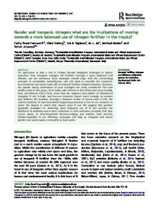
Hemoglobin
100 90 85
SC ST NT
80 75 70 65
% of normal control
% of normal control
95
60 15 Day
SC ST NT
80 75 70 65
21
9
WBC
180
560 460 SC ST NT
360 260 160
% of normal control
660 % of normal control
90 85
60 9
60
15 Day SC ST NT
160 140
21
Platelet
120 100 80 60
9 110
15 Day
21
9
Bone marrow
15 Day
21
Splenic
220 200
100 90 SC ST NT
80 70 60
% of normal control
% of normal control
RBC
100
95
180 160
SC ST NT
140 120 100 80 60
9
15 Day
21
9
15 Day
21
Fig. 2. Sequential changes in hematological parameters, femoral bone marrow and splenic cellularity in normal and S-180 bearing mice after treatment from day 1–7
was treated with concentrated hydrochloric acid (1.0 ml). After the initial exothermic reaction, the mixture was heated in a steam bath for 10 min and allowed to cool. It was then treated with saturated NaOAc solution and extracted with CHCl3. After washing with brine followed by drying and concentration, an oily residue was obtained which solidified on maintaining at 0 °C. It was crystallised from anhydrous ether to furnish the desired Napromustine compound 1, homogeneous in TLC and HPLC (yield 216 mg (63%), m.p. 155 °C). TLC : RF 0.87. HPLC : retention time 14.53 min (CH3CN — H2O v/v 1 : 1). 1H NMR (CDCl3) 2.2 (m, 2H, CH2), 3.3 (t, 6H, 3 × CH2N), 3.8 (t, 4H, 2 × CH2Cl), 4.3 [t, 2H, CH2N(CO)2], 7.8 (t, 2H, arom. H), 8.22 (d, 2H, arom H) and 8.6 (d, 2H, arom H). Determination of chemical alkylating activity. The procedure described earlier was essentially followed [6]. Thus a solution of Napromustine or N-di(2chloroethyl)amine [HN(CH2CH2Cl)2] in different concentrations as indicated in acetone (1 ml), distilled water (2 ml) and acetate buffer (1 ml, 0.25 M, pH 6.0) were incubated at 100 °C for 20 min with a solution of 4-(4nitrobenzyl)pyridine (5% w/v) in acetone (0.4 ml) and cooled to 25 °C. After the addition of acetone (2 ml),
EtOAc (5 ml) and sodium hydroxide solution (0.25 M, 1.5 ml), the reaction mixtures were vortexed and allowed to stand to separate the organic layers. The absorbances in the organic layers were immediately determined (within 2 min of NaOH addition) at 540 nm. The experiments were carried out in triplicate. The results were expressed in optical density values (mean ± S.E.M., n = 3 in all cases) (Table 1). In vivo screening. Closed colony bred Swiss albino male mice (6–7 weeks age, 24 ± 2 g) were obtained from the Institute’s vivarium, housed in cage and maintained on standard mouse food and tap water ad libitum. EAC and S-180 cells freshly obtained from National Centre for Cell Sciences (Pune, India) were used. Tumor cell suspensions in physiological saline were prepared as described [6] to final concentrations of 5.0 · 106 cells/ml. Mice were inoculated through intraperitoneal route with 1.0 · 106 viable cells/ Table 1. Determination of chemical alkylating activity* Compound Concentration of compound (µM/ml) 0.25 0.50 Napromustine 0.57 ± 0.07 1.07 ± 0.09 N-di(2-chloroethyl)amine 0.43 ± 0.03 0.62 ± 0.12 Blank 0.05 ± 0.01 0.05 ± 0.01 * Expressed in OD values (see Materials and Methods).
176
Experimental Oncology 24, 173-179, 2002 (September)
mouse in 0.2 ml on day 0. Mice were divided in groups as control (untreated) and treated and designated as NC — normal control; NT — normal treated; SC — S-180 control; ST — S-180 treated. At least 6 animals were used for a particular group in an experiment. Physiological saline containing 2% Tween 80 (Sigma Inc, USA) was used for drug administration in respective doses through intraperitoneal (i.p.) route to each mouse in treated groups. The drug solutions were prepared daily just prior to the injection. The control groups received an equal volume of above solvent (0.2 ml) on those days. Median survival time (MST) of drug-treated (T) and control (C) tumor bearing animals was calculated and expressed as T/C percentage value [9]. T/C percentage value > 125 was considered as significant. Cyclophosphamide and FU were used as positive controls for comparison. All experiments were performed under guidelines of Ethic Committee for animal welfare. In vivo toxicological assay. The optimum dose of 12.0 mg/kg was administered in normal and S-180 bearing mice from day 1 to 7. Various parameters were measured sequentially after 48 h (day 9) for noting immediate effects, after 192 h (day 15) for intermediate effects and after 336 h (day 21) for late effects. For hematological studies, blood samples were collected from tail veins or by cardiac puncture from recently sacrificed animals under deep exposure to ether for counting erythrocytes, thrombocytes or leukocytes in an improved Neubauer bright field counting chamber by standard procedures using freshly prepared RBC and WBC counting fluids. Differential counts of leukocytes were performed by staining of the blood smears with Leishman’s stain (pH 6.8); slides were observed under light microscope. Hemoglobin concentrations were meaSGOT
25
sured by the standard method. Sera were obtained from blood samples collected as above. Standard methods and reagents were used [10] to measure SAKP, SGOT, SGPT activities and BUN content. Femoral marrow cells were obtained by the standard procedure from mice sacrificed as stated above. The ends of the femur bone were snipped open immediately thereafter with scissors and the marrow plug was flushed out by forcefully injecting cold HBSS (Ca++ and Mg++ free) through the bone cavity by inserting a 22 gauze needle. The marrow plug was dissociated into single cell suspension by repeatedly passing this suspension through 22 gauze needles and the total volume was measured. Total number of nucleated cells per femur was counted in a hemocytometer after treating the cells with 2% glacial acetic acid. The whole spleen was minced similarly in cold HBSS and the resultant mixture was passed repeatedly through 22 gauze needles to make a single cell suspension. The total number of nucleated cells in the spleen was counted in a hemocytometer after treatment with 2% glacial acetic acid. 3 H-Thymidine and 3H-Uridine incorporation in S-180 cells in vitro. The assay was conducted according to [11, 12]. Briefly, the tumor cells aspirated from a mouse bearing S-180 at the log phase of growth (7th day after tumor transplantation) were washed twice in Hank’s balanced salt solutions and resuspended in RPMI-1640 medium supplemented with 10% heat inactivated fetal calf serum, streptomycin (100 µg/ml) and penicillin (100 units/ml) following a viable cell count. Cell suspensions taken in glass tubes were made and adjusted in such a fashion so that the final cell count became 1.0 · 106/100 µl after the addition of 3H-Thy-
a
15
IU/I
IU/I
20
10 5 0 NC
NT 9
Day 15
12
SC
SGPT
NC
ST
21
NT 9
SAKP
Day 15
30
c
b
SC
ST
21
BUN
d
25 mg/dl serum
10 KA unit
16 14 12 10 8 6 4 2 0
8 6 4 2
20 15 10 5
0
0 NC
NT 9
Day 15
SC 21
ST
NC
NT 9
Day 15
SC 21
Fig. 3. Sequential changes in biochemical parameters in normal and S-180 bearing mice after treatment from day 1–7
ST
100
Effect on DNA synthesis
a
80 60 40 20 0 0
30
60
Incorporation time (minute) Mitonafide
177 Incorporation of precursor (% control)
Incorporation of precursor (% control)
Experimental Oncology 24, 173-179, 2002 (September)
Napromustine
100
b
Effect on RNA synthesis
80 60 40 20 0 0
30
60
Incorporation time (minute) Mitonafide
Napromustine
Fig. 4. Effects of compounds on the synthesis of DNA and RNA in S-180 tumor cells. For details refer to the Experimental section
midine or 3H-uridine (activity 10 µCi each) dissolved in 10 ml sterile saline and addition of Napromustine at 8 µM concentration. For comparison, the same concentration of Mitonafide was used as the standard. The tubes were incubated at 37 °C for 30 min and 60 min. Cell viability assessed by trypan blue dye exclusion test was of the order of 95%. The cells were harvested at 0, 30 and 60 min of incubation and absorbed onto 25 mm discs of Whatman 3MM filter papers. The discs were dried, washed twice with ice-cold 10% trichloroacetic acid followed by absolute alcohol. The discs were dried in air, placed in scintillation vials containing scintillation fluid and the radioactivity was counted. For background counts, filter papers were soaked with 10 µl of 10 µCi of 3H-thymidine or 3H-uridine and washed as described before and the radioactivity counted. Actual incorporation of the isotopes in the drug-treated groups was calculated by subtracting the background count from the observed counts. The results are expressed as a percentage of the incorporation in the appropriate control without drug (Fig. 4). In vitro screening in human tumor cell lines. Napromustine was screened by sulforhodamine-B (SRB) semi-automated assay [13] in accordance with the protocol recommended by NCI, USA. The following human tumor cell lines were used for in vitro screening: PC-3 prostate, SiHa cervix and SNB-78 CNS. Each compound was tested in triplicate sets of experiments at concentrations 5 · 10–4 M. Mitomycin C, adriamycin and FU have been included as positive controls in different cell lines as indicated to ascertain authenticity of the experiment. The IC50 value < 10–5 M is considered as significant. Statistical analysis. Values were recorded as the mean ± SEM. Experimental results were analyzed by Student’s t-test. P < 0.05 was considered as the level of significance for values obtained for treated groups, compared with the normal group.
RESULTS It is hypothesized that there is a correlation between the chemical alkylating activity and anti-tumor activity [14]. Chemical alkylating activity of Napromustine was shown to exceed that of N-di(2-chloroethyl)amine as a standard alkylating compound (see Table 1). The LD50 value of Napromustine was found to be 200.0 mg/kg by single i.p. injection. The optimum dose
range for obtaining maximum survival rate of mice inoculated with S-180 and treated with Napromustine was found to be 6–12 mg/kg for the schedule 1–7 days with maximum T/C values of 232–248 (Expt. No. 1, Table 2). Slightly lower T/C values were obtained in EAC with this optimum dose range (T/C values of 192 and 198, Expt. No. 5 and 6, Table 2). The dose of 3 mg/kg showed rather marginal increase in the life span of the tumor bearing mice. It was found that under similar treatment schedule and route (days 1–7, i.p.) cyclophosphamide did not display appreciable activity in S-180 and EAC at the dose levels of 6 to 12 mg/kg, while higher doses of cyclophosphamide were toxic for the animals. On the other hand, FU has also demonstrated highly significant anticancer activity in these tumor systems having longterm survivors (T/C values in S-180 and EAC are 233 and 238 respectively, see Table 2). The compound did not exhibit cytotoxic effect in vitro against the human tumor cell lines since the IC50 value < 10–5 M was not reached. To study the hematological changes associated with drug application four parameters as hemoglobin level, RBC, WBC (total and differential) and platelet counts Table 2. In vivo screening data ComTu- Expt. Dose No. of Injec- MST Survi- T/C pound mor No. (mg/kg) inject. tion (days) vers in% days (> 60 days) 12 7 1–7 54.5 3 248 Napro- S-180 No. 1 6 7 1-7 51.0 3 232 mustine Control 22.0 – 100 No. 2 17 5 1–5 53.5 3 210 21 4 1,3,5,7 44.5 2 175 Control 25.5 – 100 No. 3 6 7 1–7 50.0 2 213 6 7 5–11* 40.0 2 170 6 7 10–16* 37.5 1 160 Control 23.5 – 100 Endoxan 6 7 1–7 33.0 – 143 No. 4 FU 20 7 1–7 53.5 3 233 Control 23.0 – 100 12 7 1–7 48.5 1 198 NaproEAC No. 5 17 5 1–5 41.0 1 167 mustine Control 24.5 – 100 6 7 1–7 48.0 2 192 No. 6 6 7 5–11* 38.0 1 152 6 7 10–16* 36.5 1 146 Control 25.0 – 100 Endoxan No. 7 12 7 1–7 31.0 – 138 FU 20 7 1–7 53.5 2 238 Control 22.5 – 100 %T/C value > 125 is considered as significant; * advanced tumor.
178 were determined in the peripheral blood of NT, ST and SC groups at the highest dose range of 12 mg/kg from day 1 to 7. Femoral bone marrow and splenic cellularity were also determined. The detailed data obtained for each parameter in these groups are given in Fig. 2 as the percentage of the control values (hemoglobin — 14.5 gm/dl; RBC–7.7 · 106 /µl; WBC — 8.2 · 103 /µl; platelets — 6.9 · 105 /µl; femoral bone marrow cell count — 12.2 · 106 /µl; splenic cell count — 15.2 · 107 /µl) obtained for NC mice. The data demonstrated slight (10–12%) decreases in the hemoglobin levels in NT and ST groups on day 9 which gradually tended to reach NC value later on. Similar trends in RBC counts were noted in NT and ST groups since the RBC counts fell by 12–15% on day 9 followed by recovery by day 21. There was only mild initial decrease (15%) in total WBC counts in the treated groups (see Fig. 2). Differential counts of leukocytes in peripheral blood smears of S-180 control mice showed a shift from lymphocytosis noted in normal mice to a neutrophilia of approximately 70–75% in accordance with earlier observations. Thus we have found the reversal of lymphoid–myeloid ratio from approximately 3 : 1 for normal mice to approximately 1 : 3 in SC mice. It was found that in the treated groups the lymphoid–myeloid ratio was in the range of (64–66% : 28–30%) which was close to that of NC ratio (71% : 23%) (data not presented). Platelet counts showed that neither thrombocytosis nor thrombocytopenia occurred in the treated groups on day 9. It is evident that thrombocytosis has occurred in SC group during the progression of tumor. The slight decrease (14%) in the femoral marrow cell count was noted in ST group, which persisted till day 15. Recovery from the bone marrow suppression was noted within 2 weeks post drug therapy in ST group. During this period, gradual decrease in this cellularity was observed in SC group (see Fig. 2). Marginal hyposplenic cellularity in NT group (decrease by 20%) was noted on day 9 followed by gradual increase reaching normal value on day 15. The average spleen weights recorded in NT and ST groups, however, did not exhibit any distinct change. Upon increasing tumor burden splenomegaly has been observed in SC group. In order to evaluate the drug-induced hepatotoxicity and nephrotoxicity, SAKP, SGPT, SGOT, and BUN values obtained for the treated groups were compared with those of NC mice (Fig. 3). It is worth noting that all the values remained within normal range in the treated groups. At the optimum dose range of Napromustine no significant decrease in body weight of treated animals was noted. Toxic symptoms were not externally observed in animals in general appearance, with respect to skin and hair texture and normal behavioral patterns. Since Napromustine has structural resemblance with Mitonafide, studies were conducted to ascertain whether drug-induced tumor growth inhibition was also due to the inhibitory effect of this compound on nucleic acid synthesis apart from its alkylating property. Hence 3 H-thymidine and 3H-uridine uptake by S-180 cells
Experimental Oncology 24, 173-179, 2002 (September) harvested from untreated mouse were evaluated after treating the cells in vitro. The untreated S-180 cells demonstrated an almost linear pattern of 3H-thymidine and 3H-uridine uptake over a period of 60 min incubation. Simultaneous exposure of tumor cells to Napromustine at the concentration of 8 µM resulted in gradual and marked inhibition of 3H-Thymidine and 3H-uridine uptake comparable to that of Mitonafide at the same concentration (8 µM). Thus at the end of 1 h of incubation, with respect to control, Mitonafide inhibited 99% of DNA synthesis inhibition while respective value for Napromustine was 98%. It was also noted that compound 1 and Mitonafide have exhibited greater inhibitory effect on DNA synthesis compared to RNA synthesis. The respective values for Mitonafide, Napromustine are 94% and 98% in respect to RNA synthesis (see Fig. 4). Those data are in agreement with the report of Brana et. al. [4] that Mitonafide preferentially blocked DNA synthesis than RNA synthesis. Exposure to lower drug concenteration (4 µM) of Napromustine and Mitonafide caused lesser inhibition than expected (data not presented).
DISCUSSION After evaluating the anticancer efficacy of Napromustine by MST study, total ascites cell count and ascitic fluid volume were also assessed with the optimum dose of the drug (data not presented). It was found that the highly significant (P < 0.001) inhibition of tumor growth was in full agreement with the excellent T/C values obtained (see Table 2). It is also noteworthy that Napromustine has displayed curative effects in different schedules with 1–3 of 6 animals having survival rates > 60 days. In order to simulate a clinical situation where a cancer chemotherapeutic agent is often used at an advanced stage of the disease, the efficacy of Napromustine was evaluated at different doses in mice bearing S-180 and EAC for 5 days and 10 days before the drug challenge. It was found that the dose of 6 mg/kg was more effective in tumor regression. It is interesting to note that the significant T/C values of 160 and 146 with 1 of 6 mice survival were obtained in the respective groups bearing highly advanced tumors for 10 days (Expt. no. 3 and 6, Table 2). It was found that in these two experimental tumors Napromustine has shown greater anticancer activities than cyclophosphamide. Meanwhile cyclophosphamide differs from other compounds requiring a multistep activation [15]. It is also worthy to note that compound 1 has displayed a comparable T/C value with FU. Growth of the experimental tumors is known to be accompanied with the gradual decrease in hemoglobin content, RBC count and bone marrow cellularity, the gradual increase in leukocytes and thrombocytes and splenic cellularity and the reversal of lymphoid– myeloid ratio in the differential WBC count [16]. Different parameters sequentially measured in the ST group reflected almost similar picture of NC mice. The slight initial decreases in respect of various parameter counts
Experimental Oncology 24, 173-179, 2002 (September) in the treated groups were elevated to the normal range within day 21. It was also evident that neither leukocytosis and thrombocytosis nor leukopenia and thrombocytopenia occured in ST group. Hence the results indicate that Napromustine at the optimum dose did not affect hematopoiesis. It is well known that liver disorders as well as hepatocellular damages caused by a number of agents are accompanied by the significant increase in SAKP and SGPT levels [10]. An increase in SGOT level is also observed in patients with cardiac damage due to myocardial infarction and with liver disorders. An increase in BUN level is noted [10] in cases of renal diseases and damage (BUN level > 30.0 mg/dl is considered as significant for toxicity). Since the values remained within the normal range in treated group, Napromustine has not displayed hepatotoxicity or nephrotoxicity. It was also worthy to note that the data obtained (see Fig. 4) supported our presumption that Napromustine would significantly inhibit the synthesis of the DNA and RNA. In vitro screening negative results do not exclude the possible anticancer activity of Napromustine toward other cell lines and the necessity of in vivo activation leading to the active metabolites.
ACKNOWLEDGEMENTS We wish to express our sincere thanks to Dr. Utpala Chattopadhyay, Director of CNCI, for her constant encouragement and Prof. M. F. Brana, Vice Chancellor, University of San Pablo CEU, Madrid, Spain, for the generous gift of Mitonafide.
REFERENCES 1. Tew KD, Colvin OM, Chabner BA. Alkylating agents. In: Cancer chemotherapy and biotherapy — principles and practice. Chabner BA, Longo DL, eds. Philadelphia: Lippincot Williams & Wilkins, 2001: 373 p. and the references cited therein. 2. Tercel M, Lee AE, Hogg A, Anderson RF, Lee HH, Siim BG, Denny WA, Wilson WR. Hypoxia-selective antitumor agents. 16. Nitroarylmethyl quaternary salts as bioreductive prodrugs of the alkylating agent mechlorethamine. J Med Chem 2001; 44: 3511–22 and the references cited therein. 3. Brana MF, Sanz AM, Castellano JM, Roldan CM, Roldan C. Synthesis and cytostatic activity of benz(de)isoquinolin-1,3-diones. Structure activity relationships. Eur J Med Chem 1981; 16: 207–12. 4. Brana MF, Castellano JM, Roldan CM, Santos A, Vazquez D, Jimenez A. Synthesis and mode(s) of action of a new series of imide derivatives of 3-nitro-1,8-naphthalic acid. Cancer Chemother Pharmacol; 1980; 4: 61–6. 5. Rosell R, Carles J, Abad A, Ribelles N, Barnadas A, Benavides A, Martin M. Phase I study of Mitonafide in 120 hour continuous infusion in non-small cell lung cancer. Invest New Drugs 1992; 10: 171–5.
179 6. Samanta S, Pain A, Dutta S, Sanyal U. Evaluation of Napro-NU, a 2-chloroethylnitrosourea derivative of naphthalimide as a rationally designed mixed–function anticancer agent. Exp Oncol 2001; 23: 260–6. 7. Wade PC. Use of 2-(hydroxyalkyl)-1H-benz[de] isoquinoline-1,3(2H)-diones as antiallergy agents. US Patent 4,006,238 through Chem Abs 1977; 86: P145941t. 8. Quingchang M, Abdelmalik S, Sumanas R. Preparation of 5-alkoxytryptamine derivatives as migraine treating drugs. US Patent 5,654,431 through Chem Abs 1997; 127: P205473u. 9. Geran RI, Greenberg NH, MacDonald MM, Schumacher AM, Abbott BJ. Protocols for screening chemical agents and natural products against animal tumors and other biological systems. Cancer Chemother Rep 1972; 3: 1–103. 10. Mauck JC, Davis JE. Clinical Enzymology. In: Gradwohl’s clinical laboratory methods and diagnosis. Sonnewirth AC, Jarett L, eds. St. Louis: CV Mosby, 1980: p. 305–23. 11. Panda CK, Chowdhury K, Sanyal U, Chakraborti SK. Mechanism of action of alpha-methylene-gamma-lactone derivatives of substituted nucleic acid bases in tumour cells. Chemotherapy 1989; 35: 174–80. 12. Dasgupta PS, Lahiri T. Antitumor effect of i.p. dopamine in mice bearing Ehrlich ascites carcinoma. J Cancer Res Clin Oncol 1987; 113: 363–8. 13. Skehan P, Storeng R, Scudiero D, Monks A, Mcmahon J, Vistica D, Warren JT, Bokesch H, Kenney S, Boyd MR. New colorimetric cytotoxicity assay for anti-cancer drug screening. J Natl Cancer Inst 1990; 82: 1107–12. 14. Wheeler GP, Chumley S. Alkylating activity of 1,3-bis(2-chloroethyl)-1-nitrosourea and related compounds. J Med Chem 1967; 10: 259–61. 15. Grochow LB. Covalent DNA-Binding Drugs. Cyclophosphamide. In: The chemotherapy source book. 2nd edition. Perry MC, ed. Baltimore: Williams & Wilkins, 1996: p.297–9. 16. Ghosh M, Sadhu U, Bhattacharya S, Dutta S, Bhattacharya B, Sanyal U. Evaluation of toxicity of β-tethymustine, a new anticancer compound, in mice. Cancer Lett 1999; 138: 107–14 and the references cited therein.
CORRECTION In the article of Samanta S, Pain A, Dutta S, Sanyal U “Evaluation of Napro-NU, a 2-chloroethylnitrosourea derivative of naphthalimide as a rationally designed mixed–function anticancer agent”, which appeared in Exp Oncol 2001, 23 (pp.260-6) the wrong structure of Napro-Nu was printed. The correct structure is: O
N
NHCON NO
O
Cl