Neuroscience 145 (2007) 265–278
HYPOBARIC HYPOXIA-INDUCED DENDRITIC ATROPHY OF HIPPOCAMPAL NEURONS IS ASSOCIATED WITH COGNITIVE IMPAIRMENT IN ADULT RATS A. D. J. TITUS,a B. S. SHANKARANARAYANA RAO,a H. N. HARSHA,a K. RAMKUMAR,a B. N. SRIKUMAR,a S. B. SINGH,b S. CHATTARJIc AND T. R. RAJUa*
Key words: high altitude hypoxia, Golgi-impregnation, radial arm maze, neuronal plasticity, cognitive deficits, spatial learning.
a
Department of Neurophysiology, National Institute of Mental Health and Neuro Sciences, PB # 2900, Hosur Road, Bangalore 560029, India
The reduction in barometric pressure and the consequent fall in the partial pressure of oxygen (PO2) at higher altitudes lead to hypobaric hypoxia (HBH), a unique extreme environmental condition, faced by humans at times. When HBH is mild to moderate, several physiological adaptations in the respiratory, vascular, hematological and metabolic functions ensure adequate oxygen availability in the brain. However, if the hypoxia is severe or sustained, a drop in the oxygen saturation in brain is inevitable leading to neuropsychological dysfunction. Alterations in mood, psychomotor performance, perceptive processes and cognitive functions including learning and memory have been reported to be associated with HBH (Roach and Hackett, 2001; Rodway et al., 2003; Shukitt-Hale et al., 1991). The effects of HBH on cognitive impairments in humans are well documented at different altitudes and durations (Bartholomew et al., 1999; Bolmont et al., 2000; Li et al., 2000a; Nicolas et al., 1999; 2000; Shukitt-Hale et al., 1990, 1991). The hypoxia exposure in these studies ranges from a few hours to almost a month. In humans, a 4.5-hour exposure to three levels of HBH: 500, 4200, and 4700 m, significantly affected mood and performance in an elevation-dependent fashion. The severity of the effects dramatically increases at 4700 m altitude (Shukitt-Hale et al., 1998). Further, Li et al. (2000a) have reported that exposure to acute mild HBH at 2800 m for 1 h has adverse effects on mood and anxiety of healthy individuals. The negative effects (tension, fatigue etc.) were further aggravated with the increment of altitude. In another study, exposure to HBH (altitudes of 3600 m and above) for one hour caused adverse effects on psychomotor performance (Li et al., 2000b). Performance of human short-term memory decreased following exposure to acute, mild and moderate hypoxia for 1 h at 4400 m and these effects were aggravated with an increase in altitude (Du et al., 1999). Exposure to an altitude of 4700 m for 5–7 h, adversely affected the mental performance and caused acute mountain sickness (AMS) (Shukitt-Hale et al., 1991) while shortterm exposure (7 days) at 3630 m altitude adversely affected the mood (Shukitt-Hale et al., 1990). A chronic exposure for 31-day gradual decompression in a hypobaric chamber from sea level to altitude equivalent to 8848 m,
b
Defence Institute of Physiology and Allied Sciences, DRDO, Delhi 110054, India
c
National Centre for Biological Sciences, Bangalore 560065, India
Abstract—Simulated hypobaric hypoxia (HBH), resembling high altitude hypoxia severely affects the CNS and results in several physiological changes. The hippocampus is closely associated with learning and memory and an insult to this region affects cognition. Previous studies suggest that rapid or prolonged exposures to HBH are associated with psychomotor and cognitive impairments. The defense personnel, mountain climbers and rescue teams are exposed to such harsh environment and thus it demands a systematic study emphasizing the subtle effects of such extreme environments on cognitive function. Accordingly, this study evaluated the effect of hypobaric hypoxia on structural changes in the principal neurons of the hippocampus and learning in eight-arm radial maze. Adult male Wistar rats, subjected to simulated hypobaric hypoxia equivalent to an altitude of 6000 m for a period of 2 or 7 days, in a hypoxic chamber served as hypoxic group (HY). Rats housed in a similar chamber for the same period of time, without hypoxic exposure served as sham control (SC), while normal control (NC) group of rats were housed in standard laboratory conditions. The dendritic morphology of neurons in cornu ammonis region 1 (CA1) and cornu ammonis region 3 (CA3) was studied in Golgi-impregnated hippocampal sections. Exposure for 2 days to hypobaric hypoxia had minimal deleterious effects on the CA1 pyramidal neurons, while exposure for 7 days resulted in a significant decrease in the number of branching points, intersections and dendritic length. Unlike the CA1 pyramidal neurons, the CA3 neurons exhibited dendritic atrophy following both 2 and 7 days of hypoxic exposure. Further, hippocampal-dependent spatial learning was affected marginally following 2 day exposure, while 7 day exposure severely affected learning of the partially baited radial arm maze task. Our study suggests that dendritic atrophy in the hippocampus on exposure to HBH could be one of the bases for the cognitive deficits exhibited under such conditions. © 2006 IBRO. Published by Elsevier Ltd. All rights reserved. *Corresponding author. Tel: ⫹91-080-2699-5168; fax: ⫹91-080-2656-2121. E-mail address:
[email protected] (T. R. Raju). Abbreviations: CA1, cornu ammonis region 1; CA3, cornu ammonis region 3; HBH, hypobaric hypoxia; HY, hypoxia group; NC, normal control; RAM, radial arm maze; RME, reference memory error; SC, sham control; WME, working memory error.
0306-4522/07$30.00⫹0.00 © 2006 IBRO. Published by Elsevier Ltd. All rights reserved. doi:10.1016/j.neuroscience.2006.11.037
265
266
A. D. J. Titus et al. / Neuroscience 145 (2007) 265–278
significantly resulted in changes in mood (Bolmont et al., 2000; Nicolas et al., 1999). Studies in mountaineers and in volunteers subjected to simulated HBH have suggested cognitive impairment to be associated with high altitude hypoxia (Bakharev, 1981; Nelson and Gutmann, 1982; West, 1986; Hornbein et al., 1989; Kramer et al., 1993; Bartholomew et al., 1999). For instance, Hornbein et al. (1989) have reported a decline in both visual and verbal long-term memory in mountaineers exposed to HBH for 1–30 days at altitudes ranging from 5488 m to 8848 m and in volunteers exposed to simulated altitudes of the same range. Nelson et al. (1990) found a learning impairment with intact retrieval function in a group of mountaineers returning from an ascent to an altitude of 6000 m. Further a significant impairment of spatial memory in mountaineers has been reported at altitudes greater than 5000 m during a 35 day ascent (Nelson and Gutmann, 1982). Persistent learning impairments up to 75 days following a high altitude ascent have also been reported (Cavaletti et al., 1990). However, the neurobiological mechanisms that underlie these impairments are poorly understood. A few studies have been attempted to understand the effect of HBH in animal models. At the behavioral level, postnatal exposure to high altitude hypoxia at 7000 m for 19 days impaired the spatial memory (Simonova et al., 2003), whereas in the adult rats, 2 and 6 h exposure to HBH significantly affected working and reference memory performance in the Morris water maze task (Shukitt-Hale et al., 1994). In contrast, the spatial memory was not impaired following 4 days exposure at an altitude of 6400 m (Shukitt-Hale et al., 1996). On the other hand, a retrieval deficit in an instrumental conditioning task was found in rats, 6 days after a 4-day exposure to HBH (Chleide et al., 1991). Both learning and retrieval impairments in Morris water maze tasks have been reported in adult rats exposed to 14 days of chronic intermittent hypoxia in a model of obstructive sleep apnea. However, hypoxia in this model is cyclical with 90 s periods of hypoxia alternating between 90 s of normoxia and may not therefore be a good model for HBH (Row et al., 2003). Thus, a few behavioral studies in animal models of HBH suggest the association of hippocampal-dependent memory impairment with exposure to intermittent or brief HBH. However, the effects of prolonged exposure of animals to HBH on learning are not known and the present study addresses this issue. Among the brain structures affected by hypoxia, the hippocampus appears to be particularly susceptible (Brierley, 1977; Reed et al., 1999). The role of the hippocampal formation in explicit memory, particularly spatial learning and memory is well established (Scoville and Milner, 1957; Douglas, 1967; Olton et al., 1978a,b; Olton and Werz, 1978; O’Keefe et al., 1998). It is susceptible to several insults including hypoxia, stress and elevated glucocorticoids. Not many studies have explored the morphological basis for the learning and memory impairment often observed in HBH. A histological study showed cell degeneration and death in the cornu ammonis 3 (CA3)
region of the hippocampus of adult rats exposed to HBH at 3500 m or 6400 m for 4 days (Shukitt-Hale et al., 1996). An increase in the apoptotic cell death has been reported in the cornu ammonis 1 (CA1) region of hippocampus after 2 days of exposure to intermittent hypoxia (Gozal et al., 2001). Postnatal HBH in rats delays the maturation of neurons and substantially affects microglia in the cortex and the hippocampus (Pokorny and Trojan, 1986; Simonova et al., 2003). Acute hypobaric hypoxic exposure showed increase in the neuronal nitric oxide synthase (nNOS), nitrotyrosine, reactive oxygen species (ROS) and decreased acetylcholine level in the hippocampus (Castro-Blanco et al., 2003; Jayalakshmi et al., 2005; Shukitt-Hale et al., 1993). Acute exposure caused spatial memory impairment, whereas 4 days’ exposure resulted in cell degeneration or death but not spatial memory impairment (Shukitt-Hale et al., 1994, 1996). Hippocampal dendritic atrophy produced by other factors such as stress and glucocorticoids has been associated with impairments in hippocampal dependent learning and memory (McEwen, 1999; Shankaranarayana Rao et al., 2001b; Sunanda et al., 2000; Volpe et al., 1989). Such dendritic atrophy may be a more likely alteration in mild to moderate HBH compared with the outright death and degeneration of cells. It is therefore possible that dendritic atrophy is a cause for the behavioral impairments observed in HBH. Intermittent HBH in neonate rats for 18 days has been shown to reduce apical dendritic branching and spine density in the CA1 and CA3 regions of the hippocampus (Langmeier et al., 1989; Langmeier and Maresova, 1998). The factors that contribute to cognitive impairments in HBH are not clearly understood. Many of our earlier studies suggest that changes in dendritic morphology could contribute to both ‘progressive’ and ‘regressive’ plasticity in terms of learning and memory (Shankaranarayana Rao and Raju, 2004a). Since dendritic atrophy could occur before cell death, a systematic study to understand the changes in the dendritic morphology following exposure to HBH could provide some insight into the disease progression. Further, since human studies as described earlier were done following varied exposure duration and altitudes, we chose two time points that would reflect the sub-acute (2 days) and short-term (7 days) exposure to hypoxia. The effect of HBH on dendritic morphology of hippocampal pyramidal neurons in adult rats has received scant attention. Behavioral studies suggest learning impairment in animals exposed to HBH for as less as 6 h (Shukitt-Hale et al., 1994), while a few morphological studies report neuronal degeneration following 2– 4 days of exposure (Gozal et al., 2001; Shukitt-Hale et al., 1996). There is a paucity of studies that have examined the possible relationship between cognitive impairment and hippocampal dendritic changes in animal models of HBH. The present study was therefore aimed at assessing learning and memory and the dendritic morphology of CA1 and CA3 pyramidal neurons in rats exposed to 2 or 7 days of
A. D. J. Titus et al. / Neuroscience 145 (2007) 265–278
267
simulated high altitude HBH equivalent to an altitude of 6000 m.
EXPERIMENTAL PROCEDURES Experimental animals Adult male Wistar rats about 220 –250 g (2–2.5 months old) obtained from Central Animal Research Facility, National Institute of Mental Health and Neuro Sciences, Bangalore, India, were used in the study. Rats were housed two per cage in polypropylene cages (22.5⫻35.5⫻15 cm), in a temperature- (25⫾2 °C), humidity- (50 –55%) and light-controlled (12-h light/dark cycle) environment with food and water ad libitum. The experiments were carried out in accordance with the National Institutes of Health Guide for the Care and Use of Laboratory Animals (NIH Publications No. 80 –23) revised 1996 and the institutional animal ethics committee approved the experimental protocols. All efforts were made to minimize both the suffering and the number of animals used.
Experimental groups The experiments included the following groups, hypoxia (HY) group, in which rats were placed in a hypoxic chamber for 22 h/day for a period of 2 or 7 days. The chamber was left open for two hours per day to enable cleaning and replenishment of food and water (Chavez et al., 2000; Pichiule et al., 1996). The pressure was gradually decreased to 349.2 mm Hg atmospheric barometric pressure and oxygen partial pressure of 73 mm Hg, equivalent to an altitude of 6000 m (Shukitt-Hale et al., 1996). Sham control (SC) animals were placed in a similar chamber, but were not subjected to HBH. The normal control (NC) rats were placed in their home cages, with food and water ad libitum. Each group consisted of six animals.
Golgi histology After the completion of the experiment, all groups of rats were killed under deep anesthesia (sodium pentobarbitone 50 mg/kg b.w., i.p.). The hippocampus from each hemisphere was quickly dissected out and processed for rapid Golgi staining technique as described earlier (Shankaranarayana Rao et al., 2001a; Shankaranarayana Rao and Raju, 2004b). Transverse sections (120 m thick) of the dorsal hippocampus were obtained with a microtome. Sections were collected serially, dehydrated in absolute alcohol, cleared in xylene, and coverslipped. Slides were coded during processing and decoded on the completion of analysis. The neurons which satisfied the following criteria were chosen for analysis in each of the experimental groups: (1) presence of untruncated dendrites, (2) consistent and dark impregnation along the entire extent of all of the dendrites and (3) relative isolation from neighboring impregnated neurons to avoid interference with analysis (Shankaranarayana Rao et al., 1993, 2001a).
Quantification of dendritic arborization For morphological quantification, the 10 CA1 and CA3 pyramidal neurons from the area of the dorsal hippocampus were taken (Fitch et al., 1989) and their camera lucida tracings (625⫻) were obtained (Leitz Orthoplan, Wetzlar, Germany). The number of dendritic branching points and intersections was counted in successive radial segments of 50 m by Sholl’s method using the center of the soma as reference point. The number of branching points in each segment and the points at which dendrites cross the concentric circles (dendritic intersections) were analyzed in Sholl’s analysis (Shankaranarayana Rao et al., 1993, 1994, 1998a, 2001a). The neuronal tracings
Fig. 1. Effect of 2 day exposure to HBH on hippocampal CA1 dendritic morphology. The number of apical dendritic branching points (A), dendritic length (B), and intersections (C) for each successive 50 m segments from the soma of NC (n⫽40 neurons), SC (n⫽44 neurons) and HY (n⫽46 neurons) groups of rats are shown. Each value represents the mean⫾S.E.M.; * P⬍0.05 NC vs. HY or NC vs. SC, one-way ANOVA followed by LSD post hoc test.
were scanned and the digital images were analyzed using custom-designed macros embedded in Object Image software
268
A. D. J. Titus et al. / Neuroscience 145 (2007) 265–278
Table 1. Effect of exposure to 2 and 7 days of HBH on total number of dendritic branching points and dendritic length of hippocampal CA1 pyramidal neurons 7 Days
Evaluation criteria
17.75⫾0.39 15.43⫾1.31** 15.37⫾1.55** F2,127⫽4.10; P⬍0.01
17.75⫾0.39 16.29⫾0.50** 10.07⫾0.37***$$$ F2,126⫽89.07; P⬍0.001
1003.08⫾28.57 984.72⫾72.23 1013.22⫾69.31 F2,127⫽0.13; P⬎0.05
1003.08⫾28.57 983.00⫾34.22 687.02⫾23.80***$$$ F2,126⫽35.83; P⬍0.001
Data from four trials were averaged and expressed as blocks. The data were analyzed for percentage correct choice, reference, and working memory errors (WMEs). An entry into an unbaited arm was considered a reference memory error (RME) and any re-entry was considered as a WME. A re-entry into a baited arm or an unbaited arm was considered as WME correct or WME incorrect, respectively.
2 Days Branching points NC SC HY ANOVA Dendritic length NC SC HY ANOVA
entries out of five). Ten days after acquisition, rats were evaluated for retention of the task. Rats were given two trials and the average was taken for analysis.
Values are mean⫾S.E.M. Analysis of data was carried out by oneway ANOVA followed by LSD post hoc test. ** P⬍0.01, NC vs. HY or NC vs. SC. *** P⬍0.001, NC vs. HY or NC vs. SC. $$$ P⬍0.001, SC vs. HY.
(ftp://simon.bio.uva.nl/pub/, an extended version of NIH Image; National Institutes of Health, Bethesda, MD, USA) to determine the dendritic length of the neurons (Vyas et al., 2002). The dendritic branching points, intersections and length were quantified up to 300-m distances from the soma. The total branching points and dendritic length were calculated by adding all the values from segments 0-300-m to summarize the segmentwise data.
Behavioral study After completion of 2 or 7 days HBH exposure, the animals were assessed for spatial memory in a partially baited radial arm maze (RAM). Learning and memory in the RAM were assessed in a separate set of animals as described earlier (Bindu et al., 2005; Srikumar et al., 2004, 2006). Briefly, the eight arm radial maze consisted of a computer-monitored plexiform maze (Columbus Instruments, Columbus, OH, USA), with equally spaced arms (42⫻11.4⫻11.4 cm) radiating from an octagonal central platform and the maze was kept 80 cm elevated from the ground. Prior to the training, the animals were kept on a restricted diet and body weight was maintained at 85% of their free feeding weight, with water available ad libitum.
Statistical analysis The segmental-wise dendritic branching points, intersections and length data were analyzed by two-way ANOVA with repeated measures on one-factor (segments). The total number of branching points and dendritic length were analyzed by one-way ANOVA followed by LSD post hoc test at a significance level of P⬍0.05. For behavioral data, either two-way or one-way ANOVA followed by Tukey’s post hoc test was used to compare the means. P⬍0.05 was considered statistically significant.
RESULTS Structural changes in the CA1 pyramidal neurons following 2 and 7 days of HBH Dendritic branching points. The number of dendritic branching points was counted in each segment of Sholl’s grid. The total branching points were calculated by adding all the values from segments 0-300-m. Segment-wise analysis of apical dendritic branching points following 2
Training After completion of the experiments and prior to the acquisition, all the arms were baited and rats were allowed to explore the maze for 10 min and were subjected to two such acclimatization sessions on consecutive days.
Acquisition Rats were given two trials a day. At the beginning of each trial, the maze was thoroughly cleaned with 70% ethanol and four of the arms (2, 3, 5 and 7) were baited with food reinforcement (Kellogg’s Planets and Stars™, Kellogg India Ltd., Mumbai, India). The rat was placed in the center of the octagon and was allowed a free choice. An arm choice was recorded when a rat ate a bait or reached the end of an arm. The maze arms were not rebaited, so only the first entry into the baited arm was recorded as a correct choice. The trial continued until the rat entered all the four baited arms or 5 min had elapsed. At the end of the trial, the rat was returned to the home cage and was given the second trial after an inter-trial interval of 1 h. Training was continued until the rats attained the criteria of 80% correct choice (at least four correct
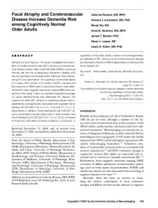
Fig. 2. Representative photomicrographs of Golgi-impregnated CA1 pyramidal neurons from NC (A) and 7 day HY (B) groups of rats. Note a decrease in the dendritic arbor in B compared with A. Scale bar⫽50 m.
A. D. J. Titus et al. / Neuroscience 145 (2007) 265–278
269
Fig. 3. Camera lucida tracings of Golgi-impregnated CA1 pyramidal neurons from NC (A) and 7 day HY (C) groups of rats depicted in Fig. 3. Note a decrease in the dendritic arbor in C compared with A and SC (B). Scale bar⫽50 m.
days of exposure to HBH revealed a significant difference at distal 300-m segment (F2,127⫽4.15; P⬍0.01). SC rats also showed a reduction in the number of branching points in the 300-m segment (P⬍0.01) (Fig. 1A). There was a significant reduction in the total number of branching points of the HY and SC groups compared with NC (F2,127⫽4.10; P⬍0.01). However, no significant difference between SC and HY groups was observed (Table 1). Following 7 days of exposure to hypoxia, the apical dendritic branching points showed a significant (P⬍0.001) reduction in all the segments when compared with NC and SC groups (Figs. 2, 3 and 4A). The SC group showed a reduction in branching points only at 300-m segment compared with NC group. The total number of branching points in HY group showed 43% and 38% reduction compared with the NC and SC (F2,125⫽89.07; P⬍0.001), respectively. The SC group showed a 8% reduction in the total branching points in comparison to NC (P⬍0.01) (Table 1). Dendritic length. The segmental-wise dendritic length was analyzed by using the NIH image software and the total dendritic length was calculated by adding all the values from segments 0-300-m. In 2 days, the segmental-wise or total length of the apical dendrites of HY group was not significantly different from SC and NC groups (Fig. 1B, Table 1). However, following 7 days of exposure, the dendritic length was decreased in all segments except the first segment (0 –50 m) of the HY group compared with NC and SC groups (Fig. 4B). The HY group showed a 31% and 30% reduction in the total dendritic length compared with NC and SC (F2,126⫽35.83; P⬍0.001), respectively, but no significant difference between NC and SC groups was observed (Table 1).
Dendritic intersections. The points at which dendrites cross the concentric circles of the Sholl’s grid are defined as the dendritic intersections (Shankaranarayana Rao et al., 2001b). Following 2 days of hypoxia exposure, the number of intersections in apical dendrites did not show any significant difference between the groups (Fig. 1C). Whereas, 7 day exposure to hypoxia significantly reduced the number of intersections in all segments of apical dendrites in comparison to NC group and segments 50 (P⬍0.001), 100 (P⬍0.05) and 150-m (P⬍0.01) compared with SC. The SC rats showed a significant reduction in dendritic intersections in 150 (P⬍0.001), 200 (P⬍0.05) and 250-m (P⬍0.01) segments compared with NC group (Fig. 4C). Structural changes in the CA3 pyramidal neurons following 2 and 7 days of HBH Dendritic branching points. Unlike the CA1 neurons, the number of branching points of CA3 neurons in the 2 day exposure group was decreased significantly. The HY group showed a significant reduction in the dendritic branching points in segments 100 (P⬍0.05), 200 (P⬍0.001), 250 (P⬍0.001) and 300-m (P⬍0.001) in comparison to NC, and segment 250 (P⬍0.05) and 300-m (P⬍0.001) compared with SC. The SC group of rats also showed a significant (P⬍0.001) reduction of branching points in 100, 200, 250 and 300-m segments compared with NC group (Fig. 5A). The total number of branching points in HY group was reduced by 29% and 13% when compared with NC and SC groups (F2,134⫽17.81, P⬍0.001), respectively. The total number of branching points was reduced by 18% in SC group compared with
270
A. D. J. Titus et al. / Neuroscience 145 (2007) 265–278
Fig. 4. HBH for 7 days produces dendritic atrophy of CA1 pyramidal neurons. The number of apical dendritic branching points (A), dendritic length (B), and intersections (C) for each successive 50 m segments from the soma of NC (n⫽40 neurons), SC (n⫽46 neurons) and HY (n⫽43 neurons) groups of rats are shown. Each value represents the mean⫾S.E.M.; * P⬍0.05, ** P⬍0.01, *** P⬍0.001 NC vs. HY or NC vs. SC; $ P⬍0.05, $$ P⬍0.01, $$$ P⬍0.001 SC vs. HY, one-way ANOVA followed by LSD post hoc test.
Fig. 5. Exposure to 2 days of HBH produces dendritic atrophy of CA3 hippocampal pyramidal neurons. The number of apical dendritic branching points (A), dendritic length (B), and intersections (C) for each successive 50 m segment from the soma of NC (n⫽40 neurons), SC (n⫽49 neurons) and HY (n⫽48 neurons) groups of rats are shown. Each value represents the mean⫾S.E.M.; * P⬍0.05, ** P⬍0.01, *** P⬍0.001 NC vs. HY or NC vs. SC; $ P⬍0.05, $$$ P⬍0.001 SC vs. HY, one-way ANOVA followed by LSD post hoc test.
NC group (P⬍0.001; Table 2). Exposure for 7 days to hypoxia decreased the dendritic branching points in all the
segments (except the initial segment 50-m) of the hypoxic group compared with NC and SC groups (Figs. 6, 7
A. D. J. Titus et al. / Neuroscience 145 (2007) 265–278 Table 2. Effect of exposure to 2 and 7 days of HBH on total number of dendritic branching points and dendritic length of hippocampal CA3 pyramidal neurons
Branching points NC SC HY ANOVA Dendritic length NC SC HY ANOVA
2 Days
7 Days
22.33⫾0.35 18.27⫾1.55*** 15.92⫾1.38***$ F2,134⫽17.81; P⬍0.001
22.33⫾0.35 18.35⫾0.38*** 14.07⫾0.49***$$$ F2,121⫽97.83; P⬍0.001
1912.98⫾42.60 1714.07⫾76.34** 1567.48⫾100.76***$ F2,134⫽10.65; P⬍0.001
1912.98⫾42.60 1602.81⫾51.63*** 1294.26⫾32.14***$$$ F2,121⫽53.58; P⬍0.001
Values are mean⫾S.E.M. Analysis of data was carried out by oneway ANOVA followed by LSD post hoc test. ** P⬍0.01, NC vs. HY or NC vs. SC. *** P⬍0.001, NC vs. HY or NC vs. SC. $ P⬍0.05, SC vs. HY. $$$ P⬍0.001, SC vs. HY.
and 8A). The SC group also showed a reduction in branching points in 150 (P⬍0.05), 200 (P⬍0.001), 250 (P⬍0.001) and 300-m (P⬍0.001) segments compared with NC. The total number of apical dendritic branching points showed a significant decrease in HY compared with NC and SC groups by 37% and 23% (F2,121⫽97.83; P⬍0.001), respectively. The SC group showed 18% reduction in the branching points compared with NC (P⬍0.001) (Table 2).
271
(P⬍0.001), 150 (P⬍0.05), 200 (P⬍0.001) 250 (P⬍0.001) and 300-m (P⬍0.001) when compared with NC group (Fig. 5C). The number of intersections following 7 days of exposure to hypoxia showed a significant (P⬍0.001) decrease in all the segments (except in the 50 m segment) in comparison to NC group. The SC group also showed a significant reduction of intersections in 200 (P⬍0.01), 250 (P⬍0.001) and 300-m (P⬍0.01) segments when compared with NC. The intersections were decreased (P⬍0.001) in HY group in 100, 150, 200, 250 and 300-m segments when compared with SC (Fig. 8C). Effect of 2 days HBH on acquisition of the partially baited RAM task Two way-ANOVA with repeated measures analysis revealed a significant main effect of the groups (F2,182⫽7.82; P⬍0.01). The rats exposed to 2 days HBH showed a significant learning impairment in the 6th and 7th blocks onwards compared with NC (P⬍0.01) and SC (P⬍0.05) rats, respectively. The NC and SC groups were able to achieve 90.76⫾2.0 and 85.42⫾2.57% correct choice, respectively by 16 days (8th block) of training. Whereas, HY group failed to attain the criterion of learning and this was evident in the block 8 (P⬍0.001). Even after 16 days of training, the percentage of correct choice was only 69.03⫾1.28 (Fig. 9A). The number of RMEs was more in the HY group compared with NC and SC groups (F2,182⫽9.15; P⬍0.001). Post hoc comparisons indicated that both NC (P⬍0.001)
Dendritic length. Exposure to hypoxia for 2 days significantly decreased the apical dendritic length in segments 50 (P⬍0.001), 200 (P⬍0.001), 250 (P⬍0.001) and 300-m (P⬍0.01) compared with NC. SC group also showed a significant reduction of dendritic length in 50 (P⬍0.01), 150 (P⬍0.01) and 200-m (P⬍0.05) segments compared with NC (Fig. 5B). Analysis of total dendritic length also showed a significant decrease in HY (P⬍0.001) and SC (P⬍0.01) groups when compared with NC group (F2,134⫽10.65; P⬍0.001). However, the difference between SC and NC groups was negligible (Table 2). Following 7 days of hypoxic exposure, the apical dendritic length was significantly decreased in 150 (P⬍0.05), 200 (P⬍0.001), 250 (P⬍0.001) and 300-m (P⬍0.001) segments in comparison with both normal and SC. The SC group also showed a significant reduction in dendritic length in the 200, 250 and 300-m segments (P⬍0.001) compared with NC (Fig. 8B). The total apical dendritic length was also significantly reduced in HY and SC groups by 32% and 16% compared with NC (F2,117⫽53.58, P⬍0.001), respectively. There was 19% reduction in the total apical dendritic length in HY group compared with SC group (P⬍0.001; Table 2). Dendritic intersections. Two days of hypoxic exposure significantly reduced the number of intersections in all segments of the HY group compared with NC and in segments 200 (P⬍0.001), 250 (P⬍0.001) and 300-m (P⬍0.001) in comparison to SC group. The SC group also showed a reduction at segments 50 (P⬍0.05), 100
Fig. 6. Representative photomicrographs of Golgi-impregnated CA3 pyramidal neurons from NC (A) and 7 day HY (B) groups of rats. Note a decrease in the dendritic arbor in B compared with A. Scale bar⫽50 m.
272
A. D. J. Titus et al. / Neuroscience 145 (2007) 265–278
Fig. 7. Camera lucida tracings of Golgi-impregnated CA3 pyramidal neurons from NC (A) and 7 day HY (C) groups of rats depicted in Fig. 6. Note a decrease in the dendritic arbor in C compared with A and SC (B). Scale bar⫽50 m.
and SC (P⬍0.001) groups exhibited significantly less RMEs in blocks 7 and 8 compared with the HY group. Performance was not significantly different between the NC and SC groups (Fig. 9B). The WMEs correct were comparable between NC and SC groups. Although there was an increase in the WMEs correct in some blocks in the HY group, it was not statistically significant. Similarly, the number of WMEs incorrect was not different between the groups and was zero in all the groups in block 8 (data not shown).
(P⬍0.05) (Fig. 9D). The number of WMEs correct in HY group showed significant difference only in the first three blocks compared with NC group (F2,196⫽11.56; P⬍0.001). The number of WMEs correct in block 8 was nil in NC and SC groups and 0.22⫾0.08 in the HY group. The number of WMEs incorrect was not statistically significant across the groups and was zero in all groups in block 8 (data not shown).
Effect of 7 days of HBH on acquisition of the partially baited RAM task
The 2 days’ exposure of HBH significantly affected the retrieval (F2,26⫽22.89; P⬍0.001). The percentage correct choice showed a significant impairment in the HY (55.32⫾3.6) group compared with NC (80.0⫾1.62) and SC (70.72⫾2.73) groups (Fig. 10A). The number of RMEs was less in the NC group compared with HY group. There was no significant difference between NC and SC or SC and HY groups (Fig. 10B). The WMEs correct and incorrect were comparable between groups. In addition to learning impairment, the 7 days HBHexposed group showed significant memory impairment in the retention. There was a significant difference between the groups (F2,29⫽17.40; P⬍0.001). The percentage correct choice (P⬍0.001), RMEs (P⬍0.001), WMEs correct (P⬍0.01) and incorrect (P⬍0.001) of HY group were significantly different from NC and SC groups. Although the SC group showed a reduction in the percentage of correct choice (P⬍0.01); RMEs and WMEs correct and incorrect were comparable with the NC (P⬎0.05) (Fig. 10C and 10D).
The short-term exposure of hypoxia (7 days) significantly impaired learning performance. Two-way ANOVA with repeated measures revealed a significant interaction (F14,203⫽3.22; P⬍0.001) and main (group) effects (F2, 203⫽33.42; P⬍0.001). The NC group attained the learning criterion (80% correct choice) by the 6th block. At the 32nd trial, the NC and SC groups were able to achieve 90.76⫾1.2 and 82.56⫾1.12% correct choice, respectively, whereas, the HY group failed to attain the criterion and started to plateau at 64.15⫾2.99% after 24 trials. Although the SC group showed a slight decline in the percentage of correct choices compared with NC group, it was not statistically significant (P⬎0.05) (Fig. 9C). In the NC and SC groups of rats, the number of RMEs declined steadily right from the first day of acquisition. While the HY group also showed a decline, it started to plateau after the 7th block. In the 8th block, the HY group committed more number of RMEs than NC and SC groups (F2,29⫽36.77; P⬍0.001). Although the SC group showed a decline in the RMEs, it was not comparable with NC group of rats. In block 8, the number of RMEs in SC (0.92⫾0.06) group was significantly higher than NC (0.29⫾0.1) group
The effect of HBH on retention
DISCUSSION The present study demonstrates that exposure to HBH alters the dendritic arborization of hippocampal neurons
A. D. J. Titus et al. / Neuroscience 145 (2007) 265–278
Fig. 8. HBH for 7 days produces dendritic atrophy of CA3 pyramidal neurons. The number of apical dendritic branching points (A), dendritic length (B), and intersections (C) for each successive 50 m segment from the soma of NC (n⫽40 neurons), SC (n⫽39 neurons) and HY (n⫽41 neurons) groups of rats are shown. Each value represents the mean⫾S.E.M.; * P⬍0.05, ** P⬍0.01, *** P⬍0.001 NC vs. HY or NC vs. SC; $ P⬍0.05, $$ P⬍0.01, $$$ P⬍0.001 SC vs. HY, one-way ANOVA followed by LSD post hoc test.
273
and impairs the spatial learning and memory. Exposure to 2 days of HBH had a marginal effect on the dendritic morphology of the CA1 neurons. On the other hand, exposure to 7 days of HBH resulted in a significant reduction in branching points, intersections and dendritic length in most of the segments in the HY group compared with both the NC and SC groups. It may be noted that SC animals also showed a moderate reduction in hippocampal dendritic arbors in comparison to the NC animals. This might be due to the stress associated with relative restriction of movement in the hypobaric chamber. However, the significant differences between HY and SC groups suggest that dendritic atrophy observed in HY is not because of restricted movement alone but is attributable to the effects of HBH as well. In contrast to the CA1 neurons, CA3 neurons appear to be affected more severely even by the subacute 2-day exposure to HBH. Dendritic length, intersections and branching points were all reduced in the HY group compared with both NC and SC groups. Exposure to 7 days of HBH was associated with more extensive dendritic atrophy in almost all segments with the exception of proximal segments. The presence of significant differences between HY and SC groups, particularly in the distal segments, suggests a specific effect of HBH even after accounting for the moderate dendritic dearborization in the SC animals. Interestingly, the behavioral results directly correlate with the HBH induced hippocampal dendritic atrophy. Two days of exposure to HBH impaired learning and memory and this was more pronounced in the 7-day exposure group. There was a significant correlation between acquisition of the RAM task and dendritic morphology of CA1 (r⫽0.69) and CA3 (r⫽0.89) pyramidal neurons. Therefore, our study suggests that the HBH induced dendritic atrophy might underlie cognitive impairments seen at high altitudes. Exposure of 18-day-old rats to HBH has been reported to reduce the length and spine density of apical dendritic branches of pyramidal neurons in the hippocampal CA1 area (Pokorny and Trojan, 1986). The present study demonstrates that long-term exposure to HBH significantly alters the morphology of CA1 and CA3 pyramidal neurons of the hippocampus even in adult rats. Previous studies have indicated an increased firing of CA1 neurons after ischemic hypoxia (Suzuki et al., 1983). The consequent elevated extracellular glutamate levels may mediate the deleterious effects of this form of hypoxia on the CA1 region (Benveniste et al., 1984). Increase in intracellular Ca2⫹ levels plays a role in the excitotoxicity of glutamate (Choi, 1985). The relatively high expression of NMDA receptors in the CA1 region may also make this region susceptible to hypoxic injury (Monaghan and Cotman, 1985). NMDA-mediated action of excessive extracellular glutamate has been suggested to play an important role in bringing about hippocampal dendritic atrophy in models of chronic stress (Magarinos and McEwen, 1995; Sunanda et al., 1997). Similar mechanisms may be involved in the neuronal morphological changes observed in this model of HBH. The greater degree of dearborization in the 7 day group com-
274
A. D. J. Titus et al. / Neuroscience 145 (2007) 265–278
Fig. 9. The effect of 2 and 7 days of HBH on percentage of correct choice (A and C) and the number of RMEs (B and D) in the partially baited RAM task. The top panel (A and B) shows the effect of exposure to 2 days of hypoxia; NC (n⫽12), SC (n⫽8) and HY (n⫽9) group of rats. The bottom panel (C and D) shows the effect of 7 days of hypoxia on the acquisition of the RAM task; NC (n⫽12), SC (n⫽10) and HY (n⫽10) groups. Data are represented as mean⫾S.E.M.; * P⬍0.05, ** P⬍0.01, *** P⬍0.001 NC vs. HY or NC vs. SC; $ P⬍0.05, $$ P⬍0.01, $$$ P⬍0.001 SC vs. HY, two-way ANOVA followed by Tukey’s post hoc test.
pared with the 2 day group may be due to the delayed excitotoxic effects of glutamate (Choi, 1988). The present study also demonstrates that CA3 and CA1 neurons differ in their susceptibility to hypoxic insult, CA3 neurons being more vulnerable. This is in agreement with the observation of greater degree of neuronal degeneration observed in the CA3 region in rats exposed to HBH in a previous study (Shukitt-Hale et al., 1996). The presence of a Ca2⫹ binding protein, calbindin D28k, distinguishes CA1 from CA3 pyramidal cells (Thal and Schlote, 1994; Shukitt-Hale et al., 1996). This protein has been shown to buffer the levels of intracellular calcium in CA1 neurons during reperfusion following experimental global forebrain ischemia (Freund et al., 1990). However, signif-
icantly elevated levels of calcium (Dienel, 1984) over a period and a persistent block of protein synthesis may eventually lead to delayed neuronal death in the CA1 region after ischemic hypoxia (Mies et al., 1993). In addition, the CA3 cells have a higher level of metabolic activity than the CA1 cells (Thal and Schlote, 1994) and this may make them more vulnerable to hypoxia. Another phenomenon associated with increased intracellular calcium concentration is the accumulation of the lipofuscin, a cytoplasmic pigment of lysosomal origin, selectively in the CA3 region of the hippocampus. Lipofuscin accumulation has been linked to neuronal atrophy and degeneration. Calbindin D28k in CA1 neurons may reduce the free Ca2⫹ content and subsequently limit lipofuscin formation
A. D. J. Titus et al. / Neuroscience 145 (2007) 265–278
275
Fig. 10. The effect of 2 and 7 days of HBH on percentage correct choice (A and C) and the number of RMEs (B and D) in the retention test of the partially baited RAM task. The top panel (A and B) shows the effect of 2 days and the bottom panel shows the effect of 7 days of hypoxia on the retention of the RAM task. Data are represented as mean⫾S.E.M.; * P⬍0.05, *** P⬍0.001 NC vs. HY or NC vs. SC; $ P⬍0.05, $$ P⬍0.01, $$$ P⬍0.001 SC vs. HY, one-way ANOVA followed by Tukey’s post hoc test.
(Brizzee et al., 1974) rendering them less susceptible to acute exposure to hypoxia. However, following long-term exposure, these protective mechanisms may not be efficient enough, thus leading to dendritic atrophy. The few morphological changes reported in animal models of HBH are generally focused on neuronal degeneration and apoptotic death (Langmeier et al., 1989; Shukitt-Hale et al., 1996; Simonova et al., 2003). The possibility that dendritic dearborization resulting from HBH may be linked to the cognitive impairment in such models has received little attention. Chronic stress-induced dendritic atrophy in the hippocampus has been documented over a range of species including mammals and non-human primates (Bennur et al., 2007; Bremner, 1999; Govindarajan et al., 2006; Pawlak et al., 2005; Sapolsky et al., 1986; Shankaranarayana Rao et al., 2001b; Sunanda et al., 1995, 1997) and in turn affects the hippocampal dependent memory (Shankaranarayana Rao et al., 2001b; Sunanda et al., 1995, 2000). In contrast, increased dendritic arbors following intracranial self-stimulation rewarding behavior are found to be
long-lasting and are associated with enhanced cognitive function (Shankaranarayana Rao et al., 1993, 1994, 1998a,b, 1999; Yoganarasimha et al., 1998). Corticosterone, the predominant glucocorticoid in rats secreted during stress, increases the hippocampal neuronal vulnerability to excitotoxins, hypoxic-ischemia, and hypoglycemia in an energy-dependent manner (Elliott et al., 1993; Sunanda et al., 1998). An exposure to high levels of corticosteroids was shown to enhance extracellular glutamate levels (Magarinos and McEwen, 1995), Ca2⫹ currents (Kerr et al., 1992), cytosolic Ca2⫹ concentrations and result in cellular damage (Elliott et al., 1993; Elliott and Sapolsky, 1993). An increase in the concentration of corticosterone has been demonstrated following 2 days of HBH (Bjerknes et al., 1990). It has been recently demonstrated that hypoxia induces oxidative stress and causes degeneration of hippocampal neurons in vitro (Jayalakshmi et al., 2005). Corticosteroids bind to hippocampal glucocorticoid (GR) and mineralocorticoid (MR) receptors, thereby affecting behavior and neurochemical transmis-
276
A. D. J. Titus et al. / Neuroscience 145 (2007) 265–278
sion. HBH could also recruit these pathways to bring about dendritic atrophy in the hippocampus.
CONCLUSION In summary, exposure to HBH for 2 and 7 days alters the dendritic morphology of the hippocampal CA1 and CA3 pyramidal neurons and induces learning and memory deficits. Such altered dendritic arborization induced by hypoxia may result in hippocampal dependent cognitive dysfunctions. Understanding the neuronal changes occurring due to exposure to HBH may provide insight into the pathophysiology and could lead to the identification of newer targets for the pharmacotherapy of high altitude disorders. Acknowledgments—Financial support by Defence Research and Developmental Organisation (DRDO), Government of India and National Institute of Mental Health and Neuro Sciences (NIMHANS) is greatly acknowledged.
REFERENCES Bakharev VD (1981) Investigation of memory during adaptation to high mountain conditions. Hum Physiol 7:409 – 414. Bartholomew CJ, Jensen W, Petros TV, Ferraro FR, Fire KM, Biberdorf D, Fraley E, Schalk J, Blumkin D (1999) The effect of moderate levels of simulated altitude on sustained cognitive performance. Int J Aviat Psychol 9:351–359. Bennur S, Shankaranarayana Rao BS, Pawlak R, Strickland S, McEwen BS, Chattarji S (2007) Stress-induced spine loss in the medial amygdala is mediated by tissue-plasminogen activator. Neuroscience 144:8 –16. Benveniste H, Drejer J, Schousboe A, Diemer NH (1984) Elevation of the extracellular concentrations of glutamate and aspartate in rat hippocampus during transient cerebral ischemia monitored by intracerebral microdialysis. J Neurochem 43:1369 –1374. Bindu B, Rekha J, Kutty BM (2005) Post insult enriched housing improves the 8-arm radial maze performance but not the Morris water maze task in ventral subicular lesioned rats. Brain Res 1063:121–131. Bjerknes R, Neslein IL, Myhre K, Andersen HT (1990) Impairment of rat polymorphonuclear neutrophilic granulocyte phagocytosis following repeated hypobaric hypoxia. Aviat Space Environ Med 61:1007–1011. Bolmont B, Thullier F, Abraini JH (2000) Relationships between mood states and performances in reaction time, psychomotor ability, and mental efficiency during a 31-day gradual decompression in a hypobaric chamber from sea level to 8848 m equivalent altitude. Physiol Behav 71:469 – 476. Bremner JD (1999) Does stress damage the brain? Biol Psychiatry 45:797– 805. Brierley JB (1977) Experimental hypoxic brain damage. J Clin Pathol Suppl (R Coll Pathol) 11:181–187. Brizzee KR, Ordy JM, Kaack B (1974) Early appearance and regional differences in intraneuronal and extraneuronal lipofuscin accumulation with age in the brain of a nonhuman primate (Macaca mulatta). J Gerontol 29:366 –381. Castro-Blanco S, Encinas JM, Serrano J, Alonso D, Gomez MB, Sanchez J, Rios-Tejada F, Fernandez-Vizarra P, Fernandez AP, Martinez-Murillo R, Rodrigo J (2003) Expression of nitrergic system and protein nitration in adult rat brains submitted to acute hypobaric hypoxia. Nitric Oxide 8:182–201. Cavaletti G, Garavaglia P, Arrigoni G, Tredici G (1990) Persistent memory impairment after high altitude climbing. Int J Sports Med 11:176 –178.
Chavez JC, Agani F, Pichiule P, LaManna JC (2000) Expression of hypoxia-inducible factor-1alpha in the brain of rats during chronic hypoxia. J Appl Physiol 89:1937–1942. Chleide E, Bruhwyler J, Mercier M (1991) Enhanced resistance effect of piracetam upon hypoxia-induced impaired retention of fixedinterval responding in rats. Pharmacol Biochem Behav 40:1– 6. Choi DW (1985) Glutamate neurotoxicity in cortical cell culture is calcium dependent. Neurosci Lett 58:293–297. Choi DW (1988) Calcium-mediated neurotoxicity: relationship to specific channel types and role in ischemic damage. Trends Neurosci 11:465– 469. Dienel GA (1984) Regional accumulation of calcium in postischemic rat brain. J Neurochem 43:913–925. Douglas RJ (1967) The hippocampus and behavior. Psychol Bull 67:416 – 422. Du JY, Li XY, Zhuang Y, Wu XY, Wang T (1999) [Effects of acute mild and moderate hypoxia on human short memory]. Space Med Med Eng (Beijing) 12:270 –273. Elliott EM, Mattson MP, Vanderklish P, Lynch G, Chang I, Sapolsky RM (1993) Corticosterone exacerbates kainate-induced alterations in hippocampal tau immunoreactivity and spectrin proteolysis in vivo. J Neurochem 61:57– 67. Elliott EM, Sapolsky RM (1993) Corticosterone impairs hippocampal neuronal calcium regulation-possible mediating mechanisms. Brain Res 602:84 –90. Fitch JM, Juraska JM, Washington LW (1989) The dendritic morphology of pyramidal neurons in the rat hippocampal CA3 area. I. Cell Types Brain Res 479:105–114. Freund TF, Buzsaki G, Leon A, Baimbridge KG, Somogyi P (1990) Relationship of neuronal vulnerability and calcium binding protein immunoreactivity in ischemia. Exp Brain Res 83:55– 66. Govindarajan A, Shankaranarayana Rao BS, Nair D, Trinh M, Mawjee N, Tonegawa S, Chattarji S (2006) Transgenic brain-derived neurotrophic factor expression causes both anxiogenic and antidepressant effects. Proc Natl Acad Sci U S A 103:13208 –13213. Gozal D, Daniel JM, Dohanich GP (2001) Behavioral and anatomical correlates of chronic episodic hypoxia during sleep in the rat. J Neurosci 21:2442–2450. Hornbein TF, Townes BD, Schoene RB, Sutton JR, Houston CS (1989) The cost to the central nervous system of climbing to extremely high altitude. N Engl J Med 321:1714 –1719. Jayalakshmi K, Sairam M, Singh SB, Sharma SK, Ilavazhagan G, Banerjee PK (2005) Neuroprotective effect of N-acetyl cysteine on hypoxia-induced oxidative stress in primary hippocampal culture. Brain Res 1046:97–104. Kerr DS, Campbell LW, Thibault O, Landfield PW (1992) Hippocampal glucocorticoid receptor activation enhances voltage-dependent Ca2⫹ conductances: relevance to brain aging. Proc Natl Acad Sci U S A 89:8527– 8531. Kramer AF, Coyne JT, Strayer DL (1993) Cognitive function at high altitude. Hum Factors 35:329 –344. Langmeier M, Maresova D (1998) [Intermittent hypobaric hypoxia during development-morphologic changes in the neocortex and hippocampus]. Cesk Fysiol 47:62– 66. Langmeier M, Pokorny J, Mares J, Trojan S (1989) Changes of the neuronal structure produced by prolonged hypobaric hypoxia in infant rats. Biomed Biochim Acta 48:S204 –S207. Li XY, Wu XY, Fu C, Shen XF, Wu YH, Wang T (2000a) Effects of acute mild and moderate hypoxia on human mood state. Space Med Med Eng (Beijing) 13:1–5. Li XY, Wu XY, Fu C, Shen XF, Yang CB, Wu YH (2000b) Effects of acute exposure to mild or moderate hypoxia on human psychomotor performance and visual-reaction time. Space Med Med Eng (Beijing) 13:235–239. Magarinos AM, McEwen BS (1995) Stress-induced atrophy of apical dendrites of hippocampal CA3c neurons: involvement of glucocorticoid secretion and excitatory amino acid receptors. Neuroscience 69:89 –98.
A. D. J. Titus et al. / Neuroscience 145 (2007) 265–278 McEwen BS (1999) Stress and hippocampal plasticity. Annu Rev Neurosci 22:105–122. Mies G, Kawai K, Saito N, Nagashima G, Nowak TS Jr, Ruetzler CA, Klatzo I (1993) Cardiac arrest-induced complete cerebral ischaemia in the rat: dynamics of postischaemic in vivo calcium uptake and protein synthesis. Neurol Res 15:253–263. Monaghan DT, Cotman CW (1985) Distribution of N-methyl-D-aspartate-sensitive L-[3H]glutamate-binding sites in rat brain. J Neurosci 5:2909 –2919. Nelson J, Gutmann L (1982) Dementia: an overview. WV Med J 78:219 –225. Nelson TO, Dunlosky J, White DM, Steinberg J, Townes BD, Anderson D (1990) Cognition and metacognition at extreme altitudes on Mount Everest. J Exp Psychol Gen 119:367–374. Nicolas M, Thullier-Lestienne F, Bouquet C, Gardette B, Gortan C, Joulia F, Bonnon M, Richalet JP, Therme P, Abraini JH (1999) An anxiety, personality and altitude symptomatology study during a 31-day period of hypoxia in a hypobaric chamber (experiment ‘Everest-Comex 1997’). J Environ Psychol 19:407– 414. Nicolas M, Thullier-Lestienne F, Bouquet C, Gardette B, Gortan C, Richalet JP, Abraini JH (2000) A study of mood changes and personality during a 31-day period of chronic hypoxia in a hypobaric chamber (Everest-Comex 97). Psychol Rep 86:119 –126. O’Keefe J, Burgess N, Donnett JG, Jeffery KJ, Maguire EA (1998) Place cells, navigational accuracy, and the human hippocampus. Philos Trans R Soc Lond B Biol Sci 353:1333–1340. Olton DS, Branch M, Best PJ (1978a) Spatial correlates of hippocampal unit activity. Exp Neurol 58:387– 409. Olton DS, Walker JA, Gage FH (1978b) Hippocampal connections and spatial discrimination. Brain Res 139:295–308. Olton DS, Werz MA (1978) Hippocampal function and behavior: spatial discrimination and response inhibition. Physiol Behav 20:597– 605. Pawlak R, Shankaranarayana Rao BS, Melchor JP, Chattarji S, McEwen B, Strickland S (2005) Tissue plasminogen activator and plasminogen mediate stress-induced decline of neuronal and cognitive functions in the mouse hippocampus. Proc Natl Acad Sci U S A 102:18201–18206. Pichiule P, Chavez JC, Boero J, Arregui A (1996) Chronic hypoxia induces modification of the N-methyl-D-aspartate receptor in rat brain. Neurosci Lett 218:83– 86. Pokorny J, Trojan S (1986) The development of hippocampal structure and how it is influenced by hypoxia. Acta Univ Carol Med Monogr 113:1–79. Reed LJ, Marsden P, Lasserson D, Sheldon N, Lewis P, Stanhope N, Guinan E, Kopelman MD (1999) FDG-PET analysis and findings in amnesia resulting from hypoxia. Memory 7:599 – 612. Roach RC, Hackett PH (2001) Frontiers of hypoxia research: acute mountain sickness. J Exp Biol 204:3161–3170. Rodway GW, Hoffman LA, Sanders MH (2003) High-altitude-related disorders, part I: Pathophysiology, differential diagnosis, and treatment. Heart Lung 32:353–359. Row BW, Liu R, Xu W, Kheirandish L, Gozal D (2003) Intermittent hypoxia is associated with oxidative stress and spatial learning deficits in the rat. Am J Respir Crit Care Med 167:1548 –1553. Sapolsky RM, Krey LC, McEwen BS (1986) The neuroendocrinology of stress and aging: the glucocorticoid cascade hypothesis. Endocr Rev 7:284 –301. Scoville WB, Milner B (1957) Loss of recent memory after bilateral hippocampal lesions. J Neurol Neurosurg Psychiatry 20:11–21. Shankaranarayana Rao BS, Desiraju T, Meti BL, Raju TR (1994) Plasticity of hippocampal and motor cortical pyramidal neurons induced by self-stimulation experience. Indian J Physiol Pharmacol 38:23–28. Shankaranarayana Rao BS, Desiraju T, Raju TR (1993) Neuronal plasticity induced by self-stimulation rewarding experience in rats: a study on alteration in dendritic branching in pyramidal neurons of hippocampus and motor cortex. Brain Res 627:216 –224.
277
Shankaranarayana Rao BS, Govindaiah, Laxmi TR, Meti BL, Raju TR (2001a) Subicular lesions cause dendritic atrophy in CA1 and CA3 pyramidal neurons of the rat hippocampus. Neuroscience 102: 319 –327. Shankaranarayana Rao BS, Madhavi V, Sunand, Raju TR (2001b) Complete reversal of dendritic atrophy in CA3 neurons of the hippocampus by rehabilitation in restraint stressed rats. Curr Sci 80:653– 659. Shankaranarayana Rao BS, Raju TR (2004a) Neuronal plasticity and brain remodeling. In: Brain and behavior (Raju TR, Kutty BM, Sathyaprabha TN, Shankaranarayana Rao BS, eds), pp 1–13. Bangalore: NIMHANS. Shankaranarayana Rao BS, Raju TR (2004b) The Golgi techniques for staining neurons. In: Brain and behavior (Raju TR, Kutty BM, Sathyaprabha TN, Shankaranarayana Rao BS, eds), pp 108 –111. Bangalore: NIMHANS. Shankaranarayana Rao BS, Raju TR, Meti BL (1998a) Alterations in the density of excrescences in CA3 neurons of hippocampus in rats subjected to self-stimulation experience. Brain Res 804:320 –324. Shankaranarayana Rao BS, Raju TR, Meti BL (1998b) Long-lasting structural changes in CA3 hippocampal and layer V motor cortical pyramidal neurons associated with self-stimulation rewarding experience: a quantitative Golgi study. Brain Res Bull 47:95–101. Shankaranarayana Rao BS, Raju TR, Meti BL (1999) Increased numerical density of synapses in CA3 region of hippocampus and molecular layer of motor cortex after self-stimulation rewarding experience. Neuroscience 91:799 – 803. Shukitt-Hale B, Banderet LE, Lieberman HR (1991) Relationships between symptoms, moods, performance, and acute mountain sickness at 4700 meters. Aviat Space Environ Med 62:865– 869. Shukitt-Hale B, Banderet LE, Lieberman HR (1998) Elevation-dependent symptom, mood, and performance changes produced by exposure to hypobaric hypoxia. Int J Aviat Psychol 8:319 –334. Shukitt-Hale B, Kadar T, Marlowe BE, Stillman MJ, Galli RL, Levy A, Devine JA, Lieberman HR (1996) Morphological alterations in the hippocampus following hypobaric hypoxia. Hum Exp Toxicol 15: 312–319. Shukitt-Hale B, Rauch TM, Foutch R (1990) Altitude symptomatology and mood states during a climb to 3,630 meters. Aviat Space Environ Med 61:225–228. Shukitt-Hale B, Stillman MJ, Levy A, Devine JA, Lieberman HR (1993) Nimodipine prevents the in vivo decrease in hippocampal extracellular acetylcholine produced by hypobaric hypoxia. Brain Res 621:291–295. Shukitt-Hale B, Stillman MJ, Welch DI, Levy A, Devine JA, Lieberman HR (1994) Hypobaric hypoxia impairs spatial memory in an elevation-dependent fashion. Behav Neural Biol 62:244 –252. Simonova Z, Sterbova K, Brozek G, Komarek V, Sykova E (2003) Postnatal hypobaric hypoxia in rats impairs water maze learning and the morphology of neurones and macroglia in cortex and hippocampus. Behav Brain Res 141:195–205. Srikumar BN, Bindu B, Priya V, Ramkumar K, Shankaranarayana Rao BS, Raju TR, Kutty BM (2004) Methods of assessment of learning and memory in rodents. In: Brain and behavior (Raju TR, Kutty BM, Sathyaprabha TN, Shankaranarayana Rao BS, eds), pp 145–151. Bangalore: NIMHANS. Srikumar BN, Raju TR, Shankaranarayana Rao BS (2006) The involvement of cholinergic and noradrenergic systems in behavioral recovery following oxotremorine treatment to chronically stressed rats. Neuroscience 143:679 – 688. Sunanda, Meti BL, Raju TR (1997) Entorhinal cortex lesioning protects hippocampal CA3 neurons from stress-induced damage. Brain Res 770:302–306. Sunanda, Rao MS, Raju TR (1995) Effect of chronic restraint stress on dendritic spines and excrescences of hippocampal CA3 pyramidal neurons: a quantitative study. Brain Res 694:312–317.
278
A. D. J. Titus et al. / Neuroscience 145 (2007) 265–278
Sunanda, Shankaranarayana Rao BS, Raju TR (1998) Corticosterone attenuates zinc-induced neurotoxicity in primary hippocampal cultures. Brain Res 791:295–298. Sunanda, Shankaranarayana Rao BS, Raju TR (2000) Chronic restraint stress impairs acquisition and retention of spatial memory task in rats. Curr Sci 79:1581–1584. Suzuki R, Yamaguchi T, Li CL, Klatzo I (1983) The effects of 5-minute ischemia in Mongolian gerbils: II. Changes of spontaneous neuronal activity in cerebral cortex and CA1 sector of hippocampus. Acta Neuropathol (Berl) 60:217–222. Thal DR, Schlote W (1994) Ultrastructural morphometric analysis of lipofuscin in pyramidal cells of the human Ammon’s horn. Arch Gerontol Geriatr 18:59 –77.
Volpe BT, Davis HP, Colombo PJ (1989) Preoperative training modifies radial maze performance in rats with ischemic hippocampal injury. Stroke 20:1700 –1706. Vyas A, Mitra R, Shankaranarayana Rao BS, Chattarji S (2002) Chronic stress induces contrasting patterns of dendritic remodeling in hippocampal and amygdaloid neurons. J Neurosci 22: 6810 – 6818. West JB (1986) Do climbs to extreme altitude cause brain damage? Lancet 2:387–388. Yoganarasimha D, Shankaranarayana Rao BS, Raju TR, Meti BL (1998) Facilitation of acquisition and performance of operant and spatial learning tasks in self-stimulation experienced rats. Behav Neurosci 112:725–729.
(Accepted 18 November 2006) (Available online 12 January 2007)