Neuron, Vol. 27, 301–312, August, 2000, Copyright 2000 by Cell Press
Fission and Uncoating of Synaptic Clathrin-Coated Vesicles Are Perturbed by Disruption of Interactions with the SH3 Domain of Endophilin Helge Gad,* Niels Ringstad,† Peter Lo¨w,* Ole Kjaerulff,* Jenny Gustafsson,* Markus Wenk,† Gilbert Di Paolo,† Yasuo Nemoto,†k John Crum,‡ Mark H. Ellisman,‡ Pietro De Camilli,† Oleg Shupliakov,* and Lennart Brodin*§ * The Nobel Institute for Neurophysiology Department of Neuroscience Karolinska Institutet S-171 77 Stockholm Sweden † Howard Hughes Medical Institute and Department of Cell Biology Yale University School of Medicine New Haven, Connecticut 06510 ‡ Center for Research on Biological Structure and National Center for Microscopy and Imaging Research University of California, San Diego School of Medicine La Jolla, California 92093
Summary Coordination between sequential steps in synaptic vesicle endocytosis, including clathrin coat formation, fission, and uncoating, appears to involve protein– protein interactions. Here, we show that compounds that disrupt interactions of the SH3 domain of endophilin with dynamin and synaptojanin impair synaptic vesicle endocytosis in a living synapse. Two distinct endocytic intermediates accumulated. Free clathrincoated vesicles were induced by a peptide-blocking endophilin’s SH3 domain and by antibodies to the proline-rich domain (PRD) of synaptojanin. Invaginated clathrin-coated pits were induced by the same peptide and by the SH3 domain of endophilin. We suggest that the SH3 domain of endophilin participates in both fission and uncoating and that it may be a key component of a molecular switch that couples the fission reaction to uncoating. Introduction Clathrin-mediated endocytosis is a major pathway for selective internalization of plasma membrane components (Schmid, 1997). In the nerve terminal, this pathway takes part in the recycling of synaptic vesicles (Heuser, 1989; Cremona and De Camilli, 1997). The clathrin-mediated budding process is thought to begin with recruitment of the clathrin adaptor complex AP2 to the membrane, which serves as a template for assembly of the clathrin lattice. The clathrin coat grows progressively and invaginates to form a bud with a narrow neck. The neck is then cut off to form a free vesicle that rapidly § To whom correspondence should be addressed (e-mail: lennart.
[email protected]). k Present address: Brain Research Institute, The Institute of Physical and Chemical Research, Wako, Saitama 351-0198, Japan.
loses its coat (Schmid, 1997; Kirchhausen, 1999). An array of proteins, including dynamin, amphiphysin, synaptojanin, and endophilin, have been implicated as accessory factors in the endocytic reaction (Schmid et al., 1998; Brodin et al., 2000). The GTPase dynamin is thought to act at the fission step, as nerve terminals in the Drosophila dynamin mutant shibire (Chen et al., 1991; Van Der Bliek and Meyerowitz, 1991) show an accumulation of invaginated endocytic pits with necks surrounded by ring-like structures (Koenig and Ikeda, 1989). Moreover, clathrin-coated pits with a dynamin-coated tubular neck are observed when synaptic membranes are incubated in cytosol with GTP␥S and ATP (Takei et al., 1995). Dynamin alone can polymerize into rings (Hinshaw and Schmid, 1995), and it can evaginate spherical liposomes into narrow tubules surrounded by polymerized dynamin (Sweitzer and Hinshaw, 1998; Takei et al., 1998; Sever et al., 2000). One of dynamin’s binding partners, the SH3 domain–containing protein amphiphysin, can also induce tubulation of liposomes, and it can copolymerize with dynamin on a lipid template (Takei et al., 1999). The polyphosphoinositide phosphatase synaptojanin (McPherson et al., 1996; Guo et al., 1999) has been proposed to function as a negative regulator of interactions between coat proteins and membrane phospholipids (Cremona et al., 1999; see also Beck and Keen, 1991; Hao et al., 1997; Martin, 1998; Arneson et al., 1999; Corvera et al., 1999). In a cell-free clathrin coat recruitment assay, brain cytosol from synaptojanin-deficient mice was found to be more potent than cytosol of wildtype mice in the generation of coated intermediates on liposomes. Moreover, nerve terminals in synaptojanin 1–deficient mice exhibited an increased proportion of clathrin-coated vesicles, suggesting that synaptojanin 1 may facilitate the shedding of the coat (Cremona et al., 1999). Endophilin, an SH3 domain–containing protein that is the preferred binding partner of synaptojanin (Sparks et al., 1996; de Heuvel et al., 1997; Ringstad et al., 1997), is required for the generation of synaptic-like vesicles in permeabilized PC12 cells (Schmidt et al., 1999). Disruption of endophilin function by antibody microinjection in the lamprey reticulospinal synapse inhibits endocytosis at the stage of shallow coated pits, indicating a role for this protein in the invagination of the clathrin-coated membrane (Ringstad et al., 1999). Endophilin was reported to exhibit lysophosphatidic acid acyl transferase (LPAAT) activity and has been proposed to modify the properties of the lipid bilayer through this catalytic activity (Schmidt et al., 1999). Different accessory endocytic proteins thus appear to exert their functions at distinct steps of the clathrinmediated budding process. It can be assumed that these functions are coordinated through a hierarchy of sequential protein–protein interactions, but the role of these interactions is as yet poorly understood. The most detailed studies have focused on the interaction between dynamin and amphiphysin. Amphiphysin binds the clathrin heavy chain, AP2, dynamin, and synaptojanin, respectively, and it has therefore been hypothesized to function as a multifunctional adaptor (David et al., 1996; Ramjaun and McPherson, 1998; Slepnev et al., 1998; Wigge and McMahon, 1998). The SH3 domain
Neuron 302
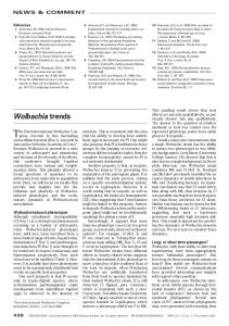
Figure 1. Endophilin SH3 Domain Interactions with Dynamin and Synaptojanin Are Conserved in Lamprey (A) The deduced amino acid sequence of the COOH-terminal of a lamprey synaptojanin ortholog, aligned with rat and human synaptojanin. Boxes indicate regions of sequence identity. The endophilin binding site corresponding to PP-19 (Ringstad et al., 1999; E2⬘ of Cestra et al., 1999) and the amphiphysin (A1 and A2) binding sites (Cestra et al., 1999) have been marked. (B) Protein extracts from lamprey spinal cord and brain analysed by SDS–PAGE and Western blot using synaptojanin antibodies (LSJ-1). (C) Immunoprecipitation of lamprey synaptojanin with LSJ-1 antibodies. The synaptojanin bands probably include breakdown products of the two bands seen in Figure 1C.
Endophilin SH3 Domain Interactions in Endocytosis 303
of amphiphysin binds to a specific site in the prolinerich domain (PRD) of dynamin (Grabs et al., 1997; Owen et al., 1998; Cestra et al., 1999), and this interaction is phylogenetically conserved, as it also occurs in lamprey (Shupliakov et al., 1997). Microinjection studies in the lamprey reticulospinal synapse showed that disruption of this interaction inhibits endocytosis at the stage of invaginated clathrin-coated pits (Shupliakov et al., 1997). These results suggest that the dynamin–amphiphysin interaction is required for fission, possibly in the assembly of a putative fission complex (Shupliakov et al., 1997; Owen et al., 1998). Several other SH3 domains have been found to affect vesicle formation in permeabilized cells, but their precise functions remain unclear (Schmidt et al., 1999; Simpson et al., 1999). Recently, the binding specificity of endophilin’s SH3 domain was characterized. This SH3 domain binds dynamin and synaptojanin at sites that are distinct from those that mediate amphiphysin binding (Micheva et al., 1997; Cestra et al., 1999; Ringstad et al., 1999). Mapping of the PRD of synaptojanin led to the identification of a peptide ligand of endophilin’s SH3 domain (Ringstad et al., 1999; see also Results). This peptide binds endophilin with high preference toward other neuronal SH3 domain– containing proteins (Ringstad et al., 1999). In the present study, we use this peptide, along with a fusion protein containing the SH3 domain of endophilin and antibodies to synaptojanin’s PRD, to probe the function of endophilin’s SH3 domain in vivo.
endophilin binding site located immediately upstream (E2 region of Cestra et al., 1999), on the other hand, was not conserved. In our affinity purification experiments using a series of truncated proteins from the rat synaptojanin PRD that defined the region of PP-19 (see Experimental Procedures), the latter region (i.e., E2⬘), but not the E2 region, was found to be necessary for endophilin binding. Thus, the PRD of synaptojanin appears to contain a single functional endophilin SH3 domain binding site that is conserved between rat and lamprey. Neither one of the two amphiphysin binding regions (Figure 1A, A1 and A2; Cestra et al., 1999) was conserved in lamprey. Polyclonal antibodies (LSJ-1) raised against the PRD of lamprey synaptojanin recognized two bands of ⵑ150 and 170 kDa, respectively (Figure 1B). Both bands were also recognized by antibodies to the lamprey 5-phosphatase domain (data not shown). The two bands may correspond to splice variants of synaptojanin 1 (McPherson et al., 1996; Ramjaun and McPherson, 1996) or to different posttranslationally modified forms of the same protein. To confirm that the catalytic actions of lamprey and mammalian synaptojanins are similar, an immunoprecipitate obtained with the LSJ-1 antibodies (Figure 1C) was incubated with radiolabeled inositol phosphates. The immunoprecipitate dephosphorylated [3H]PI(4,5)P2 to PI(4)P (Figure 1D) and [32P]PI(3,4,5)P3 to PI(3,4)P2 (data not shown), confirming the functional similarity between lamprey and mammalian synaptojanins.
Results
Peptide Inhibition of the Binding of Dynamin and Synaptojanin to Endophilin To test the effect of PP-19 on binding to the SH3 domain of endophilin, affinity purification experiments were performed. Full-length endophilin fused with glutathione S-transferase (GST) bound two major proteins, dynamin and synaptojanin, in lamprey spinal cord extract (Figure 1E, rat brain extract shown for comparison; cf. Ringstad et al., 1997, 1999). The binding of both proteins was blocked by PP-19 (Figure 1, lanes 2, 5, and 8). In contrast, a 15-mer peptide corresponding to the amphiphysin binding site in dynamin (henceforth defined as PP-15) and shown to interact specifically with the SH3 domain of amphiphysin (Grabs et al., 1997; Shupliakov et al., 1997; Cestra et al., 1999) did not affect binding (Figure 1E, lanes 3, 6, and 9). Similar, although not identical, results were obtained when affinity purification was performed with a GST fusion protein containing the isolated SH3 domain of endophilin. A prominent dynamin band was observed, whereas synaptojanin binding was somewhat weaker (Figure 1F, lanes 1 and 5). As for the fulllength protein, the binding was inhibited by PP-19 (Figure 1F, lane 6). Thus, although the preference for binding partners of the isolated SH3 domain may be slightly
Characterization of Binding Partners of Endophilin in the Lamprey The molecular characterization of lamprey endophilin and dynamin was discussed previously (Shupliakov et al., 1997; Ringstad et al., 1999; see also Experimental Procedures). As a premise to study the role of endophilin SH3 domain interactions in the lamprey reticulospinal synapse, we characterized synaptojanin in lamprey. A synaptojanin ortholog was isolated from a Lampetra fluviatilis cDNA library (see Experimental Procedures). The open reading frame encoded a protein with an overall amino acid identity of 58% to the short form (145 kDa) of rat synaptojanin 1 (McPherson et al., 1996). The two inositol phosphatase domains, the inositol-5-phosphatase (McPherson et al., 1996) and Sac1 homology domains (Guo et al., 1999), were well conserved (67% and 65% identity, respectively). The PRD was less conserved (Figure 1A; 38% identity), but the part corresponding to the endophilin binding peptide (henceforth defined as proline-rich 19-mer peptide [PP-19], underlined in Figure 1A; Ringstad et al., 1999) was well conserved (E2⬘ region of Cestra et al., 1999). The putative
(D) PI(4,5)P2 phosphatase activity of lamprey synaptojanin immunoprecipitates. Pellets from immunoprecipitate reactions were incubated with [3H]PI(4,5)P2 for 60 min at 37⬚C. The lipid products were deacylated and separated by high-pressure liquid chromatography. Typical chromatograms of a control incubation (control, rabbit IgGs) and SJ immunoprecipitates (SJ, LSJ-1 antibodies) are shown in the left panels. Quantification of three independent experiments is shown in the right panels. (E) Binding of proteins from extract of rat brain (lane 1) and lamprey spinal cord (lane 4) with a GST fusion protein of rat endophilin 1 (Coomassie blue staining). The identity of the upper band as synaptojanin was verified by Western blotting with LSJ-1 antibodies (␣-SJ), and the identity of the lower band as dynamin was confirmed using dynamin DG-1 antibodies (␣-dynamin, lane 7). Preincubation of the extract with PP-19 (lanes 2, 5, and 8), but not with PP-15 (lanes 3, 6, and 9), inhibited the binding of both synaptojanin and dynamin. (F) Affinity purification of proteins from lamprey spinal cord (lanes 1–3) with GST fusion protein containing the SH3 domain of endophilin (endoSH3, lane 1) or amphiphysin (amphSH3, lane 2), or with GST alone (lane 3) stained with Coomassie blue. Preincubation with PP-19 inhibited the endoSH3 interactions but not the amphSH3 interactions (lanes 5–8).
Neuron 304
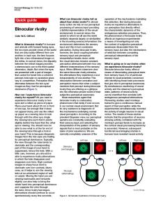
Figure 2. Inhibition of Synaptic Vesicle Recycling after Presynaptic Microinjection of PP-19 at the Lamprey Reticulospinal Synapse (A) Electron micrograph of a synapse in an axon stimulated with action potentials at 5 Hz for 30 min after injection of the PP-19 peptide (“high” peptide concentration). The synaptic vesicle cluster (sv) is depleted, and the plasma membrane has expanded (curved arrows). Note the accumulation of free coated vesicles (arrowheads), coated pits, and cytoskeletal filaments (asterisk). Open arrows indicate the postsynaptic density. (B–D) The boxed area in (A), shown at higher magnification in three consecutive serial sections. The free coated vesicles (arrowheads) lack visible connections with the plasma membrane. (E) A synapse in an axon microinjected with PP-19 and thereafter maintained at rest in low-Ca2⫹ solution.
Endophilin SH3 Domain Interactions in Endocytosis 305
Table 1. Quantification of the Ultrastructural Effects Observed after Injection of Different Compounds
PP-19 PP-15 Anti-SJ EndoSH3 Uninjected GST IgG
Number of Synapses with CCVs
Coated Pits
15 0 12 0 0 0 0
15.9 29.1 1.7 12.8 0.8 1.1 1.5
of of of of of of of
15 6 12 14 18 13 10
⫾ ⫾ ⫾ ⫾ ⫾ ⫾ ⫾
4.3 3.3 1.6 6.4 1.0 0.5 0.8
Synaptic Vesicles 45 156 139 47 270 234 251
⫾ ⫾ ⫾ ⫾ ⫾ ⫾ ⫾
35 30 25 20 58 26 36
Curvature Index 0.46 ⫾ 0.10 0.09 ⫾ 0.17 0.26 ⫾ 0.08 0.06 ⫾ 0.04 0.02 ⫾ 0.01 0.04 ⫾ 0.02
All synapses were stimulated at 5 Hz for 30 min before fixation. All values represent mean ⫾ SD. The number of synaptic vesicles and coated pits was quantified on the center section and normalized to the length of the active zone (n ⫽ 6). The number of free coated vesicles per synapse in PP-19-injected synapses was 19.8 ⫾ 7.7 (low concentration, mean ⫾ SD) and 163.0 ⫾ 56.7 (high concentration), respectively. For synaptojanin antibody- (anti-SJ) injected synapses, the number of free coated vesicles per synapse was 5.3 ⫾ 3.4. The curvature index, a measure of the plasma membrane expansions, was calculated as described by Wickelgren et al. (1985). The reduction in the number of synaptic vesicles was significant for PP-19, anti-SJ, and GSTendoSH3 as compared with uninjected as well as GST- or control antibody(IgG) injected synapses. The increase in the number of coated pits was significant for PP-19- and GSTendoSH3-injected synapses, but not for anti-SJ, as compared with uninjected or GST-injected synapses. There was a significant increase in the curvature index in PP-19- and anti-SJ-injected synapses as compared with uninjected, GST-injected, or IgG-injected synapses. In GSTendoSH3-injected synapses, the plasma membrane expansions were difficult to quantify due to the presence of endosome-like membrane infoldings.
different, it binds dynamin and synaptojanin at the same binding site as full-length endophilin does. As expected, GSTamphSH3 bound lamprey dynamin (Shupliakov et al., 1997), but no binding of synaptojanin could be detected (Figure 1F, lane 7), consistent with the lack of conserved amphiphysin binding sites in the synaptojanin PRD. Binding of dynamin to GSTamphSH3 was not affected by PP-19 (Figure 1F, lanes 7 and 8). The effect of PP-19 was also tested in an LPAAT assay (Schmidt et al., 1999). The LPAAT activity of extract of bacteria expressing endophilin was similar in the absence and presence of PP-19 (data not shown). These results, together with our previous affinity purification studies (Ringstad et al., 1999), indicate that PP19 can be used in lamprey neurons to inhibit specifically synaptojanin and dynamin binding with the SH3 domain of endophilin. Free Coated Vesicles and Invaginated Coated Pits Accumulate after Microinjection of a Peptide Blocking the Endophilin SH3 Domain The peptide PP-19 was microinjected in reticulospinal axons, which were then subjected to stimulation at 5 Hz for 30 min to induce vesicle cycling. Electron microscopy showed that the peptide caused a severe disruption of the synaptic structure, in contrast to only minor changes in stimulated control synapses (see below). The synaptic vesicle pool was greatly reduced (Figure 2A; Table 1). The plasma membrane showed large pocket-like expansions (curved arrows in Figure 2A; curvature index in Table 1), and tubular and flat infoldings of the plasma
membrane were present. A large increase in the number of clathrin-coated endocytic intermediates was also evident. Most strikingly, numerous free clathrin-coated vesicles occurred in the synaptic regions (Figure 2A, arrowheads). Free coated vesicles are normally not seen in this synapse (Shupliakov et al., 1997; Gad et al., 1998; Table 1), presumably due to a rapid uncoating (see Discussion). To verify that these structures were indeed free clathrin-coated vesicles, two methods were used. First, they were traced in serial sections (Figures 2B–2D) to ascertain that they were not associated with other membrane structures, such as endosomes and plasma membrane infoldings. Second, they were examined in 0.3–0.5 m thick sections using intermediate voltage electron microscopy (IVEM; Figures 2H and 2H⬘; see also supplemental movie at http://www.neuron.org.cgi/ content/full/27/2/301/DC1). This method, in addition to confirming the anatomical isolation of the vesicles, allowed a description of nerve terminal clathrin-coated vesicles at a level of resolution much improved over that obtained by conventional electron microscopy (see legend of Figure 2). The number of free coated vesicles at individual synapses was variable. Quantitative analysis of two groups of synapses that had been exposed to “low” and “high” PP-19 concentrations, respectively (as judged from the fluorescence intensity of the injection marker; see Experimental Procedures), indicated that this variability was due to a relation between the number of free coated vesicles and the relative peptide concentration (see legend of Table 1). At the higher peptide concentration, the coated vesicles occurred at
(F) Low-power electron micrograph showing free clathrin-coated vesicles in a reticulospinal axon at a long distance from the nearest synaptic region (open arrow). The section is adjacent to the one shown in (A). Inset shows the boxed area at high magnification. Free coated vesicles (arrowheads) occur in the center of the axon (the center of the axon typically contains large mitochondria [mt]). (G) Accumulation of invaginated clathrin-coated pits in a PP-19 injected synapse (stimulated as in [A]). (H and H⬘) Stereo pair of a free clathrin-coated vesicle visualized by intermediate voltage electron microscopy. The synapse was treated as in (A). Examination of free coated vesicles fully contained within a section at different tilt angles showed that they were completely surrounded by a coat. Partially coated vesicles were not detected in the examined synapses (n ⫽ 3). The free coated vesicles had a slightly ellipsoid shape with largest and smallest diameters of 85 ⫾ 4 and 93 ⫾ 4 nm, respectively (n ⫽ 10, mean ⫾ SD). The vesicle incorporated in the coat had largest and smallest diameters of 45 ⫾ 3 and 53 ⫾ 3 nm, thus giving a coat thickness of about 20 nm. The supplemental movie shows a three-dimensional reconstruction of a coated vesicle obtained from a series of electron micrographs taken at tilt angles ⫾ 60⬚. Scale bars, 0.5 m (A and E); 0.2 m (B–D and G); 2 m (F); 0.05 m H and H⬘).
Neuron 306
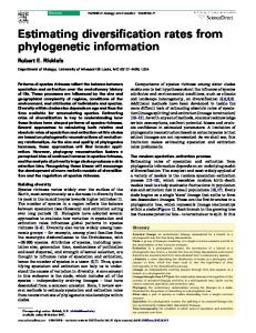
Figure 3. Effects of PP-19 on Synaptic Transmission and Protein Binding to Liposomes (A) Amplitude of EPSPs evoked by low-frequency stimulation during PP-19 loading of the presynaptic axon. PP-19 accumulation at the synaptic site was monitored as a rise in fluorescence emitted by coinjected Texas red–linked dextran (open diamonds). Closed circles, total EPSP amplitude; closed triangles, amplitude of the electrical component (arrow in raw EPSPs above amplitude plot). (B) Recovery of the chemical EPSP from 50 Hz stimulation before (open circles) and after (closed circles) injection of PP-19. The chemical EPSP was estimated by subtracting the electrotonic EPSP from the total EPSP (dashed line in raw EPSPs to the right of amplitude plots). (C) Western blot of proteins bound to liposomes after incubation with rat brain cytosol. Note the strong reduction of synaptojanin binding after preincubation of the cytosol with PP-19. One fortieth (5 g) of the starting material (SM) and one tenth of the bound material, respectively, were loaded. The endophilin antibody also reacted with a larger band in the starting material corresponding to endophilin 2. (D) Quantification of protein binding from autoradiograms as shown in (C). The data are expressed as mean ⫾ SD (n ⫽ 3) and have been normalized to the values obtained in the absence of peptide pretreatment (synaptojanin 1, closed bar; endophilin, open bar; dynamin 1, shaded bar; tubulin, stippled bar).
greater distances from synaptic regions, and they could even be detected in the center of the giant axon (Figure 2F). Thus, the value obtained in these synapses, which include coated vesicles within 2 m from active zones, is an underestimation of the actual number. In addition to the free coated vesicles and plasma membrane expansions, clathrin-coated pits accumulated at the plasmalemma (Figure 2G; Table 1; see also below). Moreover, a prominent filament matrix occurred around the synaptic area and filled much of the membrane expansions around the active zones (Figure 2A, asterisk). Different controls were used: axons injected with an inactive compound (GST) and stimulated at 5 Hz, uninjected axons adjacent to those injected with PP-19 and therefore subjected to otherwise identical conditions, and axons injected with PP-15 at a concentration similar to the “high” concentration of PP-19 and stimulated at 5 Hz. We did not detect a single free coated vesicle in serially sectioned synapses in any of these control axons (Table 1; for other controls, see inactive IgG, below, and dynamin antibodies, Discussion). Different mechanisms can lead to accumulation of free coated vesicles, including “reversed” coating of synaptic vesicles independent of vesicle cycling, enhanced vesicle turnover, and impaired uncoating (Faundez et al., 1998; Cremona et al., 1999). To further explore the effect of PP-19, we first examined its effect on the ultrastructure of synapses maintained at rest. Following microinjection, the specimen was maintained in lowCa2⫹ solution without stimulation. After this treatment, PP-19-injected synapses had a normal appearance, with large synaptic vesicle clusters (Figure 2E). The plasma membrane around active zones remained flat, and neither free clathrin-coated vesicles nor clathrin-coated
pits were observed. These results speak against a reversed coating of synaptic vesicles. If the accumulation of free coated vesicles had been due to enhanced vesicle turnover, PP-19 would be expected to alter the level of transmitter release. To examine this possibility, excitatory postsynaptic potentials (EPSPs) evoked in spinal neurons were monitored before and during injection of PP-19 in the stimulated axon. The reticulospinal EPSP consists of an electrotonic component (mediated by a synaptic gap junction) and a chemical component (Rovainen, 1979). Under conditions of low-frequency stimulation (Figure 3A; 0.2 Hz), neither the total EPSP amplitude (Figure 3A, closed circles) nor the amplitude of the electrotonic component (Figure 3A, triangles) was affected by PP-19 (Figure 3A, diamonds indicate the relative PP-19 concentration at the presynaptic site). The EPSP amplitude at the peak PP-19 concentration relative to the amplitude before injection was 95.8% ⫾ 8.0% (mean ⫾ SD) for the total component and 95.7% ⫾ 0.5% for the electrotonic component (n ⫽ 3, p ⬎ 0.05 for both components). We then tested whether PP-19 affects the EPSP after intense stimulation, as would be expected if the peptide impairs synaptic vesicle recycling. Following a 10 min stimulation train at 50 Hz, the recovery of the EPSP amplitude was monitored during stimulation at 0.2 Hz (chemical component; see Experimental Procedures). Before injection, the EPSP showed a rapid recovery (Figure 3B, open circles), while after injection of PP-19, the recovery was markedly suppressed (Figure 3B, closed circles). In contrast, control injection of the injection marker Texas red–dextran alone had no effect on recovery (data not shown). The reduction of the average amplitude of the recovering chemical component (over a period of 0–5 min) was 37.1% ⫾ 10.6% after injection of PP-19 and
Endophilin SH3 Domain Interactions in Endocytosis 307
Figure 4. Inhibition of Synaptic Vesicle Recycling by Presynaptic Microinjection of Synaptojanin Antibodies (A) Electron micrograph of a synapse in an axon injected with synaptojanin antibodies (LSJ-1) followed by stimulation at 5 Hz for 30 min. Note the reduction in the size of the synaptic vesicle cluster (sv) and the plasma membrane expansion (curved arrows) filled with cytoskeletal filaments (asterisk). A free clathrin-coated vesicle is indicated with an arrowhead. Open arrows indicate the postsynaptic density. (B–D) The boxed area in (A) shown at higher magnification in three consecutive serial sections. Note that the free coated vesicle (arrowhead) lacks connections with the plasma membrane. (E) Synapse in an axon microinjected with inactive control IgG followed by stimulation at 5 Hz for 30 min. Scale bars, 0.5 m (A and E) and 0.2 m (B–D).
1.8% ⫾ 8.6% after injection of the injection marker alone. The difference in reduction between PP-19 and Texas red–dextran was significant (p ⬍ 0.05, n ⫽ 4). The electrotonic component was not affected. Thus, PP-19 delays the recovery of synaptic transmission after high-frequency stimulation, but it has no effect during low-frequency stimulation. This argues against effects that could cause enhanced vesicle turnover. Based on our microscopic analysis, PP-19 inhibits synaptic vesicle recycling at more than one stage. One of its sites of action appears to be uncoating. Possible Role of Synaptojanin in the Accumulation of Free Coated Vesicles As synaptojanin has been implicated in the regulation of coat protein–lipid membrane interactions (see Introduction), it seemed plausible that the uncoating defect could be due to disruption of the synaptojanin–endophilin interaction. This interaction could possibly mediate the recruitment of synaptojanin to the membrane. A blockade of the endophilin SH3 domain would then be expected to inhibit dephosphorylation of phosphoinositides, which could retard the shedding of the coat. The possible involvement of endophilin in the membrane recruitment of synaptojanin was examined with a liposome binding assay (Takei et al., 1998). In this assay, the liposomes are used as a membrane template to study binding of proteins from brain cytosolic extract. Prominent binding of synaptojanin, endophilin, and dynamin, as determined by Western blotting, was observed after incubation with control cytosol (Figure 3C). When the cytosol had been preincubated with PP-19, the binding of endophilin was not affected, but that of synaptojanin was drastically reduced (Figures 3C and
3D). Dynamin binding was only slightly decreased, consistent with previously demonstrated interactions of this protein with membranes independent of other proteins (Sweitzer and Hinshaw, 1998; Takei et al., 1998). Neither a control peptide corresponding to a different part of synaptojanin (Figures 3C and 3D) nor PP-15 (data not shown) had any obvious effects on synaptojanin binding. A direct test of the effect of PP-19 on the membrane recruitment of synaptojanin, e.g., by immunolocalization of the synaptojanin pool, was difficult to perform, as available methods do not give a sufficient spatial resolution in intact synapses. Instead, we sought further evidence for a role of endophilin–synaptojanin interactions by microinjecting the synaptojanin antibodies (LSJ-1). Synapses in antibody-injected axons subjected to stimulation (5 Hz for 30 min) showed a significant reduction in the number of synaptic vesicles (Figure 4A). The plasma membrane showed pocket-like expansions (Table 1) with a proliferation of a filament matrix similar to that seen after PP-19 injection. Free clathrin-coated vesicles were present (Figures 4A–4D), and, although they were comparatively few, they occurred in every synapse examined (n ⫽ 12; Table 1). In axons injected with inactive control IgG and subjected to the same stimulation protocol (Figure 4E), synapses had a structure similar to that found in stimulated GST-injected and uninjected control axons (see above), and no free coated vesicles were detected (Table 1). Synapses in axons that had been maintained at rest (in low-Ca2⫹ solution) following LSJ-1 antibody injection had a structure similar to that of PP19-injected axons maintained at rest, again with no detectable free coated vesicles (data not shown). Taken together, the effects of PP-19 and LSJ-1 indi-
Neuron 308
Figure 5. Accumulation of Deeply Invaginated Coated Pits after Microinjection of GSTendoSH3 (A) A lamprey reticulospinal synapse microinjected with GSTendoSH3 and subjected to stimulation at 5 Hz for 30 min. The synaptic vesicles (sv) were depleted, and the plasma membrane showed pocketlike expansions (curved arrow), as well as large invaginations (i). Numerous coated pits were present, and most of them had electrondense rings/spirals (small arrows). An accumulation of filaments is also evident, although this effect was variable between synapses injected with GSTendoSH3. (B–D) Examples of coated pits with tubular necks surrounded by electron-dense ring-like structures (arrows) at magnification. (D2) shows the same coated pit as in (D1) at a tilt angle of 40⬚. (D) Scale bars, 0.5 m (A); 0.1 m (B and C); 0.05 m (D1 and D2).
cate that the interaction between endophilin and synaptojanin plays a critical role in endocytic uncoating. Synaptojanin may, in addition, have other functions, as suggested by the presence of plasma membrane expansions and a prominent filament matrix. Effects at the Fission Step of Perturbing Endophilin SH3 Domain Interactions As mentioned above, another significant effect of PP19 was an accumulation of clathrin-coated pits at the plasma membrane around active zones (Figure 2G; Table 1). A classification of these coated pits into morphological stages (see Gad et al., 1998) showed that the majority were of the late invaginated type with a narrow neck (Figure 6A). The combined accumulation of coated vesicles and invaginated coated pits suggests that PP19 not only perturbs uncoating but also causes a kinetic delay at the fission step. We examined further the possibility that SH3 domain–mediated interactions of endophilin participate in fission by microinjecting GSTendoSH3 (Figure 1F). This protein as well caused marked changes of the synaptic structure in stimulated synapses (Figure 5A). As in the case of PP-19, the number of synaptic vesicles was greatly reduced (Table 1), and the plasma membrane showed expansions (Figure 5A). Clathrincoated pits, most of which were deeply invaginated, also accumulated at the plasma membrane (Figures 5
and 6A). Two differences from the effects of PP-19 were, however, observed. No free coated vesicles were detected, presumably due to a stronger inhibition of fission relative to the inhibition of uncoating. Furthermore, the majority of the coated pits induced by GSTendoSH3 had an elongated neck that was often surrounded by one or more electron-dense ring-like structures (Figures 5B–5D and 6). The length of the neck could reach up to 130 nm. The coated pits induced by GSTendoSH3 were reminiscent of the coated pits with dynamin-coated tubular necks induced in vitro by GTP␥S (Takei et al., 1995; cf. also Koenig and Ikeda, 1989). The generation of such tubular necks depends on endophilin (see Ringstad et al., 1999, and Discussion). We found that coated pits with tubular necks surrounded by rings can be induced at living synapses by microinjection of GTP␥S followed by stimulation (data not shown). The necks of these coated pits were even longer than those induced by GSTendoSH3, and the rings appeared more regular and thicker (the effects of GTP␥S will be described in further detail separately; J. G. et al., unpublished data). Taken together, the effects of PP-19, GSTendoSH3, and GTP␥S on clathrin-coated pits provide morphological support for a role of endophilin and its SH3 domain also at the fission step. Discussion Clathrin-mediated endocytosis involves a sequence of events that starts with the assembly of the clathrin coat and ends with uncoating (Figure 6B). Endophilin has been shown to have a critical role in the progression from shallow to invaginated coated pits (Ringstad et al., 1999). The present results provide evidence that endophilin also regulates late events in the endocytic sequence through its interactions with dynamin and synaptojanin. The interactions of endophilin with dynamin and synaptojanin are conserved between rat and lamprey, and in both species binding is blocked by a peptide spanning the endophilin binding site in synaptojanin (PP-19) but not by one spanning the amphiphysin binding site in dynamin (PP-15). When immobilized, PP-19 affinity purifies endophilin as the only main protein from lamprey (Ringstad et al., 1999) and rat CNS (N. R. and P. D. C., unpublished data). Although it is difficult to rule out a possible interference with other, less abundant SH3 domains, these binding data suggest that PP-19 primarily affects interactions with the SH3 domain of endophilin. The accumulation of invaginated coated pits after microinjection of either PP-19 or GSTendoSH3 provides morphological evidence for a role of endophilin SH3 domain interactions at the fission step. Such a role appears to be consistent with results obtained in in vitro systems. Schmidt et al. (1999) showed that formation of synaptic-like vesicles in permeabilized PC12 cells requires endophilin with its SH3 domain intact, and it was proposed that the endophilin–dynamin interaction was critical (Schmidt et al., 1999). Ringstad et al. (1999) showed that clathrin-coated pits generated in vitro exhibit dynamin-coated tubules when they are generated in the presence, but not in the absence, of endophilin. Moreover, the dynamin-coated tubules were found to be immunopositive to endophilin. PP-19, in contrast to GSTendoSH3, did not induce tubular necks. The difference in the effects of GSTendoSH3 and PP-19, in spite
Endophilin SH3 Domain Interactions in Endocytosis 309
Figure 6. Summary of the Effects of Compounds Interfering with Endophilin SH3 Domain Interactions (A) Quantitative analysis of clathrin-coated intermediates occurring after PP-19 and GSTendoSH3 injections. Coated intermediates were counted on synapses microinjected with PP-19 (n ⫽ 199) or GSTendoSH3 (n ⫽ 139) and divided into five groups according to their morphology, as indicated in the box. The number of coated intermediates of each stage was expressed as the percentage of the total number of coated intermediates. After injection of GSTendoSH3, the proportion of coated pits of stage 4 with a single electron-dense ring was 79%, while the remaining 21% had elongated necks with more than one ring. (B) The normal sequence of synaptic vesicle endocytosis is interrupted at late stages after disruption of endophilin SH3 domain interactions. A peptide (PP-19) binding endophilin’s SH3 domain causes accumulation of both invaginated coated pits and free coated vesicles. Under specific conditions, a similar effect can also be induced by antibodies to endophilin (see Ringstad et al., 1999). The SH3 domain of endophilin causes accumulation of coated pits with collared necks. Antibodies to synaptojanin cause accumulation of free coated vesicles.
of their shared property to disrupt the SH3 domain– mediated interactions of endophilin, is presumably due to their different properties. PP-19 is a small peptide blocking the endophilin SH3 domain, whereas GSTendoSH3 is a protein of 35 kDa that binds its binding partners. It may block by steric hindrance other interactions of dynamin and thereby prevent the fission reaction. Thus, together with previous results (Shupliakov et al., 1997), the present findings indicate that interactions of both amphiphysin and endophilin with the dynamin PRD participate at the fission step. The effect of PP-19 suggests that the SH3 domain of endophilin is involved but not critically required in the fission reaction. With regard to uncoating, the effect of PP-19 can be explained by inhibition of the interaction between endophilin and synaptojanin, which could impair the recruitment of the latter (see below). The specific contribution of synaptojanin (versus dynamin) to this effect is supported by the accumulation of free coated vesicles induced by synaptojanin antibodies. Moreover, our recent studies have shown that microinjection of dynamin antibodies inhibits synaptic vesicle endocytosis without inducing free clathrin-coated vesicles (H. G. et al., unpublished data). The synaptojanin antibodies produced fewer free coated vesicles than did PP-19, possibly due to the different nature of the two reagents with regard to, for example, diffusion and steric hindrance effects. It is also possible that synaptojanin has multiple functions in the synaptic vesicle cycle and that the antibodies generally impaired these functions, whereas PP-19 interfered more selectively with the function in uncoating. The low number of clathrin-coated intermediates induced by LSJ-1 antibodies (Table 1) suggests that coat formation may have been perturbed in parallel, possibly due to depletion of available pools of coat proteins. Both PP-19 and the LSJ-1 antibodies caused proliferation of a cytoskeletal matrix in the synaptic region. This matrix is likely to consist of actin filaments, as similar filaments can be induced by actin-stabilizing toxins (O. S. et al., unpublished data). Actin polymerization is
known to be stimulated by elevated levels of phosphoinositides (Martin, 1998). A dual SH3-mediated involvement of endophilin in fission and uncoating, in addition to its early role in the endocytic reaction (Ringstad et al., 1999), is consistent with the effects of endophilin antibody injection. The predominant effect of these antibodies is an accumulation of shallow coated pits, but occasional deeply invaginated coated pits and apparently free coated vesicles were also noted (Ringstad et al., 1999). Extended analysis of our previous material using IVEM and serial sectioning has confirmed this observation. In synapses with deep infoldings of the plasma membrane, both invaginated coated pits and a low number of free coated vesicles were observed. Synapses lacking such plasma membrane infoldings only contained shallow coated pits. Thus, the “early” antibody blockade appears to be bypassed when the plasma membrane structure is disrupted by infoldings, and the endophilin antibodies then appear to inhibit fission and uncoating. A Possible Switch between Fission and Uncoating Regulated by the Endophilin SH3 Domain Numerous electron microscopic studies have concluded that free clathrin-coated vesicles are very rare or absent in nerve terminals even after massive stimulation, which suggests that uncoating in the synaptic vesicle cycle is rapid (reviewed by Heuser, 1989). Clathrincoated pits, on the other hand, are commonly seen after stimulation. It would appear that some mechanism prevents uncoating at the coated pit stage but allows its catastrophic occurrence after fission, suggesting a switch closely linked to fission. Such a switch would promote a strict vectoriality of the synaptic vesicle cycle. Our findings suggest that endophilin may be an important component of this switch. A possible scenario could be that the SH3 domain of endophilin primarily interacts with dynamin during the fission reaction and thereafter becomes available to recruit synaptojanin from a soluble pool. Alternatively, synaptojanin may already be associated with the coated
Neuron 310
pit, but its access to the membrane could be prevented. In either case, binding with the endophilin SH3 domain could give synaptojanin’s catalytic domains access to the membrane. The resulting dephosphorylation of membrane phosphoinositides could then weaken the interaction between coat proteins and the membrane (Beck and Keen, 1991; Hao et al., 1997; Cremona et al., 1999; Gaidarov and Keen, 1999) and permit the cytosolic uncoating factors (Chappell et al., 1986; Ungewickell et al., 1995) to initiate uncoating. This hypothetical model may explain the apparent coupling between fission and uncoating, and it can accommodate available data from protein perturbation studies. To examine this model further, however, a better understanding of the fission mechanism is needed (Brodin et al., 2000; Sever et al., 2000). In the context of nerve terminal function, an efficient coupling between fission and uncoating seems to be critical. After endocytosis, the newly retrieved vesicle appears to return quickly to the active zone, most likely as the result of an active transport mechanism (Betz and Angleson, 1998; Murthy and Stevens, 1998; Brodin et al., 2000). After microinjection of PP-19, free coated vesicles were observed at a long distance from the synaptic sites. This observation suggests that vesicles which maintain their coat cannot be handled by the “postendocytic” transport mechanism and may thus be lost from the circulating pool. Experimental Procedures Molecular Cloning of a Lamprey Synaptojanin Ortholog Four stretches of highly conserved amino acids were identified by alignment of human, rat, bovine, and Drosophila sequences of synaptojanin 1 orthologs. Two were located in the Sac1 homology domain, IYAGTGA and RTIQNNF, and two in the 5-phosphatase domain, VKTGMGG and FWCGDFN. Degenerate oligonucleotides were designed from the four stretches, and a random primed lamprey CNS cDNA library was screened by nested PCR. The resulting PCR product was used as a probe to isolate several clones from the same cDNA library. The 3⬘ and 5⬘ ends of the mRNA were cloned with the RACE method (Boehringer Mannheim and Clontech) using oligonucleotides from the known part of the sequence and an oligodT primer. In addition, the Advantage-GC kit (Clontech) was used for the 5⬘ RACE. Sequencing was performed by KIseq, Karolinska Institutet, and by Keck Biotechnology, Yale University. Sequence alignments were performed using Lasergene software and the GCG program package. To obtain antibodies (LSJ-1), a GST fusion protein containing amino acids 907–1226 of the PRD was expressed in a pGEX-6P-2 vector (Amersham Pharmacia) and used to immunize a rabbit. The antigen was coupled to a NHS-activated Hi-Trap column that was used to affinity purify the antibodies. They were further purified with protein A Sepharose (Amersham Pharmacia). Affinity Chromatography of Tissue Extracts Five grams of rat brain or lamprey spinal cord was minced and homogenized in 10 ml of buffer A (20 mM HEPES-KOH [pH 7.2], 100 mM KCl) plus protease inhibitors (Complete protease inhibitor cocktail, Boehringer Mannheim). A postnuclear supernatant of the homogenate was prepared by centrifugation at 2,600 rpm for 10 min at 4⬚C in an SS-34 rotor (Beckman). Triton X-100 was added to the supernatant to a final concentration of 1%, incubated at 4⬚C for 30 min, and then centrifuged for 60 min at 60,000 rpm in a Ti-70 rotor (Beckman), and the resulting high speed supernatant was saved as Triton X-100 extract. Detergent extracts were incubated for 1 hr at 4⬚C with glutathione sepharose (Amersham Pharmacia) loaded with GST fusion proteins. After incubation, the affinity matrix was collected by centrifugation and washed three times with buffer A ⫹ 1% Triton X-100. Bound proteins were eluted with SDS–PAGE sample buffer and analyzed by SDS–PAGE and Western blotting. In some
experiments, detergent extracts were preincubated for 1 hr at 4⬚C with 100 M PP-19 (VAPPARPAPPQRPPPPSGA) or 100 M PP-15 (PPPQVPSRPNRAPPG) prior to affinity chromatography. The sequence corresponding to PP-19 was initially identified (as the endophilin binding site) by affinity purification with a series of GST fusion proteins containing fragments of the rat synaptojanin PRD. Binding of endophilin was observed with the following fragments: 985–1151, 985–1128, 985–1118, 1054–1151, 1069–1151, 1106–1151, and 1113–1151, while no binding was detected with 985–1112, 1130– 1151, 1165–1307, 1264–1307, or 1277–1307. The fusion proteins were made with the pGEX-4T-2 vector. Inositol Phosphatase Activity of Lamprey Synaptojanin Synaptojanin was immunoprecipitated from a Triton X-100 extract of lamprey spinal cord (250 g) by incubation for 2 hr at 4⬚C in the presence of LSJ-1 antibodies or of control rabbit IgGs (10 g) in a 100 l volume of phosphate-buffered saline (PBS). Protein G Sepharose (Amersham Pharmacia) was added (30 l of 50% slurry), and samples were incubated for 1 hr, with rotation at 4⬚C. Following two washes with 0.5 ml of PBS/1% Triton X-100 and a third wash with 0.5 ml of PBS, beads were assessed for inositol phosphatase activity or processed for SDS–PAGE and immunoblotting after elution in sample buffer for 5 min at 95⬚C. Radiolabeled lipid substrate was either purchased ([3H]PtdIns(4,5)P2, NEN Dupont) or prepared by using PI 3-kinase (PI3K) activity immunoprecipitated from rat muscle to produce [32P]PtdIns(3,4,5)P3 . The pellet was incubated with the mixed micellar substrate for 1 hr at 37⬚C. The reaction was stopped and the lipid extracted by addition of 500 l each of ice-cold chloroform:methanol (2:1 v/v) and 1 N HCl. The aqueous phase was reextracted with 500 l of chloroform, and the organic phases were pooled and dried. Lipids were deacylated using methylamine. Samples were redissolved in water, applied to the column (Partisphere SAX 4.6 ⫻ 125 mm, Whatman), and eluted at 1 ml/min using the following gradient generated by mixing water with buffer B: 0–10 min, 0% B; 10–55 min, 0%–35% B; 55–70 min, 35%–100% B; buffer B: 1.4 M (NH4)2HPO4 (pH 3.7). Radioactivity was measured using an online liquid scintillation counter (Packard). Liposome Experiments Liposomes composed of a bovine brain extract (type I Folch fraction I, Sigma) were prepared as described (Takei et al., 1998). Rat brain cytosol (0.5 mg/ml final concentration) was preincubated for 2 hr at 4⬚C with peptides in 0.3 ml of “cytosolic” buffer (25 mM HEPESKOH [pH 7.4], 25 mM KCl, 2.5 mM magnesium acetate, 150 mM K-glutamate) supplemented with 0.2 mM GTP, 2 mM ATP, and an ATP regenerating system (Takei et al., 1998). In addition to PP-19 and PP-15, a control peptide corresponding to amino acids 1099– 1107 of rat synaptojanin was used. Liposomes (0.25 mg/ml final concentration) were added to the mixture (0.4 ml final volume), and samples were incubated for 15 min at 37⬚C. Samples were then loaded on a bed of 0.5 M/2 M sucrose and centrifuged at 150,000 ⫻ g in a TLA100.2 rotor (Beckman) for 30 min at 4⬚C. Liposomes collected at the interphase were washed in 0.5 ml of “cytosolic” buffer, resuspended in 0.2 ml of sample buffer, and processed for SDS– PAGE and Western blotting. Quantification of autoradiograms were performed by optic densitometry (Bio-Rad). Microinjection in the Lamprey Reticulospinal Synapse Lamprey spinal cords were prepared as described (Brodin et al., 1994; Shupliakov et al., 1995) and placed in a recording chamber with Ringer solution maintained at 9⬚C. The endophilin binding peptide was mixed to a concentration of 30 mM in 250 mM K acetate and 10 mM HEPES [pH 7.4] with 5 M Texas red–coupled dextran (MW, 3000; Amersham Pharmacia) added as injection marker. The synaptojanin antibodies and GSTendoSH3 were mixed 10:1 with Cy5-labeled inactive antibodies (rabbit anti-mouse IgG; Pieribone et al., 1995). GTP␥S was mixed to a concentration of 10 mM in 250 mM K acetate with 100 mM HEPES [pH 7.4] and 5 M Texas red–coupled dextran. The microinjections, including the preparation of control solutions (GST, PP-15), were performed as described (Shupliakov et al., 1997; Ringstad et al., 1999). The effects of injected compounds in resting axons were examined in unstimulated specimens maintained in a low-Ca2⫹ Ringer solution (0.1 mM Ca2⫹ and
Endophilin SH3 Domain Interactions in Endocytosis 311
4 mM Mg2⫹) for at least 40 min after the microinjection. The effects induced by stimulation were examined in specimens stimulated for 30 min (Shupliakov et al., 1997). The relative concentration of the endophilin binding peptide was estimated from the fluorescence intensity in images of axons taken immediately before the fixation. The division into “high” and “low” peptide concentration was based on an ⵑ2-fold difference in the fluorescence intensity. The specimens were fixed and prepared for electron microscopy as described (Shupliakov et al., 1997; Ringstad et al., 1999). The experiments with simultaneous recording from spinal neurons were performed as described (Brodin et al., 1994), and the fluorescence intensity in the presynaptic axon was monitored at the area of synaptic contact (Pieribone et al., 1995). To estimate the amplitude of the chemical component during stimulation at 0.2 Hz, the electrotonic EPSP (Rovainen, 1979; Brodin et al., 1994) was subtracted from the total component (Figure 3C, inset). The electrotonic EPSP (averages of 50–100) was isolated during stimulation at 50 Hz, which completely exhausts chemical transmission. Intraaxonal high-frequency stimulation was performed by reimpaling the injected axon with KCl-filled electrodes (resistance ⬎ 25 M⍀).
Electron Microscopy Serial ultrathin sections (thickness of 80–90 nm) from the area of the injection were counterstained with uranyl acetate and lead citrate, and examined and photographed in a Philips CM12 electron microscope. The number of examined synapses was as follows: PP-19 at 5 Hz stimulation: 12 (⫹3 synapses in thicker sections examined with the aid of IVEM) synapses in 3 axons, 9 of which were from an axon injected with a “high” concentration of peptide (see above); PP-19 without stimulation: 6 synapses in 3 axons (including both “high” and “low” concentrations); PP-15 at 5 Hz: 6 synapses in 3 axons (all corresponding to “high” concentration); LSJ-1 antibody at 5 Hz: 12 synapses in 3 axons; LSJ-1 antibody without stimulation: 6 synapses in 3 axons; Cy5-GST at 5 Hz: 13 synapses in 3 axons; GSTendoSH3 at 5 Hz: 14 synapses in 3 axons; and inactive antibodies (rabbit IgG) at 5 Hz: 10 synapses in 3 axons. Finally, 18 synapses in stimulated uninjected axons adjacent to those injected with PP-19 and LSJ-1, respectively, were examined. The quantitative analysis (Table 1) was performed on 6 representative synapses. The number of synaptic vesicles and the number of coated pits was counted on the center section of each synapse and normalized to the length of the active zone (Shupliakov et al., 1995). Free coated vesicles were counted in serial sections covering the entire active zone area, either on micrographs photographed at 17,000⫻ (PP-19 and LSJ-1 antibody at 5 Hz stimulation) or directly in the electron microscope (all other experiments). For each free coated vesicle, the lack of connection to other membrane structures was checked on adjacent ultrathin sections. To visualize intact coated vesicles, semithin sections (0.3 and 0.5 m) were examined using IVEM. The semithin sections were cut from the synapses of 2 injected axons, collected onto formvar-coated slot grids and stained for 20 min in 1% aqueous uranyl acetate, followed by 6 min in lead salts. Thereafter, both surfaces of the grid were coated with carbon. The sections were analyzed in a JEOL 4000EX electron microscope. Synapses were examined at magnifications of 10,000– 15,000⫻, and single coated vesicles at 100,000–200,000⫻ at an accelerating voltage of 400 kV at different tilt angles. Single structures were photographed as stereo pairs acquired at ⫾4⬚ tilt increments.
Miscellaneous Procedures Western blotting was performed as described (Shupliakov et al., 1997; Ringstad et al., 1999) using ECL (Amersham Pharmacia) or 125 I-labeled protein A (Dupont NEN). Endophilin was detected with anti-peptide antibodies (affinity chromatography in rat; Ringstad et al., 1999), or pan-endophilin antibodies (liposome experiments; Ringstad et al., 1997). Synaptojanin was detected with CAT-1 (affinity chromatography in rat; Ringstad et al., 1999), OTT (N. R., unpublished data), and LSJ-1 antibodies (affinity chromatography in lamprey) or with anti-rat synaptojanin 1 antibodies (liposome experiments; McPherson et al., 1996). Dynamin was detected with DG-1 antibodies (Grabs et al., 1997). The identity of the 100 kDa band as
dynamin (Shupliakov et al., 1997) has been confirmed by microsequencing of trypsin-cleaved peptide fragments (P. L. and L. B., unpublished data). LPAAT assays were performed according to Schmidt et al. (1999) using 1 nM bacterially expressed GST– endophilin 1 either in the presence or absence of 100 M PP-19. Acknowledgments This work was supported by grants from the Swedish Medical Research Council (11287, L. B.; 13473, O. S.), the European Commission–Biomed II (L. B.), P. and A. Hedlunds Stiftelse (L. B.), Jeanssons Stiftelse and Stiftelsen S. and E. Goljes Minne (O. S.), the National Institutes of Health (P. D. C., M. H. E.), and the United States Army Medical Research and Development Command (P. D. C.). N. R. is supported by a National Institutes of Health predoctoral training grant to the Department of Cellular and Molecular Physiology, Yale University School of Medicine. O. K. is a Marie Curie Research fellow of the European Commission, and G. D. P. is supported by a longterm European Molecular Biology Organization fellowship. We are indebted to Dr. H. T. Kao for expert advice. Received February 24, 2000; revised July 12, 2000. References Arneson, L.S., Kunz, J., Anderson, R.A., and Traub, L.M. (1999). Coupled inositide phosphorylation and phospholipase D activation initiates clathrin-coat assembly on lysosomes. J. Biol. Chem. 274, 17794–17805. Beck, K.A., and Keen, J.H. (1991). Interaction of phosphoinositide cycle intermediates with the plasma membrane–associated clathrin assembly protein AP-2. J. Biol. Chem. 266, 4442–4447. Betz, W.J., and Angleson, J.K. (1998). The synaptic vesicle cycle. Annu. Rev. Physiol. 60, 347–363. Brodin, L., Shupliakov, O., Hellgren, J., Pieribone, V., and Hill, R. (1994). The reticulospinal glutamate synapse in lamprey: plasticity and presynaptic variability. J. Neurophysiol. 72, 592–604. Brodin, L., Lo¨w, P., and Shupliakov, O. (2000). Sequential steps in clathrin-mediated synaptic vesicle endocytosis. Curr. Opin. Neurobiol. 10, 312–320. Cestra, G., Castagnoli, L., Dente, L., Minenkova, O., Petrelli, A., Mignone, N., Hoffmuller, U., Schneider-Mergener, J., and Cesarini, G. (1999). The SH3 domains of endophilin and amphiphysin bind to the proline-rich region of synaptojanin 1 at distinct sites that display an unconventional binding specificity. J. Biol. Chem. 274, 32001– 32007. Chappell, T.G., Welch, W.J., Schlossman, D.M., Palter, K.B., Schlesinger, M.J., and Rothman, J.E. (1986). Uncoating ATPase is a member of the 70 kilodalton family of stress proteins. Cell 45, 3–13. Chen, M.S., Obar, R.A., Schroeder, C.C., Austin, T.W., Poodry, C.A., Wadsworth, S.C., and Vallee, R.B. (1991). Multiple forms of dynamin are encoded by shibire, a Drosophila gene involved in endocytosis. Nature 351, 583–586. Corvera, S., D’Arrigo, S., and Stenmark, H. (1999). Phosphoinositides in membrane traffic. Curr. Opin. Cell Biol. 11, 460–465. Cremona, O., and De Camilli, P. (1997). Synaptic vesicle endocytosis. Curr. Opin. Neurobiol. 7, 323–330. Cremona, O., Di Paolo, G., Wenk, M.K., Luthi, A., Kim, W., Takei, K., Daniell, L., Nemoto, Y., Shears, S.B., Flavell, R.A., et al. (1999). Essential role of phosphoinositide metabolism in synaptic vesicle recycling. Cell 99, 179–188. David, C., McPherson, P.S., Mundigl, O., and De Camilli, P. (1996). A role of amphiphysin in synaptic vesicle endocytosis suggested by its binding to dynamin in nerve terminals. Proc. Natl. Acad. Sci. USA 93, 331–335. de Heuvel, E., Bell, A.W., Ramjaun, A.R., Wong, K., Sossin, W.S., and McPherson, P.S. (1997). Identification of the major synaptojaninbinding proteins in brain. J. Biol. Chem. 272, 8710–8716. Faundez, V., Horng, J.T., and Kelly, R.B. (1998). A function for the
Neuron 312
AP3 coat complex in synaptic vesicle formation from endosomes. Cell 93, 423–432.
Schmid, S.L. (1997). Clathrin-coated vesicle formation and protein sorting: an integrated process. Annu. Rev. Biochem. 66, 511–548.
Gad, H., Lo¨w, P., Zotova, E., Brodin, L., and Shupliakov, O. (1998). Dissociation between Ca2⫹-triggered synaptic vesicle exocytosis and clathrin-mediated endocytosis at a central synapse. Neuron 21, 607–616.
Schmid, S.L., McNiven, M.A., and De Camilli, P. (1998). Dynamin and its partners: a progress report. Curr. Opin. Cell Biol. 10, 504–512.
Gaidarov, I., and Keen, J.H. (1999). Phosphoinositide-AP-2 interactions required for targeting to plasma membrane clathrin-coated pits. J. Cell Biol. 146, 755–764. Grabs, D., Slepnev, V.I., Songyang, Z., David, C., Lynch, M., Cantley, L.C., and De Camilli, P. (1997). The SH3 domain of amphiphysin binds the proline-rich domain of dynamin at a single site that defines a new SH3 binding consensus sequence. J. Biol. Chem. 272, 13419– 13425. Guo, S., Stolz, L.E., Lemrow, S.M., and York, J.D. (1999). SAC1-like domains of yeast SAC1, INP52, and INP53 and of human synaptojanin encode polyphosphoinositide phosphatases. J. Biol. Chem. 274, 12990–12995. Hao, W., Tan, Z., Prasad, K., Reddy, K.K., Chen, J., Prestwich, G.D., Falck, J.R., Shears, S.B., and Lafer, E.M. (1997). Regulation of AP-3 function by inositides. Identification of phosphatidylinositol 3,4,5trisphosphate as a potent ligand. J. Biol. Chem. 272, 6393–6398. Heuser, J.E. (1989). The role of coated vesicles in recycling of synaptic vesicle membrane. Cell Biol. Intern. Reports 13, 1063–1076. Hinshaw, J.E., and Schmid, S.L. (1995). Dynamin self-assembles into rings suggesting a mechanism for coated vesicle budding. Nature 374, 190–192. Kirchhausen, T. (1999). Adaptors for clathrin-mediated traffic. Annu. Rev. Cell Dev. Biol. 15, 705. Koenig, J.H., and Ikeda, K. (1989). Disappearance and reformation of synaptic vesicle membrane upon transmitter release observed under reversible blockage of membrane retrieval. J. Neurosci. 9, 3844–3860. Martin, T.F. (1998). Phosphoinositide lipids as signaling molecules: common themes for signal transduction, cytoskeletal regulation, and membrane trafficking. Annu. Rev. Cell Dev. Biol. 14, 231–264. McPherson, P.S., Garcia, E.P., Slepnev, V.I., David, C., Zhang, X., Grabs, D., Sossin, W.S., Bauerfeind, R., Nemoto, Y., and De Camilli, P. (1996). A presynaptic inositol-5-phosphatase. Nature 379, 353–357. Micheva, K.D., Kay, B.K., and McPherson, P.S. (1997). Synaptojanin forms two separate complexes in the nerve terminal. Interactions with endophilin and amphiphysin. J. Biol. Chem. 272, 27239–27245. Murthy, V.N., and Stevens, C.F. (1998). Synaptic vesicles retain their identity through the endocytic cycle. Nature 392, 497–501. Owen, D.J., Wigge, P., Vallis, Y., Moore, J.D.A., Evans, P.R., and McMahon, H.T. (1998). Crystal structure of the amphiphysin-2 SH3 domain and its role in the prevention of dynamin ring formation. EMBO J. 17, 5273–5285. Pieribone, V., Shupliakov, O., Brodin, L., Czernik, A.J., HilfikerRotenfluh, S., and Greengard, P. (1995). Distinct pools of synaptic vesicles in neurotransmitter release. Nature 375, 493–497. Ramjaun, A.R., and McPherson, P.S. (1996). Tissue-specific alternative splicing generates two synaptojanin isoforms with differential membrane binding properties. J. Biol. Chem. 271, 24856–24861. Ramjaun, A.R., and McPherson, P.S. (1998). Multiple amphiphysin II splice variants display differential clathrin binding: identification of two distinct clathrin-binding sites. J. Neurochem. 70, 2369–2376. Ringstad, N., Nemoto, Y., and De Camilli, P. (1997). The SH3p4/ Sh3p8/SH3p13 protein family: binding partners for synaptojanin and dynamin via a Grb2-like Src homology 3 domain. Proc. Natl. Acad. Sci. USA 94, 8569–8574. Ringstad, N., Gad, H., Takei, K., Lo¨w, P., Brodin, L., Shupliakov, O., and De Camilli, P. (1999). Endophilin/SH3p4 is required for the transition from early to late stages in clathrin-mediated synaptic vesicle endocytosis. Neuron 24, 143–154. Rovainen, C.M. (1979). Neurobiology of lampreys. Physiol. Rev. 59, 1007–1077.
Schmidt, A., Wolde, M., Thiele, C., Fest, W., Kratzin, H., Podtelejnikov, A.V., Witke, W., Huttner, W.B., and So¨ling, H.D. (1999). Endophilin I mediates synaptic vesicle formation by transfer of arachidonate to lysophosphatidic acid. Nature 401, 133–141. Sever, S., Damke, H., and Schmid, S.L. (2000). Garrotes, springs, ratchets, and whips: putting dynamin models to the test. Traffic 1, 385–392. Shupliakov, O., Pieribone, V., Gad, H., and Brodin, L. (1995). Synaptic vesicle depletion in reticulospinal axons is reduced by 5-HT: direct evidence for presynaptic modulation of glutamatergic transmission. Eur. J. Neurosci. 7, 1111–1116. Shupliakov, O., Lo¨w, P., Grabs, D., Gad, H., Chen, H., David, C., Takei, K., De Camilli, P., and Brodin, L. (1997). Synaptic vesicle endocytosis impaired by disruption of dynamin-SH3 domain interactions. Science 276, 259–263. Simpson, F., Hussain, N.K., Qualmann, B., Kelly, R.B., Kay, B.K., McPherson, P., and Schmid, S.L. (1999). SH3-domain-containing proteins function at distinct steps in clathrin-coated vesicle formation. Nat. Cell Biol. 1, 119–124. Slepnev, V.I., Ochoa, G.C., Butler, M.H., Grabs, D., and Camilli, P.D. (1998). Role of phosphorylation in regulation of the assembly of endocytic coat complexes. Science 281, 821–824. Sparks, A.B., Hoffman, N.G., McConnell, S.J., Fowlkes, D.M., and Kay, B.K. (1996). Cloning of ligand targets: systematic isolation of SH3 domain–containing proteins. Nat. Biotechnol. 14, 741–744. Sweitzer, S.M., and Hinshaw, J.E. (1998). Dynamin undergoes a GTP-dependent conformational change causing vesiculation. Cell 93, 1021–1029. Takei, K., McPherson, P.S., Schmid, S.L., and De Camilli, P. (1995). Tubular membrane invaginations coated by dynamin rings are induced by GTP–gamma S in nerve terminals. Nature 374, 186–190. Takei, K., Haucke, V., Slepnev, V., Farsad, K., Salazar, M., Chen, H., and De Camilli, P. (1998). Generation of coated intermediates of clathrin-mediated endocytosis on protein-free liposomes. Cell 94, 131–141. Takei, K., Slepnev, V., Haucke, V., and DeCamilli, P. (1999). Functional partnership between amphiphysin and dynamin in clathrinmediated endocytosis. Nat. Cell Biol. 1, 33–39. Ungewickell, E., Ungewickell, H., Holstein, S.E., Lindner, R., Prasad, K., Barouch, W., Martin, B., Greene, L.E., and Eisenberg, E. (1995). Role of auxilin in uncoating clathrin-coated vesicles. Nature 378, 632–635. van der Bliek, A.M., and Meyerowitz, E.M. (1991). Dynamin-like protein encoded by the Drosophila shibire gene associated with vesicular traffic. Nature 351, 411–414. Wickelgren, W.O., Leonard, J.P., Grimes, M.J., and Clark, R.D. (1985). Ultrastructural correlates of transmitter release in presynaptic areas of lamprey reticulospinal axons. J. Neurosci. 5, 1188–1201. Wigge, P., and McMahon, H.T. (1998). The amphiphysin family of proteins and their role in endocytosis at the synapse. Trends Neurosci. 21, 339–344. GenBank Accession Number The GenBank accession number for the Lampetra fluviatilis synaptojanin sequence reported in this paper is AF205939.