Structure 14, 1263–1272, August 2006 ª2006 Elsevier Ltd All rights reserved
DOI 10.1016/j.str.2006.06.009
Origin of Exopolyphosphatase Processivity: Fusion of an ASKHA Phosphotransferase and a Cyclic Nucleotide Phosphodiesterase Homolog Johnjeff Alvarado,1,2 Anita Ghosh,1 Tyler Janovitz,1 Andrew Jauregui,1,3 Miriam S. Hasson,1,4 and David Avram Sanders1,* 1 Department of Biological Sciences Purdue University West Lafayette, Indiana 47907
Summary The Escherichia coli Ppx protein is an exopolyphosphatase that degrades long-chain polyphosphates in a highly processive reaction. It also hydrolyzes the terminal 50 phosphate of the modified nucleotide guanosine 50 triphosphate 30 diphosphate (pppGpp). The ˚ resolustructure of Ppx has been determined to 1.9 A tion by X-ray crystallography. The exopolyphosphatase is an ASKHA (acetate and sugar kinases, Hsp70, actin) phosphotransferase with an active site found in a cleft between the two amino-terminal domains. Analysis of the active site indicates that among the ASKHA phosphotranferases of known structure, Ppx is the closest to the ectonucleoside triphosphate diphosphohydrolases. A third domain forms a six-helix claw that is similar to the catalytic core of the eukaryotic cyclic nucleotide phosphodiesterases. Most of the 29 sulfate ions bound to the Ppx dimer occupy sites where the polyP chain likely binds. An aqueduct that passes through the enzyme provides a physical basis for the enzyme’s high processivity.
Introduction Inorganic polyphosphate (polyP) is a polymer of three to hundreds of orthophosphate (Pi) residues linked by phosphoanhydride bonds (Kornberg et al., 1999). PolyP has been found in all organisms, from prokaryotes to eukaryotes, examined. Its most prominent known function is its indispensability for bacterial survival during the stationary phase (Rao and Kornberg, 1996). In Escherichia coli, the principal hydrolysis of polyP is affected by an exopolyphosphatase (Ppx) encoded by the ppx gene. ppk, encoding the enzyme polyphosphate kinase, which synthesizes polyP, and ppx together make up the polyP operon (Akiyama et al., 1993). Ppx carries out the highly processive hydrolysis of phosphates from the ends of long-chain polyP molecules (750 6 50 phosphates) (Akiyama et al., 1993). Under optimal conditions, only Pi and pyrophosphate (PPi) are observed as products (Bolesch and Keasling, 2000). Therefore, a polyP chain is bound to Ppx throughout the hydrolysis reaction. Ppx has a strong preference for long-chain poly-
*Correspondence:
[email protected] 2 Present address: Department of Biochemistry, Albert Einstein College of Medicine, Bronx, New York 10461. 3 Present address: Graduate Program in Genetics, University of Wisconsin-Madison, Madison, Wisconsin 53706. 4 Deceased.
phosphates; ATP and short-chain polyphosphates are not hydrolyzed (Akiyama et al., 1993). E. coli Ppx, normally a dimer of 58 kDa subunits (Akiyama et al., 1993), was determined by limited proteolysis to consist of a 34 kDa N-terminal unit and a 24 kDa C-terminal unit (Bolesch and Keasling, 2000). The C-terminal region was shown to be the source of polyP binding and to be necessary for processivity, but it lacked polyP hydrolytic activity (Bolesch and Keasling, 2000). PolyP metabolism in E. coli is intimately connected to the stringent response to amino acid starvation (Rao et al., 1998). When ribosomes stall because of insufficient charged tRNAs, the RelA protein synthesizes guanosine 50 triphosphate 30 diphosphate (pppGpp) from ATP and GTP (Chatterji and Ojha, 2001). Both pppGpp and ppGpp, which is produced from pppGpp by guanosine pentaphosphate phosphohydrolase (Gpp) (Hara and Sy, 1983), regulate transcription through an interaction with the b subunit of RNA polymerase (Chatterji and Ojha, 2001). Gpp is a homolog of Ppx; each enzyme was demonstrated to possess both Gpp activity and exopolyphosphatase activity (Keasling et al., 1993; Kuroda et al., 1997). Furthermore, pppGpp was demonstrated to be an inhibitor of the exopolyphosphatase activity of Ppx with a Ki that was equivalent to its Km in the Gpp reaction (Kuroda et al., 1997). It was concluded that pppGpp was a competitive inhibitor of Ppx (Kuroda et al., 1997). Through inhibition of Ppx and Gpp, pppGpp should stimulate net polyP synthesis (Kuroda et al., 1997). It has been demonstrated that polyphosphates do accumulate in E. coli in response to amino acid starvation through a RelA-dependent mechanism (Rao et al., 1998). This accumulation is involved in protein degradation during amino acid starvation. The Lon protease, which is responsible for most of the protein degradation during amino acid starvation, forms a complex with polyP (Kuroda et al., 2001). Additionally, it was shown that in the presence of polyP, ribosomal proteins were degraded by the Lon protease. These results suggest a model in which cells starved for amino acids accumulate polyP, and that the consequent formation of a Lon:ribosomal protein:polyP complex leads to degradation of ribosomal proteins by Lon to supply amino acids for survival (Kuroda et al., 2001). Ppx, Gpp, and their numerous homologs, which include eubacterial, archaeal, and plant proteins as well as the Saccharomyces cerevisiae RTG2 (a protein involved in the retrograde response—the signaling of mitochondrial dysfunction to the nucleus; Liao and Butow, 1993), are predicted to belong to the ASKHA (acetate and sugar kinases, Hsp70, actin) superfamily (Buss et al., 2001; Holmes et al., 1993; Koonin, 1994; Reizer et al., 1993). This superfamily, which includes glycerol kinase (Hurley et al., 1993), eukaryotic hexokinase, and the glycerol dehydratase reactivase a subunit (Liao et al., 2003), utilizes a common fold for catalyzing hydrolysis of the ATP b2g phosphoanhydride (with transfer of the g-phosphoryl group to a substrate in the case of the kinases) (Hurley, 1996).
Structure 1264
Table 1. Crystallographic Data Hg MAD Data Set
Edge
Peak
Remote
Native1
Native2
Beamline Wavelength (A˚) Space group Unit cell (A˚) a b c Resolution (A˚) Reflections Total Unique Completeness (%) Average I/s Multiplicity Rmergeb (%)
14-BMD (BioCARS) 1.009
14-BMD (BioCARS) 1.008 P41212
14-BMD (BioCARS) 0.984
14-IDB (BioCARS) 1.008 P41212
14-BMC (BioCARS) 1.000 P41212
88.93 88.93 350.87 99.00–2.47 (2.56–2.47)
89.14 89.14 350.69 30.00–1.90 (1.97–1.90)
1,000,997 51,251 98.5 (92.8) 63.6 (23.8) 19.5 4.1 (8.5)
2,116,279 104,035 92.9 (73.3) 18.4 (4.9) 20.3 8.3 (24.2)
a b
88.51 88.51 351.10 35.00–3.25 (3.37–3.25)a 533,849 22,603 97.2 (80.4) 21.0 (6.1) 23.6 9.6 (22.9)
533,987 22,643 97.3 (80.9) 24.2 (8.4) 23.6 8.6 (18.1)
561,127 22,921 98.5 (90.2) 21.7 (6.7) 24.5 9.4 (21.0)
Numbers in parentheses correspond to data for the outermost resolution shell. Rmerge = SjI 2
j/SI.
Determination of the structure of E. coli Ppx demonstrates that the protein does belong to the ASKHA superfamily and also contains a domain that is similar to the catalytic core of the eukaryotic cyclic nucleotide phosphodiesterases. The numerous sulfate-ion binding sites and aqueducts that traverse the Ppx dimer suggest a model for polyphosphate binding and enzyme processivity. Results and Discussion Overall Architecture of E. coli Ppx In order to investigate the structural basis of the specificity of E. coli Ppx for long-chain polyphosphates and pppGpp and its processivity, we determined the enzyme’s three-dimensional structure by X-ray crystallography to 1.9 A˚ resolution (Tables 1 and 2). Ppx exhibits an elongated dimeric structure, with each monomer consisting of four domains, as predicted by sequence and biochemical analyses (Akiyama et al., 1993; Bolesch and Keasling, 2000; Reizer et al., 1993) (Figure 1A). The first two domains each possess the bbbababa fold characteristic of the ASKHA superfamily of phosphotransferases (Buss et al., 2001; Hurley, 1996) (Figure 1B). A similar conclusion has been made concerning the published structure of the Aquifex aeolicus homolog of Ppx (Kristensen et al., 2004), which lacks the third and fourth domains present in the E. coli Ppx. Superposition of domain I of Ppx with domain IA of the ASKHA members and superposition of Ppx domain II with their IIA domains supports the identification of Ppx as an ASKHA superfamily member (Figure S1; see the Supplemental Data available with this article online). As is true in the other ASKHA members, the interactions of helix a3 are mainly with secondary structure elements in domain II, whereas helix a30 is inserted into domain I. At characteristic positions in the sequence, the ASKHA phosphotransferases possess insertions in the core fold that play roles in oligomerization and substrate and effector binding (Buss et al., 2001; Hurley, 1996). The two insertions in Ppx domain I are found in the same topological positions as those in other ASKHA
members. A 310 helix is inserted between b3 and a1, whereas helix aa is inserted between b4 and a2. The first of two secondary structure insertions in the conserved fold of domain II, ab, is a 10 residue a helix between b30 and a10 . In the structures of the other ASKHA members, a significantly larger insertion made up of a combination of a helices and b strands, subdomain IIB, is present between b30 and a10 (Buss et al., 2001; Hurley, 1996). Subdomain IIB in the other ASKHA members functions to bind the adenine and ribose of the bound nucleotide as well as to form oligomeric contacts. Domain III is made up of six a helices (aA–aF), which form a six-helix claw. A comparison of the six-helix claw with structures deposited in the Protein Database indicates that domain III is closely related to the catalytic core of human cAMP phosphodiesterase 4B2B (PDE4B2B) (Xu et al., 2000) (Figure 2A). The six-helix claw constitutes the highly conserved catalytic pocket
Table 2. Refinement Statistics and Properties of the Refined Model Resolution range Number of reflections (F/s > 0) Rworka Rfreeb Number of atoms Protein Water Sulfate ions Average B values Overall Protein Main chain Side chain Sulfates Water Rms deviations Rmsd bond lengths Rmsd bond angles
1.9–27.7 A˚ 93,793 20.4% 24.1% 8,205 634 29 16.4 A˚2 14.5 A˚2 14.0 A˚2 15.0 A˚2 43.3 A˚2 34.4 A˚2 0.014 A˚ 1.48�
a Rwork = SjFo 2 Fcj/SjFoj, where Fo and Fc are observed and calculated structure factors, respectively. b Rfree is the crossvalidation R factor calculated with 10% of the data omitted from the refinement.
E. coli Exopolyphosphatase Processivity 1265
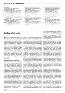
Figure 1. Architecture of the E. coli Exopolyphosphatase Structure (A) The structure of the Ppx dimer depicted as a ribbon diagram. Domains I, II, III, and IV are shown in shades of red, green, blue, and gold, respectively (darker colors designate monomer A). The monomers form an elongated dimer; the 2-fold axis of symmetry is along the z axis of the figure (Kraulis, 1991). (B) Topology diagram showing all four domains. In domains I and II, the conserved core fold characteristic of the ASKHA superfamily is shown in gray and secondary structure inserts are shown in white. The conserved secondary structures in domain I are designated b1, b2, b3, a1, b4, a2, b5, and a3, whereas those in domain II are designated b10 , b20 , etc. to emphasize the similarity of the folds.
of PDE4B2B (helices 5–7, 10, 11, and 13) and binds two metal ions, at least one of which is a zinc ion. The twometal-ion binding site contains a histidine/aspartate (HD) pair in the loop following helix 7. This HD motif is characteristic of a family of proteins that includes the SpoT (p)ppGpp pyrophosphatases (the structure of the Rel/SpoT homolog from Streptococcus dysgalactiae subsp. equisimilis has recently been solved; Hogg et al., 2004), the bacterial dGTPases, glnD PII uridylyl transferase/uridylyl hydrolase, as well as helicases, DNA repair proteins, bacterial signal transduction proteins, and many bacterial proteins of unknown function (Aravind and Koonin, 1998). Remarkably, a conserved histidine (H370) of a His-Glu pair at the end of Ppx aC superimposes precisely with the PDE4B2B H274, which is the histidine of the HD motif. We have not identified any
divalent metal ion bound to domain III of Ppx; this is not surprising given that the protein was purified in the presence of EDTA and that no divalent metal ion was included in the crystallization buffers. Domain IV forms a small a/b layer domain that consists of helix aG and strands bA, bB, and bC. Residues from bB, bC, and aG make hydrophobic contacts with residues from helix aF, thereby linking domain IV to domain III. A trio of tryptophan residues (Figure 2B), which are among the most highly conserved residues of this domain, are part of this domain interface. Unusual Features of the Ppx Dimer Dimers and filaments formed by other members of the ASKHA superfamily involve contacts between domains inserted into the core fold (Buss et al., 2001; Hurley, 1996).
Structure 1266
Figure 3. Ppx Dimer Interactions (A) Stereoview of the homotypic association of aD helices, formed by a mixture of hydrophobic and hydrogen bond interactions. (B) Stereoview of the heterotypic interactions formed between residues of domains II and IV.
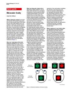
Figure 2. Structural Features of the E. coli Ppx Domains III and IV (A) Superposition of the E. coli Ppx domain III and human cAMP phosphodiesterase 4B2B (PDE4B2B) catalytic fragment structures. The Ppx domain III six-helix claw structure is shown in blue, superimposed onto the catalytic core, in purple, of PDE4B2B (helices 5–7, 10, 11, and 13; PDB code: 1F0J). Shown in gray is the PDE4B2B structure that surrounds the catalytic core. In this superposition, the a carbon positions of 44 structurally equivalent residues superimpose with a root-mean-square deviation of 1.7 A˚. The conserved HD motifs in PDE4B2B (H274/D275, pink) and Ppx (H370/E371, light blue) are shown in a ball-and-stick representation. (B) Structure of E. coli Ppx domain IV. Domain IV is an a/b domain that consists of helix aG and three strands—bA, bB, and bC. Helix aG follows bB and makes hydrophobic contacts with residues from bC. The most highly conserved residues in domain IV, W467, W493, and W499, are shown in ball-and-stick representation.
In most cases, the insert in domain II between b30 and a10 has a critical role. In contrast, in E. coli Ppx, the C-terminal domains are the central participants in the contacts between monomers. A mixture of hydrophobic and
hydrogen bond interactions form the basis of the homotypic association of aD helices (Figure 3A). It is noteworthy that some of the interactions between monomers in the PDE4B2B tetramer (Huai et al., 2003) and the Ppx dimer involve the equivalent regions of the sixhelix claws, although the details of the contacts differ. The second dimer interface is made by heterotypic interactions between residues of domains II and IV (Figure 3B). The interactions are composed of a network of hydrogen bonds and are made between residues in helix aG and the loop connecting aF and bA of domain IV in one monomer and residues in helix a10 of domain II in the other monomer. Intriguingly, there is a gap between the III-III and the IIIV dimer interfaces. This results in the existence of two aqueducts that run through the Ppx dimer perpendicular to its long axis (Figure 4A). Numerous Sulfate Ions Bound to Ppx One of the most remarkable features of the structure of the crystallized E. coli Ppx dimer is the presence of 29 bound sulfate ions (Figure 4A). The source of the sulfate ions is the 1.6 M ammonium sulfate included in the Ppx crystallization conditions. Despite the common use of
E. coli Exopolyphosphatase Processivity 1267
Figure 4. Sulfate Ions Bound to Ppx and the Polyphosphatase Active Site (A) A surface representation of the Ppx dimer is shown with the 29 bound sulfate ions represented as CPK models. To monomer A, shown in darker shades of color, 12 sulfates are bound (SA1–SA12), whereas in monomer B, shown in lighter shades of color, 16 sulfates are bound (SB1–SB16). A single sulfate ion is bound at a site of symmetry between the two monomers (SAB1). The sulfate ions SB5, SA6, and SB7 and SA5, SB6, and SA7 are bound in the two aqueducts indicated by the arrows. See Figures S2C and S2D for a clearer view of the aqueducts. (B) Superposition of domains II and IIA of E. coli Ppx and Thermotoga maritima FtsA (PDB code: 1E4G), respectively. The ATP nucleotide bound in FtsA is shown with the sulfate ions (SA1–SA3) bound in the active site of Ppx. (C) The active site of monomer B is shown with the residues contacting the bound sulfate ions. Residues that contact the g-phosphate of bound ATP in the other ASKHA members originate from loops that are structurally equivalent to those whose residues contact sulfate SB1 in Ppx. (D) The ATP nucleotide as bound in the active site of Thermotoga maritima FtsA (PDB code: 1E4G, ball-and-stick representation) is shown superimposed on the active site of Ppx monomer B (light green and red). If a nucleotide were present in the active site of Ppx, the side chains of residues N21, C169, and R267 (ball-and-stick representation) would clash with the ribose and adenine. These residues would therefore occlude ATP from the Ppx active site and allow binding only of a polyP chain.
ammonium sulfate as a precipitant in X-ray crystallographic protocols, no previous structure in the Protein Database contains more than six sulfate ions bound per dimer. We hypothesize that most of the sulfate binding sites on Ppx represent the sites at which phosphate moieties of the substrate polyP chain would bind. A striking feature of the sulfate ions bound in the Ppx active site (identified through analogy with the active sites of other ASKHA members) is the spacing of the sulfate ions. The distance between SA1 and SA2 is 6.4 A˚, and the distance between SA2 and SA3 is 7.0 A˚, as measured
from the sulfur atoms. These distances are consistent with the distance between the g- and a-phosphate moieties in the triphosphate of an ATP nucleotide. Even more noteworthy is the fact that a superposition of the active site of FtsA bound to ATP upon Ppx indicates that SA1 and SA2 correspond to the g- and a-phosphoryl groups of the ATP molecules bound by the other ASKHA members (Figure 4B). A surface electrostatic potential diagram of Ppx displays a central depression consisting of regions of positive potential (Figure S2). Two aqueducts that lie
Structure 1268
between the Ppx dimer interfaces are populated by trios of sulfate ions (Figure 4A). If these sulfate ions represent phosphate moieties of bound polyP, then it can be concluded that the substrate chain actually traverses an enclosed aqueduct in the enzyme. The sulfate ions are bound to residues (R166, K197, H378, and R413) that represent a subset of the positively charged residues that line the channel and its outlets. We also observe sulfate ions that are unlikely to represent phosphate moieties of polyphosphate. These include SA12 and SB12, which bind in domain IV. The binding of sulfate ions SA12 and SB12 suggests that a phosphate-bearing compound might bind in these regions, although the residues that bind the sulfate ions are not well conserved. Exopolyphosphatase Active Site The putative Ppx active site is located in a cleft between domains I and II. Three sulfate ions are bound in this region, which resembles the active sites of the other ASKHA members (Figure 4C). Two sulfate ions (SA1, SA2, SB1, and SB2) in each monomer are bound directly or through bridging water molecules to the backbone amides and side chains of residues in the loop between strands b1 and b2 (D17, S20, N21, and S22). This loop contains the (D/N)XG motif that is characteristic of the ASKHA phosphotransferases and provides a critical phosphate binding region in the other ASKHA members. The ASKHA phosphohydrolases (Hsp70, actin, FtsA [van den Ent and Lowe, 2000], MreB [van den Ent et al., 2001], component A of 2-hydroxyglutaryl-CoA dehydratase [Locher et al., 2001] and Ppx) possess a second (D/N)XG motif in a loop between b10 and b20 that distinguishes them from the ASKHA kinases. Residues in this loop (G146, G147, and S148) also contact sulfates SA1 and SB1. The two DXG aspartate residues, D17 and D143, bind to SA1 through intervening water molecules and are predicted to act as ligands for the catalytic magnesium ion by analogy to the equivalent aspartate residues in other ASKHA members (Hurley, 1996). The two residues that are absolutely conserved in the ASKHA superfamily are both glycines. The first is the glycine in the (D/N)XG motif (G19 in Ppx). The second is present at the N terminus of helix a2A0 and binds the aphosphate of ATP through its backbone amide in the other ASKHA members. The equivalent residues in the Ppx dimer (G218) bind SA2 and SB2 through their backbone amides. If one considers the triphosphate of ATP as the terminal triphosphate of polyP, then the phosphoryl group of polyP that is hydrolyzed is equivalent to the g-phosphate of ATP, whereas the a-phosphate of ATP is equivalent to the g-phosphate of polyP. The conserved interactions in the Ppx active site are consistent with the identification of SA1/SB1 and SA2/SB2 as representing, respectively, the terminal and g-phosphates of polyP. As is true for the conserved a-phosphate binding glycine residues in the other ASKHA members (Buss et al., 2001), the residue preceding G218, S217, is in the epsilon conformation (f = 78.3� and J = 2173.0� in monomer A), which essentially permits the transition from a b strand to an a helix to occur with just 1 intervening residue. Such transitions have been observed in other protein structures, but the evolutionary conservation of a residue in this conformation is frequently overlooked.
Analysis of the active site residues that are conserved within the enzymes in the Ppx family provides important information on the structural basis of their substrate specificity. T89, E121, and E150 are conserved residues whose side chains bind to SA1 and SB1 either directly or through bridging water molecules. E121 is at the N terminus of a3 and is equivalent to residues with Glx residues that bind divalent cations in Hsp70 and actin through intervening water molecules. The highly conserved R93 is hydrogen bonded to E121. The equivalent residues to T89, E121, and R93 are conserved in the eukaryotic ectonucleoside triphosphate diphosphohydrolases (E-NTPDases), also known as ectoapyrases, which are predicted to be members of the ASKHA superfamily. Mutant E-NTPDase3 proteins bearing substitutions for the E121 equivalent (E182) are completely inactive (Yang et al., 2001). The eukaryotic E-NTPDases can sequentially remove both the g- and b-phosphates of NTPs. The Ppx active site may have advantageous properties as a framework for the evolution of enzymes that can hydrolyze the terminal phosphates of either NTPs or NDPs. The Ppx residues whose side chains contact SA3 and SB3 (R44, S264, and R267) all originate from loops that are parts of insertions into the ASKHA core fold (Figure 4C). Multifarious and conserved R267 not only binds SA2 and SB2 through its guanidinium group, but it also occludes the binding of a nucleotide base in the active site. Superposition of ATP as bound in the active site of FtsA on the Ppx structure suggests that if there were a nucleotide present in the active site of Ppx, the side chain of residues N21, C169, and R267 would clash with the ribose and adenine (Figure 4D). These residues would therefore prevent ATP from binding to Ppx. Ppx is the only ASKHA member to have an asparagine residue at an equivalent site to N21 in the loop between b1 and b2; the other members have different small residues. C169 originates from the secondary structure insert ab. It can be concluded that the preference for polyP and the exclusion of nucleotides can be attributed mainly to the inserts into the core ASKHA fold. The exopolyphosphatase active site was confirmed through the analysis of the activity of mutant Ppx proteins by using a coupled enzyme assay that measured the release of inorganic phosphate from polyP. Deletion of domains I and II (PpxD 1–304) resulted in a loss of exopolyphosphatase activity, whereas the deletion of the last two domains (PpxD 321–513) resulted in substantial retention of activity. Mutant Ppx proteins bearing substitutions for conserved domain I/II residues E121, D143, and E150 possessed only residual activity (<5% of the activity of the wild-type protein), whereas a mutant protein bearing a substitution for a conserved domain III residue (E371A) retains substantial activity (Table 3). These data strongly support the identification of the cleft between domains I and II as the exopolyphosphatase active site. Evolution of the Exopolyphosphatase Active Site The ASKHA superfamily is a paradigm for understanding enzyme evolution (Buss et al., 2001; Hurley, 1996). The divergent members of the ASKHA phosphotransferases share virtually no sequence identity; only two glycine residues are absolutely conserved, and a third residue
E. coli Exopolyphosphatase Processivity 1269
Table 3. Exopolyphosphatase Activity of Ppx
Ppx D (321–513)b D (1–304)c E121Ad D143Ae E150Ae E371Af
Pi Release (mM/min)a
Percent Pi Releaseda
25.50 6 2.50 8.988 6 1.34 0.118 6 0.05 0.288 6 0.18 1.098 6 0.05 0.605 6 0.05 9.461 6 0.86
100 35.2 0.46 1.12 4.30 2.37 37.1
a
Pi release was measured by using the EnzChek Phosphate Assay Kit from Molecular Probes. The samples containing 0.6 mM P75+ (50 mM Pi) were preincubated with 0.2 mM MESG and 1 U PNP in 50 mM tricine/KOH (pH 8.0), 175 mM KCl, and 1 mM MgCl2. Pi release was monitored at 360 nm for 2.5 min after the addition of 0.2 mg protein. b Domains I and II. c Domains III and IV. d Mutation in domain I. e Mutation in domain II. f Mutation in domain III.
is conserved as either an aspartate or an asparagine residue. Nevertheless, these enzymes possess identical core folds and carry out identical core reactions, phosphoryl transfers. The first two domains of Ppx share commonalities with the other ASKHA members and also possess some distinctive characteristics. Ppx possesses the ASKHA core-repeated bbbababa structure that is believed to have arisen as a duplication of the RNaseH fold (Artymiuk et al., 1993). As compared with the other ASKHA phosphotransferases, the inserts in Ppx into the core fold are relatively small. In the other ASKHA enzymes, the inserts contribute to the binding of the adenosine base of ATP. In Ppx, the inserts provide the specificity for polyP by providing both polyP binding sites and side chains that would preclude the binding of nucleotide. Basis of Exopolyphosphatase Processivity The sulfate ions bound in the active site probably represent every other phosphate moiety of the terminus of the polyP chain. This is consistent with the overall architecture of the cleft between domains I and II. It is likely that other sulfate ions also represent phosphate moieties of polyP. We propose a model of polyP binding in which the phosphate chain follows a path marked by the sulfates bound to the protein (Figure 5). This model immediately suggests a mechanism for the high processivity of Ppx. The polyP chain is not only bound at multiple points to the surface of the protein, but it is also actually enclosed in an aqueduct between the heterotypic domain II-domain IV and the homotypic domain III-domain III dimer interfaces. A prediction of this model is that destabilization of the dimer would disrupt processivity. The path of bound polyP predicted by the Ppx structure is thoroughly consistent with data obtained concerning the effects of high salt concentrations and carboxyterminal deletions upon Ppx processivity (Bolesch and Keasling, 2000). Processive hydrolysis of polyP by E. coli Ppx was disrupted by high salt concentrations and carboxy-terminal deletions (Bolesch and Keasling, 2000). The dimer interface between domain IV and domain II is constituted exclusively of hydrogen bonds and might be destabilized by high salt concentrations,
Figure 5. Model of PolyP Binding Monomer A is depicted in a surface representation; a black line connecting the sulfates shows the suggested polyP binding path. The polyP chain exits the monomer A active site (SA1–SA3), passes on the surface of the enzyme through the binding site occupied by SA4, traverses the protein through the channel occupied by sulfates SB5, SA6, and SB7, and finally crosses over the protein surface to reach the binding site occupied by sulfates SA8–SA11.
resulting in disruption of processivity. Even relatively small carboxy-terminal deletions (>16 residues) were found to reduce processivity (Bolesch and Keasling, 2000). The sizes of the polyP chain intermediates obtained under suboptimal conditions of quasiprocessivity (P50, P14, and P3) are also consistent with the binding sites predicted by our model. The proposed structural basis of processivity of the exopolyphosphatase can be compared with that of the enzymes that degrade cellulose, a polymer of glucose residues. Insertions into a core fold lead to a feature that distinguishes processive exoglucanase from their endoglucanase relatives; the exoglucanases possess a tunnel through which the cellulose chain can be threaded (Divne et al., 1998). Our interpretation of the structure furthermore indicates that there is a ball-and-cup-game mechanism for the feeding of the terminus of the polyP chain into the active site. The chain is tethered to the enzyme, and after each hydrolysis event the terminus is released from the active site so that the liberated phosphate ion can dissociate. The new terminus can then bind in the active site for the next round of hydrolysis to proceed. Alternatively, it is possible that the inorganic phosphate could escape from the active site with the rest of the chain still bound through an opening of the cleft between domains I and II. Until the polyphosphate chain is smaller than w50 inorganic phosphate moieties, there is no need for the bulk of the chain to dissociate from the enzyme under these scenarios. Smaller chains would make fewer contacts with Ppx. No component of this model or the determined structure precludes the simultaneous hydrolysis of two polyphosphate chains by the Ppx dimer. A Cyclic Nucleotide Phosphodiesterase Fold in Ppx Domain III possesses the six-helix claw fold that is the core of the eukaryotic cyclic nucleotide phosphodiesterases. H370 of Ppx corresponds in space precisely with the conserved histidine residue in the phosphodiesterases that binds a zinc ion. Our identification of
Structure 1270
the six-helix claw as a conserved structural unit suggests a reinterpretation of the PDE4 structure (Xu et al., 2000) in which helices 5, 6, 7, 10, 11, and 13 form the core of the domain and helices 8, 9, and 12 are insertions into the core. Domain III of Ppx and the core of the PDEs belong to the HD phosphohydrolase superfamily, which, among many others, includes the SpoT (p)ppGpp pyrophosphatases and the bacterial dGTPases (Aravind and Koonin, 1998). The sequence of Gpp indicates that it will also have a domain III belonging to this superfamily. We predict that all of these proteins will share the six-helix claw core. Other proteins, such as E. coli RelA and the glycine tRNA synthetase b subunit, that are predicted to be members of the HD family will also likely have a six-helix claw core, although they lack the HD zinc binding residues (Aravind and Koonin, 1998; Wolf et al., 1999). It is noteworthy that whereas the plant Ppx homologs contain an HD domain with the critical conserved histidine residue, homologs from the archaeal Sulfolobus solfataricus (Cardona et al., 2002) and Sulfolobus tokodaii and the homologous Saccharomyces cerevisiae RTG2 protein (Koonin, 1994; Liao and Butow, 1993) do not. Indeed, the divergent sequences in RTG2 suggest that it utilizes a substrate that differs from polyP (Pierce et al., 2001). It is of interest that the sequence of the Thermotoga maritima homolog of Ppx (TM0195) encodes only the first two domains and lacks the sequences corresponding to some of the inserts. It thereby represents nearly a minimal ASKHA fold and may have quite divergent properties, including a different substrate. In summary, determination of the structure of the E. coli exopolyphosphatase has given us insight into how the specificity of a member of a superfamily of ATP-utilizing phosphotransferases, the ASKHA superfamily, can be modified so that polyP is the substrate. Passage of the polyP chain through an aqueduct that traverses the protein between two dimer interfaces provides a structural explanation for the high processivity of the exopolyphosphatase. The third domain originates from a transition-metal binding phosphohydrolase fold. It provides the major dimer interface and provides residues that probably bind the polyphosphate substrate. Experimental Procedures Plasmid Cloning The source of the Escherichia coli ppx gene was the pBC6 plasmid, kindly provided by Arthur Kornberg (Akiyama et al., 1993). The ppx gene was subcloned into the bacterial expression vector pET30a(+) (Novagen) behind the T7 RNA polymerase promoter, resulting in the expression plasmid pEcPpx. This was constructed by digesting the pBC6 plasmid with the restriction enzyme NruI. This yielded a DNA fragment of 2650 bp, which was subsequently digested with BsiEI to yield a fragment of 1547 bp that encodes amino acids 16–513 and the wild-type stop codon of Ppx. In order to encode for the first 15 amino acids of Ppx, a double-stranded oligonucleotide (ppx[aa1–15]+/2) possessing NdeI and BsiEI overhangs was constructed (Integrated DNA Technologies, Inc.) by annealing together the single-stranded oligonucleotides: 50 -TATGCCAATACAC GATAAATCCCCTCGTCCGCAGGAGTTTGCTGCGGT-30 and 50 -CG CAGCAAACTCCTGCGGACGAGGGGATTTATCGTGTATTGGCA-30 . The plasmid pET-30a(+)was digested with NdeI and EcoRV, yielding a DNA fragment of 5280 bp, which was ligated to (ppx[aa1–15]+/2) and the 1547 bp fragment from pBC6, resulting in plasmid pEcPpx. For cloning of the PpxD 1–304 expression plasmid, the relevant sequence of the pEcPpx plasmid was amplified by using the primers
50 -GAAGGCGTACTGTATACC ATGGAAGGA-30 and 50 -GTTGCAAAG CTTTTAAGCGGCGATTTC-30 . The PCR product was digested with NcoI and HindIII and ligated to pET30a(+) to generate the plasmid expressing PpxD 1–304 with an N-terminal His6 sequence. The plasmid encoding the soluble N-terminal Ppx fragment (PpxD 321–513) was constructed by digesting pEcPpx with BamHI and NdeI to yield a 1599 bp fragment and a 5275 bp fragment. The 1599 bp fragment was digested with DrdI to yield a 928 bp fragment and a 671 bp fragment. The 928 bp fragment was isolated. A doublestranded oligonucleotide encoding amino acids 310–320 that was compatible for ligation to a DrdI- and BamHI-digested plasmid was constructed by annealing together the single-stranded oligonucleotides: 50 -CCGTCATCAGGATGTGCGTAGTCGCACCGCCAGCA GCTGAG-30 and50 -GATCCTCAGCTGCTGGCGGTGCGACTACGCA CATACTAATGACGGAA-30 . The 5275 bp fragment, the 928 bp fragment, and the annealed oligonucleotide were ligated to generate the plasmid encoding PpxD 321–513. The expression plasmids encoding the E121A, D143A, E150A, and E371A mutant Ppx proteins were constructed through use of the polymerase chain reaction. All mutations were confirmed by DNA sequencing. Protein Expression The pEcPPX plasmid and those encoding mutant Ppx proteins were transformed into the BL21(DE3) bacterial host strain (Novagen). A single colony was inoculated into 1 liter of LB medium containing 30 mg/ml kanamycin and was grown to an OD600 of 0.6 at 37� C. Protein expression was induced by adding IPTG to the culture to a final concentration of 1 mM. Induction was carried out for 4 hr at 37� C. Cells were harvested by centrifugation at 6,000 rpm (5,855 3 g) at 4� C, flash frozen in liquid nitrogen, and stored at 280� C. Protein Purification Purification of wild-type Ppx and Ppx bearing single amino acid substitutions was carried out according to the protocol of Akiyama et al. (1993), with some modifications. All purification steps for wild-type and mutant Ppx were carried out at 4� C. Cells were suspended in lysis buffer (50 mM Tris-Cl [pH 7.5], 1 mM EDTA, 2 mM DTT) and lysed in a French Press cell at 18,000 psi. Cell debris was removed by centrifugation at 51,000 rpm (184,692 3 g) for 1 hr, and the soluble fraction was dialyzed (three times, 1 hr each) against a solution of 0.2 M potassium phosphate (pH 7.0), 10% (w/v) glycerol, 1 mM DTT, 1 mM EDTA. The protein solution was dialyzed further against Buffer A (50 mM HEPES/KOH [pH 7.5], 10% [w/v] glycerol, 1 mM DTT, 1 mM EDTA) before its application to a High-Performance SP-Sepharose column (Pharmacia). Bound Ppx protein was eluted with Buffer A containing 0.2 M NaCl by using a step gradient. The fractions containing Ppx protein were pooled and dialyzed against Buffer A containing 50 mM NaCl. The protein was loaded onto a HiTrap Q column (Pharmacia) and eluted with a linear gradient from 0.05 to 0.7 M NaCl. The final yield of purified Ppx protein was 15–20 mg per liter of cells. When the mutant Ppx proteins were purified, EDTA was omitted from all buffers. Purified protein was concentrated to 11–12 mg/ml, frozen in liquid nitrogen, and stored at 280� C. Cells expressing the domain III/IV fragment (PpxD 1–304) were suspended in 50 mM sodium phosphate, 10% (w/v) glycerol, 300 mM NaCl, 10 mM imidazole (pH 8.0) and lysed in a French Press cell. After centrifugation at 51,000 rpm (184,692 3 g) for 1 hr, the cleared lysate was loaded onto an Ni-NTA Superflow column (Qiagen) and eluted with 50 mM sodium phosphate, 10% (w/v) glycerol, 300 mM NaCl, 200 mM imidazole. The fractions containing the protein were pooled and dialyzed against Buffer A containing 0.2 M NaCl, concentrated to 10–12 mg/ml, flash frozen in liquid nitrogen, and stored at 280� C. Cells expressing the domain I/II fragment (PpxD 321–513) were suspended in lysis buffer (50 mM Tris-Cl [pH 7.5], 2 mM DTT) and lysed in a French Press cell. The cell debris was removed by centrifugation at 51,000 rpm (184,692 3 g) for 1 hr. The cell lysate was then dialyzed three times in 0.2 M potassium phosphate (pH 7.0), 10% (w/ v) glycerol, and 1 mM DTT. This was followed by further dialysis in Buffer B (50 mM HEPES/KOH [pH 7.0], 10% [w/v] glycerol, and 1 mM DTT). The protein was applied to a Heparin Sepharose 6 Fast Flow column (Pharmacia) and eluted with Buffer B containing
E. coli Exopolyphosphatase Processivity 1271
0.2 M NaCl. This protein was concentrated and loaded onto a Superdex 75 10/300 GL gel filtration column (Pharmacia) and eluted with Buffer B containing 0.2 M NaCl. The protein thus obtained was concentrated to 10–12 mg/ml, flash frozen in liquid nitrogen, and stored at 280� C. Exopolyphosphatase Assay Polyphosphate (P75+) was obtained from Sigma-Aldrich and dissolved in 50 mM tricine/KOH (pH 8.0) to a final concentration of 20 mM. Exopolyphosphatase activity of wild-type and mutant Ppx enzymes was assayed by using the EnzChek Phosphate Assay Kit from Molecular Probes (Webb, 1992). In the presence of Pi, the enzyme purine nucleoside phosphorylase (PNP) converts 2-amino-6mercapto-7-methylpurine riboside (MESG) to ribose 1-phosphate and 2-amino-6-mercapto-7-methylpurine. The reaction causes a spectrophotometric shift in maximum absorbance from 330 nm for the substrate to 360 nm for the product. The exopolyphosphatase reaction was set up by adding 0.6 mM P75+ to 0.2 mM MESG and 1 U PNP in 50 mM tricine/KOH (pH 8.0), 175 mM KCl, and 1 mM MgCl2. This was incubated for 5 min to remove contamination due to inorganic phosphates in solution. The reaction was initiated by the addition of 0.2 mg Ppx (or the mutants) and was followed at 360 nm as a function of time for 2.5 min in a Cary 100 UV-Visible Spectrophotometer. The initial slopes were then determined in terms of absorbance/minute and converted to Pi obtained per minute by using a phosphate standard curve. Protein Crystallization Crystallization of the E. coli Ppx dimer was accomplished by using the hanging-drop vapor diffusion technique. A frozen aliquot of 11–12 mg/ml purified protein was thawed on ice and centrifuged at 14,000 rpm (16,000 3 g) for 10 min to remove any particulate matter. Crystals were grown at 20� C from a 1:1 mixture of protein:reservoir solution (unbuffered 1.6 M ammonium sulfate). E. coli Ppx crystals typically grew in 2–6 weeks and were, on average, 750 3 350 3 150 mm in size. The crystals formed in space group P41212 with two monomers per asymmetric unit. Flash freezing was carried out by immersing the crystals in a cryoprotectant solution (unbuffered 2.1 M lithium sulfate, 10% [v/v] glycerol) for less than 30 s and placing the crystals in a nitrogen stream at 100 K. Data Collection and Structure Determination All diffraction data were collected at the Advanced Photon Source (APS), Argonne National Laboratory at the 14-IDB, 14-BMC, and 14-BMD beamlines at BioCARS. All data were processed and scaled by using the HKL package (Otwinowski and Minor, 1996); Table 1 displays the quality of the diffraction data. The very large I/s values of the native1 crystal are due to the fact that data could only be collected to 2.47 A˚ (although it diffracted to 1.7 A˚) in order to prevent reflections from overlapping, given the 351.0 A˚ axis of the unit cell. Data from the native2 crystal were collected to 1.9 A˚ with minimal reflection overlap due to the beam optics in beamline 14-BMC, which are optimized for crystals possessing large unit cell dimensions. The structure was solved by MAD methods by using the anomalous scattering of Hg from a crystal soaked in ethyl mercury phosphate. Six mercury sites were located by using the Patterson-solving algorithm of SOLVE (Terwilliger, 2002). The experimental phases were improved by solvent flattening with RESOLVE (Terwilliger, 2002), and a 3.25 A˚ electron density map was calculated. A continuous main chain model of the Ppx dimer, which includes 97% of the total main chain for each monomer, was built by using the solventflattened 3.25 A˚ electron density map and the graphics program O (Jones et al., 1991). The phases and structure factors of the 3.25 A˚ data and structure factors from a 2.47 A˚ native data set were used in combination to carry out solvent flattening, histogram matching, 2-fold NCS averaging, multicrystal averaging, and phase extension with DMMULTI (Cowtan, 1994). The resulting 2.47 A˚ electron density map was used to build side chains into the model. Model Refinement The initial model was subjected to four cycles of simulated annealing or positional refinement and unrestrained grouped B factor refinement followed by manual model rebuilding. Refinement was carried out with the program CNS (Brunger et al., 1998) by including all re-
flections (F/s > 0) in the 2.47 A˚ native data set. The model was improved by increasing the resolution to 1.9 A˚ with data from a second native crystal. Refinement was continued by carrying out six rounds of restrained positional, individual B factor, and TLS refinement by using Refmac5 (Winn et al., 2001), with model rebuilding after each round. Model bias was removed from the final model by using simulated annealed omit maps to correct any errors, and a model with excellent refinement statistics resulted (Table 2). The final model includes residues 12–509 and 12–511 for monomers A and B, respectively. Supplemental Data Supplemental Data include a movie showing the proposed binding of the polyphosphatase chain on Ppx (Movie S1), Supplemental Experimental Procedures, and additional figures and are available at http://www.structure.org/cgi/content/full/14/8/1263/DC1/. Acknowledgments This research was supported by National Institutes of Health (NIH) grant RO1-GM57056 and by David and Lucille Packard Foundation Fellowship 99-1463 to M.S.H., National Science Foundation (NSF) CAREER award 99-84919-MCB to D.A.S., NSF Minority Fellowship and NIH Institutional Training Award GM-08296 to J.A., and NIH Cancer Center support at Purdue University. Facilities shared by the Structural Biology group at Purdue have been developed and supported by grants from the NIH, the NSF, the Lucille P. Markey Foundation, the Keck Foundation, and the office of the university executive vice president for academic affairs. Use of the Advanced Photon Source was supported by the U.S. Department of Energy, Basic Energy Sciences, Office of Science, under contract no. W-31-109Eng-38. Use of the BioCARS Sector 14 was supported by the NIH, National Center for Research Resources, under grant number RR07707. We thank Rodney McPhail and Jeff Bolin for help with figures and Dr. Bolin for critical reading of the manuscript. We also thank Dr. Mitchel S. Berger for providing M.S.H. with the opportunity to conduct this research. Received: February 18, 2005 Revised: June 5, 2006 Accepted: June 9, 2006 Published: August 15, 2006 References Akiyama, M., Crooke, E., and Kornberg, A. (1993). An exopolyphosphatase of Escherichia coli. The enzyme and its ppx gene in a polyphosphate operon. J. Biol. Chem. 268, 633–639. Aravind, L., and Koonin, E.V. (1998). The HD domain defines a new superfamily of metal-dependent phosphohydrolases. Trends Biochem. Sci. 23, 469–472. Artymiuk, P.J., Grindley, H.M., Kumar, K., Rice, D.W., and Willett, P. (1993). Three-dimensional structural resemblance between the ribonuclease H and connection domains of HIV reverse transcriptase and the ATPase fold revealed using graph theoretical techniques. FEBS Lett. 324, 15–21. Bolesch, D.G., and Keasling, J.D. (2000). Polyphosphate binding and chain length recognition of Escherichia coli exopolyphosphatase. J. Biol. Chem. 275, 33814–33819. Brunger, A.T., Adams, P.D., Clore, G.M., DeLano, W.L., Gros, P., Grosse-Kunstleve, R.W., Jiang, J.S., Kuszewski, J., Nilges, M., Pannu, N.S., et al. (1998). Crystallography & NMR system: a new software suite for macromolecular structure determination. Acta Crystallogr. D Biol. Crystallogr. 54, 905–921. Buss, K.A., Cooper, D.R., Ingram-Smith, C., Ferry, J.G., Sanders, D.A., and Hasson, M.S. (2001). Urkinase: structure of acetate kinase, a member of the ASKHA superfamily of phosphotransferases. J. Bacteriol. 183, 680–686. Cardona, S.T., Chavez, F.P., and Jerez, C.A. (2002). The exopolyphosphatase gene from Sulfolobus solfataricus: characterization of the first gene found to be involved in polyphosphate metabolism in archaea. Appl. Environ. Microbiol. 68, 4812–4819.
Structure 1272
Chatterji, D., and Ojha, A.K. (2001). Revisiting the stringent response, ppGpp and starvation signaling. Curr. Opin. Microbiol. 4, 160–165. Cowtan, K. (1994). Joint CCP4 and ESF-EACBM Newsletter on Protein Crystallography 31, 34–38. Divne, C., Stahlberg, J., Teeri, T.T., and Jones, T.A. (1998). High-resolution crystal structures reveal how a cellulose chain is bound in the 50 A˚ long tunnel of cellobiohydrolase I from Trichoderma reesei. J. Mol. Biol. 275, 309–325. Hara, A., and Sy, J. (1983). Guanosine 50 -triphosphate, 30 -diphosphate 50 -phosphohydrolase. Purification and substrate specificity. J. Biol. Chem. 258, 1678–1683. Hogg, T., Mechold, U., Malke, H., Cashel, M., and Hilgenfeld, R. (2004). Conformational antagonism between opposing active sites in a bifunctional RelA/SpoT homolog modulates (p)ppGpp metabolism during the stringent response. Cell 117, 57–68. Holmes, K.C., Ander, C., and Valencia, A. (1993). A new ATP-binding fold in actin, hexokinase and Hsc70. Trends Cell Biol. 3, 53–59. Huai, Q., Colicelli, J., and Ke, H. (2003). The crystal structure of AMPbound PDE4 suggests a mechanism for phosphodiesterase catalysis. Biochemistry 42, 13220–13226. Hurley, J.H. (1996). The sugar kinase/heat shock protein 70/actin superfamily- implications of conserved structure for mechanism. Annu. Rev. Biophys. Biomol. Struct. 25, 137–162. Hurley, J.H., Faber, H.R., Worthylake, D., Meadow, N.D., Roseman, S., Pettigrew, D.W., and Remington, S.J. (1993). Structure of the regulatory complex of Escherichia coli IIIGlc with glycerol kinase. Science 259, 673–677. Jones, T.A., Zou, J.Y., Cowtan, S.W., and Kjeldgaard, M. (1991). Improved methods for binding protein models in electron density maps and the local of errors in these models. Acta Crystallogr A 47, 110–119. Keasling, J.D., Bertsch, L., and Kornberg, A. (1993). Guanosine pentaphosphate phosphohydrolase of Escherichia coli is a long- chain exopolyphosphatase. Proc. Natl. Acad. Sci. USA 90, 7029–7033. Koonin, E.V. (1994). Yeast protein controlling inter-organelle communication is related to bacterial phosphatases containing the Hsp 70-type ATP-binding domain. Trends Biochem. Sci. 19, 156–157. Kornberg, A., Rao, N.N., and Ault-Riche, D. (1999). Inorganic polyphosphate: a molecule of many functions. Annu. Rev. Biochem. 68, 89–125. Kraulis, P.J. (1991). MOLSCRIPT: A program to produce both detailed and schematic plots of protein structures. J. Appl. Crystallogr. 24, 946–950. Kristensen, O., Laurberg, M., Liljas, A., Kastrup, J.S., and Gajhede, M. (2004). Structural characterization of the stringent response related exopolyphosphatase/guanosine pentaphosphate phosphohydrolase protein family. Biochemistry 43, 8894–8900. Kuroda, A., Murphy, H., Cashel, M., and Kornberg, A. (1997). Guanosine tetra- and pentaphosphate promote accumulation of inorganic polyphosphate in Escherichia coli. J. Biol. Chem. 272, 21240–21243. Kuroda, A., Nomura, K., Ohtomo, R., Kato, J., Ikeda, T., Takiguchi, N., Ohtake, H., and Kornberg, A. (2001). Role of inorganic polyphosphate in promoting ribosomal protein degradation by the Lon protease in E. coli. Science 293, 705–708. Liao, D.I., Reiss, L., Turner, I., and Dotson, G. (2003). Structure of glycerol dehydratase reactivase: a new type of molecular chaperone. Structure 11, 109–119. Liao, X., and Butow, R.A. (1993). RTG1 and RTG2: two yeast genes required for a novel path of communication from mitochondria to the nucleus. Cell 72, 61–71. Locher, K.P., Hans, M., Yeh, A.P., Schmid, B., Buckel, W., and Rees, D.C. (2001). Crystal structure of the Acidaminococcus fermentans 2-hydroxyglutaryl-CoA dehydratase component A. J. Mol. Biol. 307, 297–308. Otwinowski, Z., and Minor, W. (1996). Processing of X-ray diffraction data collected in oscillation mode. Methods Enzymol. 276, 307–326. Pierce, M.M., Maddelein, M.L., Roberts, B.T., and Wickner, R.B. (2001). A novel Rtg2p activity regulates nitrogen catabolism in yeast. Proc. Natl. Acad. Sci. USA 98, 13213–13218.
Rao, N.N., and Kornberg, A. (1996). Inorganic polyphosphate supports resistance and survival of stationary-phase Escherichia coli. J. Bacteriol. 178, 1394–1400. Rao, N.N., Liu, S., and Kornberg, A. (1998). Inorganic polyphosphate in Escherichia coli: the phosphate regulon and the stringent response. J. Bacteriol. 180, 2186–2193. Reizer, J., Reizer, A., Saier, M.H., Jr., Bork, P., and Sander, C. (1993). Exopolyphosphate phosphatase and guanosine pentaphosphate phosphatase belong to the sugar kinase/actin/hsp 70 superfamily. Trends Biochem. Sci. 18, 247–248. Terwilliger, T.C. (2002). Automated structure solution, density modification and model building. Acta Crystallogr. D Biol. Crystallogr. 58, 1937–1940. van den Ent, F., and Lowe, J. (2000). Crystal structure of the cell division protein FtsA from Thermotoga maritima. EMBO J. 19, 5300– 5307. van den Ent, F., Amos, L.A., and Lowe, J. (2001). Prokaryotic origin of the actin cytoskeleton. Nature 413, 39–44. Webb, M.R. (1992). A continuous spectrophotometric assay for inorganic phosphate and for measuring phosphate release kinetics in biological systems. Proc. Natl. Acad. Sci. USA 89, 4884–4887. Winn, M.D., Isupov, M.N., and Murshudov, G.N. (2001). Use of TLS parameters to model anisotropic displacements in macromolecular refinement. Acta Crystallogr. D Biol. Crystallogr. 57, 122–133. Wolf, Y.I., Aravind, L., Grishin, N.V., and Koonin, E.V. (1999). Evolution of aminoacyl-tRNA synthetases—analysis of unique domain architectures and phylogenetic trees reveals a complex history of horizontal gene transfer events. Genome Res. 9, 689–710. Xu, R.X., Hassell, A.M., Vanderwall, D., Lambert, M.H., Holmes, W.D., Luther, M.A., Rocque, W.J., Milburn, M.V., Zhao, Y., Ke, H., and Nolte, R.T. (2000). Atomic structure of PDE4: insights into phosphodiesterase mechanism and specificity. Science 288, 1822–1825. Yang, F., Hicks-Berger, C.A., Smith, T.M., and Kirley, T.L. (2001). Site-directed mutagenesis of human nucleoside triphosphate diphosphohydrolase 3: the importance of residues in the apyrase conserved regions. Biochemistry 40, 3943–3950.
Accession Numbers Coordinates have been deposited in the RCSB Protein Data Bank with the PDB code 1U6Z.