Psychopharmacology © Springer-Verlag 2005 10.1007/s00213-005-2264-9
Original Investigation
Plasma level-dependent effects of methylphenidate on task-related functional magnetic resonance imaging signal changes Ulrich Müller1, 2, 5 , J. Suckling1, 3, F. Zelaya3, G. Honey1, 3, H. Faessel4, S. C. R. Williams3, C. Routledge4, J. Brown4, T. W. Robbins2 and E. T. Bullmore1, 3, 6 (1) Department of Psychiatry, University of Cambridge, Cambridge, UK (2) Department of Experimental Psychology, University of Cambridge, Cambridge, UK (3) Institute of Psychiatry, King s College London, London, UK (4) GlaxoSmithKline, Clinical Research Unit, Addenbrooke s Centre for Clinical Investigation, Cambridge, UK (5) MRC Centre for Behavioural and Clinical Neuroscience, University of Cambridge, Downing Site, Cambridge, CB2 3EB, UK (6) Brain Mapping Unit, Department of Psychiatry, Addenbrooke s Hospital, University of Cambridge, Cambridge, CB2 2QQ, UK Ulrich Müller Email:
[email protected] Phone: +44-1223-333535 Fax: +44-1223-764760 URL: http://research.psychol.cam.ac.uk/~bcnc E. T. Bullmore Email:
[email protected] Phone: +44-1223-336583 Fax: +44-1223-336581 URL: http://www-bmu.psychiatry.cam.ac.uk
Received: 8 November 2004 Accepted: 5 March 2005 Published online: 14 April 2005 Abstract Rationale Methylphenidate (MPH) is a dopamine and noradrenaline enhancing drug used to treat attentional deficits. Understanding of its cognition-enhancing effects and the neurobiological mechanisms involved, especially in elderly people, is currently incomplete. Objectives The aim of this study was to investigate the relationship between MPH plasma levels and brain activation during visuospatial attention and movement preparation.
Methods Twelve healthy elderly volunteers were scanned twice using functional magnetic resonance imaging (fMRI) after oral administration of MPH 20 mg or placebo in a within-subject design. The cognitive paradigm was a four-choice reaction time task presented at two levels of difficulty (with and without spatial cue). Plasma MPH levels were measured at six time points between 30 and 205 min after dosing. FMRI data were analysed using a linear model to estimate physiological response to the task and nonparametric permutation tests for inference. Results Lateral premotor and medial posterior parietal cortical activation was increased by MPH, on average, over both levels of task difficulty. There was considerable intersubject variability in the pharmacokinetics of MPH. Greater area under the plasma concentration-time curve was positively correlated with strength of activation in motor and premotor cortex, temporoparietal cortex and caudate nucleus during the difficult version of the task. Conclusion This is the first pharmacokinetic/pharmacodynamic study to find an association between plasma levels of MPH and its modulatory effects on brain activation measured using fMRI. The results suggest that catecholaminergic mechanisms may be important in brain adaptivity to task difficulty and in task-specific recruitment of spatial attention systems.
Keywords Attention - Dopamine - fMRI - Methylphenidate Pharmacokinetics - Pharmacodynamics
Introduction The development of neuroimaging techniques such as positron emission tomography (PET) and, more recently, functional magnetic resonance imaging (fMRI) has revolutionised the field of cognitive neuropharmacology. These techniques allow the visualisation and quantification of the effects of drugs on regional blood flow and metabolism in the brain. During the last 5 years, an increasing number of studies used fMRI in conjunction with placebo-controlled pharmacological challenges to investigate the neurotransmitter modulation of brain activation during cognitive and motor tasks (Braus et al. 2003; Honey and Bullmore 2004). Although drugs like caffeine may have global effects on cerebral blood flow at baseline and blood oxygenation level dependent (BOLD) signals during cognitive activation (Mulderink et al. 2002; Laurienti et al. 2003; Liu et al. 2004), other drugs have anatomically and cognitively more specific effects on activation signals related to performance of motor or cognitive tasks in fMRI experiments (Furey et al. 2000; Mattay et al. 2003; Bullmore et al. 2003). Methylphenidate (MPH) is a psychostimulant drug that is widely used to treat children and adults with attention deficit hyperactivity disorder (ADHD) and other neuropsychiatric disorders (Murray and Cassem 1998; Solanto 1998; Greenhill et al. 2002; Biederman et al. 2004; Leonard et al. 2004). Mehta et al. (2000) studied ten healthy male volunteers (mean age, 34.8 years) using PET in a placebo-controlled design. They found reduced regional cerebral blood flow (rCBF), associated with improved behavioural performance on a self-ordered spatial working memory task, in the left dorsolateral prefrontal cortex, supplementary motor area (SMA) and medial posterior parietal cortex after MPH 40 mg orally. Silveri et al. (2004) found decreased transverse relaxation times (T2) in the putamen, indicating increased rCBF, after a single oral dose of MPH 40 mg in a pharmacological MRI study of healthy volunteers (mean age, 24.9 years). Vaidya et al. (1998) used fMRI to investigate 6 healthy children and ten children with ADHD aged 8–13 years. They found increased activation by a go/no-go task in several frontal regions of interest (ROIs) in both groups after oral MPH (10 mg for healthy children and 7.5–30 mg for children with ADHD). Healthy children demonstrated reduced activation of the caudate nucleus and putamen, whereas children with ADHD showed increased activation of striatal
regions after MPH. Rao et al. (2000) used fMRI to measure brain activation by a finger-tapping task in six healthy volunteers (age range, 20–45 years); they found no significant effects of MPH 20 mg orally. Shafritz et al. (2004) used fMRI to demonstrate a restoration to normal of reduced striatal activation by a divided attention task after MPH 15–25 mg given to adolescents (age range, 12–17 years) with ADHD in a placebo-controlled, crossover design. Schweitzer et al. (2004), using PET in a group of 13 young men (mean age, 31.5 years) with ADHD, found reduced rCBF in the prefrontal cortex and increased rCBF in the right thalamus and precentral gyrus after more than 3 weeks of treatment with MPH. Volkow et al. (2004) showed in a recent PET study with [11C]raclopride that MPH 20 mg enhanced the salience of a cognitive task by increasing endogenous dopamine release to a similar extent as motivational factors. In a series of experiments performed in a group of healthy elderly volunteers, we used fMRI to measure effects of MPH and other drugs on brain activation induced by an object-location learning task performed at two levels of difficulty (Bullmore et al. 2003). Both dopaminergic drugs selectively attenuated activation in a fronto-striatal system that was normally load responsive, or differentially activated by the task at the more demanding level of difficulty, in data acquired after placebo. This observation was compatible with an inverted-U model of dopamine effects on neurophysiological function (Goldman-Rakic et al. 2000; Cools et al. 2001; Mattay et al. 2003). However, MPH also significantly enhanced load response (and sulpiride significantly attenuated it) in premotor, cingulate and parietal regions putatively comprising a spatial attention network (Gitelman et al. 1999; Mesulam 2000). This is a complex set of prior results from which some heuristic generalisations can be drawn. Effects of MPH on brain activation seem to be related to the type and difficulty of the cognitive task presented during scanning. Effects have been most consistently demonstrated in frontal and striatal regions, which are known to receive a major dopaminergic input from midbrain nuclei, and parietal regions which are reciprocally connected to the prefrontal or premotor cortex in large-scale networks for visuospatial attention and working memory (Mesulam 2000). Observations of reduced rCBF after MPH have mainly been observed in PET studies (Mehta et al. 2000; Schweitzer et al. 2004), whereas pharmacological fMRI studies generally show enhanced cortical and subcortical BOLD responses after MPH (Vaidya et al. 1998; Bullmore et al. 2003; Shafritz et al. 2004). Effects of MPH may be modulated by subject characteristics including age (Volkow et al. 2002) and neuropsychiatric disorders implicating dopamine systems (Vaidya et al. 1998). By analogy to behavioural and imaging studies on other dopaminergic agents (Kimberg and D Esposito 2003; Mattay et al. 2003; Knutson et al. 2004; Völlm et al. 2004) one might further anticipate that individual differences in baseline cognitive capacity, in turn perhaps related to allelic variation in genes relevant to dopamine metabolism or transmission, would predict individual differences in MPH effects on fMRI measures of neurocognitive activation (Bilder et al. 2004). It also seems reasonable to expect that variation in administered dose of MPH, or variation in bioavailability or effective dose of MPH, might impact on its neurocognitive effects. In short, previous brain imaging studies of MPH provide some convergent results concerning neurobiological mechanisms for its cognitive effects; they also suggest many possibly relevant individual and experimental differences that might account for variation of MPH effects both within and between studies. Age is likely to be an important subject characteristic modifying the effects of MPH. Elderly people are known to need lower doses of most psychopharmacological drugs and demonstrate greater variability of individual pharmacokinetic parameters. However, most pharmacological fMRI studies investigating the effects of psychostimulants have been conducted in young adults, adolescents or children. We currently know little about the potential of this method to detect drug effects on neurocognitive functions in elderly human participants. The current study is part of a longer term research strategy using fMRI to investigate age-related differences in
drug effects on cognition and brain function (Bullmore et al. 2003). An important aspect of pharmacological fMRI studies in this context is the selection of tasks that are drug sensitive and not too complicated for elderly participants. We have used a visuospatial attention task that was easy enough to be performed by elderly volunteers and sensitive to dopaminergic deficits in patients with Parkinson s disease (Jahanshahi et al. 1992). The main aims of this study were (1) to test the prediction that MPH would enhance brain activation engendered by a visuospatial attention task in healthy elderly volunteers and (2) to test the hypothesis that activation-enhancing effects of MPH would be positively correlated with effective dose of MPH, indexed by individually variable areas under the plasma concentration–time curve.
Methods Subjects Twelve, right-handed, healthy, elderly volunteers were recruited from the local community by word of mouth and newspaper advertising: four men, eight women; mean age, 69.8 years, age range, 61–80 years. All participants passed a medical examination and a psychiatric screening interview as well as supplementary investigations including ECG and urine drug screen to exclude any current or past neurological, cardiovascular or psychiatric disorder, any contraindication to MRI, or any undeclared drug use. Specifically to exclude any subclinical neurodegenerative disorder, participants were required to have a radiologically normal anatomic MRI examination and a Mini-Mental State Examination (MMSE) score >26, as previously described (Bullmore et al. 2003; Honey et al. 2003). All participants provided informed consent in writing. The study was approved by the Ethics (Research) Committee of the Bethlem Royal and Maudsley NHS Trust, London, UK. Study design We used a randomized, single-blind, placebo-controlled design: MPH 20 mg or placebo was administered orally before each scanning session. For ethical and safety reasons, one member of the study team, who was not in direct contact with the volunteers, was not blind to treatment. Participants were scanned using fMRI on two separate occasions with at least 14 days between sessions. To avoid confounding effects of task practice between sessions, the order of drug treatments was counterbalanced across subjects. To habituate participants to the MR scanning environment and to acquire anatomic MRI data, participants were also briefly scanned as part of their screening assessment before starting the fMRI study. MPH and placebo were given 90 min before scanning. All drugs were well tolerated at these doses and all participants successfully completed the scanning protocol. Pharmacokinetics Blood samples (approximately 5 ml into EDTA tubes) were collected for MPH assay pre-dose and nominally at 30, 60, 90, 170 and 205 min after single oral doses. The exact times of sampling differed slightly from the protocol due to operational priorities of fMRI scanning. Blood samples were centrifuged at 3000 rpm for 10 min at approximately +4°C; plasma was transferred to uniquely labelled plain polypropylene tubes and stored frozen at approximately –20°C or colder until transported in the frozen state to Pharma-Bio Research (BJ Assen, the Netherlands). Most of the plasma samples were analysed within 48 h of sample receipt, with the remaining samples analysed within 2 weeks of their receipt. Plasma samples were analysed for MPH by liquid–liquid extraction of drug from plasma followed by liquid chromatography/tandem mass spectrometry (LC/MS/MS) analysis employing negative ion electrospray ionisation (Applied Biosystems, Foster City, USA). The lower limit of quantification (LLQ) was 0.5 ng/ml for a 100 l aliquot of human plasma. Quality control samples were analysed at appropriate concentrations for each method to assess the day-to-day performance of the assays.
Individual plasma concentration–time data for MPH were analysed by noncompartmental methods using the pharmacokinetic software package WinNonlin Professional (Version 2.1) (T. Smith, August 1999). The reported sample times are the actual sample times of collection. Pharmacokinetic parameters determined for all four compounds included the observed maximum plasma concentration (Cmax) and observed time to reach Cmax (Tmax). The area under the plasma concentration–time curve from time zero to the last quantifiable plasma concentration [AUC0–T] was determined using the linear trapezoidal rule up to Cmax and the log trapezoidal rule for each trapezoid following Cmax (Chou 1978). The partial AUC from time zero to the end of the scanning session [AUC0–scan] was also estimated in the same way. Cognitive activation paradigm A four-choice motor reaction task with specific and non-specific visuospatial cueing of attention (Posner 1980; Jahanshahi et al. 1992) was adapted for use in the MRI scanner in a version that could be performed satisfactorily by healthy elderly volunteers (see Fig. 1). Task instructions and a short practice session outside the scanner preceded the scanning.
Fig. 1 Schematic illustrating cued (easy) and uncued (difficult) trials of the choice reaction time task. Top, cued trial. A cross-hair was centrally presented for 200 ms; a single grey box appeared for 800 ms in one of the four possible positions around the cross-hair; the box changed color to light blue, cueing the subject to move a handheld joystick in the direction of the colored box. Accuracy of performance was coded by another change in color of the box (green indicating movement in the correct direction, red indicating movement in an incorrect direction) and visual feedback was maintained until the end of the trial, 2000 ms after the (imperative) cue to action. Bottom, uncued trial. A cross-hair was centrally presented for 200 ms; a set of grey boxes appeared for 800 ms in all four possible positions around the cross-hair; one of the boxes changed color, cueing the subject to move a hand-held joystick in the direction of the colored box. Accuracy of performance was color-coded and visual feedback was sustained until the end of the trial, 2000 ms after the cue to action, as in the cued trial. The uncued trials are more difficult because there is no opportunity to prepare a movement in the direction indicated by the imperative cue
The task was presented in a graded block periodic design with 24-s epochs of fourchoice motor reaction alternating with 24-s epochs of a low-level baseline condition (cross-hair fixation); this cycle was repeated six times so the total duration of the experiment was 4 min 48 s (or 144 images). The difficulty of the four-choice reaction time task was manipulated by presenting it as epochs or blocks of spatially cued or uncued trials in randomised order within each set of two consecutive activation
epochs. In each trial of the easier cued version of the task, presentation of a central cross-hair for 200 ms was followed by display of a single grey square, which could appear in one of four possible locations relative to the cross-hair (up, down, left, right). After display of this spatially specific preliminary cue for 800 ms, it was replaced by a white square in the same location, which indicated that the participants should immediately move a joystick in the corresponding direction using their right hand. Participants were naturally able to use the spatially specific preliminary cue (grey square) to prepare the correct hand movement in response to the imperative cue (white square). In each trial of the more difficult uncued version of the task, presentation of a central cross-hair for 200 ms was followed by presentation for 800 ms of four grey squares in all four possible locations around the central crosshair. This spatially non-specific preliminary cue was followed by an imperative cue (the appearance of a single white square), which indicated that the participant should immediately move the joystick in the corresponding direction. For both cued and uncued trials, participants were able to respond at will in a 2,000 ms period immediately after appearance of the imperative cue; thus, trial duration was identically 3,000 ms in total for both versions of the task. Participants were informed by visual feedback (colour changes of the imperative cue) whether their response to each trial was correct or incorrect. Accuracy and latency of joystick movement were recorded during scanning. Additional experiments were conducted in the same scanning sessions with the same group of participants including an object-location learning experiment that has been previously reported elsewhere (Bullmore et al. 2003; Honey et al. 2003). The order of experiments within each scanning session was randomised between subjects. fMRI data acquisition Gradient-echo echo-planar imaging (EPI) data were acquired at 1.5 T using a GE LX-NV/CV system (Maudsley Hospital, London, UK) equipped with ultrafast SR150 field gradients allowing a maximum gradient amplitude of 40 mT/m (General Electric, Milwaukee, WI, USA). Sixteen near-axial slices of fMRI data depicting BOLD contrast were acquired with the following parameters: repetition time (TR)=2,000 ms; echo time (TE)=40 ms; flip angle=70°; slice thickness=7 mm; interslice gap=0.7 mm; in-plane resolution=3.75 mm; matrix size=64×64. To facilitate later registration of fMRI data in standard space, a higher resolution EPI data set composed of 43 near-axial slices was also acquired with the following parameters: TR=6,000 ms; TE=40 ms; inversion time (TI)=1500 ms; flip angle=90°; slice thickness=3 mm; interslice gap=0.3 mm; matrix size=128×128. FMRI data analysis All fMRI data sets were analysed using the Brain Activation and Morphology Mapping (BAMM) software package developed for nonparametric statistical analysis of structural and functional MR images of the human brain (http://www-bmu.psychiatry.cam.ac.uk/software). The use of nonparametric permutation tests has been shown to support more powerful inference on a wider range of possible test statistics than parametric alternatives (see Bullmore et al. 1999; Suckling and Bullmore 2004, for detailed rationale and validation). After correction of head-movement-related effects in the fMRI time series at each voxel, a linear regression model was fitted by least squares to estimate experimentally induced signal changes in the context of residual noise with 1/f-like or long memory autocorrelation (Bullmore et al. 2001). The design matrix was constructed of two columns: one coding the response to blocks of cued trials and one coding the response to uncued trials. Before model fitting, each column of the design matrix was convolved with a pair of Poisson kernels ( =4 or 8 s) to model locally variable haemodynamic response functions. Maps of the standardised response to each version of the task were coregistered in the standard space of Talairach and Tournoux (1988) using affine transforms (Brammer et al. 1997) and a template image constructed from high-resolution EPI scans of the subjects mapped into standard space with landmark-based coregistration (AFNI, Cox 1994). Group activation maps indicating areas of significant
median response to each version of the task following placebo were created by a permutation test of suprathreshold voxel clusters (Brammer et al. 1997; Bullmore et al. 1999). To investigate the effect of drug treatment on brain activation, we fitted a two-way analysis of variance (ANOVA) model to the observed response maps, modelling a main effect of task difficulty (cued vs uncued versions of the task), a main effect of drug treatment (placebo vs MPH), and the interaction between task difficulty and drug treatment. The resulting set of three F maps was tested for statistical significance using a cluster-level permutation test described in detail by Suckling and Bullmore (2004). To explore relationships between bioavailability of MPH and activation in response to each version of the task, we simply regressed the standardised responses to cued or uncued versions of the task at each voxel on the area under the plasma concentration–time curve, AUC0–scan. The resulting map of standardised regression coefficients was tested for statistical significance by a cluster-level permutation test (Suckling and Bullmore 2004). Stringent levels of statistical significance were adopted for all the hypothesis tests reported on imaging data. For generic activation maps, ANOVA and regression analysis, the cluster-level threshold was set at one-tailed P<0.005, thereby controlling the expected number of false-positive tests at less than one cluster per whole-brain map. Behavioral data analysis Repeated measures ANOVA was used to estimate main effects of cue and drug treatment and the interactions between these factors. Correlational data analysis Auxiliary correlations between pharmacokinetic, behavioural and fMRI data were estimated using Pearson s rho (after inspection for normal distribution).
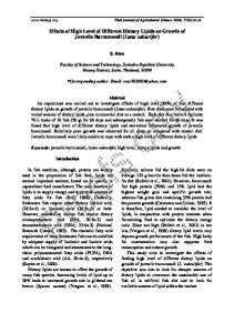
Results Pharmacokinetic data Single-dose plasma concentration–time curves obtained from all 12 subjects are shown in Fig. 2. The maximal plasma concentration (Cmax) after 20 mg of MPH was 10.1±5.0 ng/ml (mean±SD; range, 4.5–17.9 ng/ml) and the median elapsed time between drug administration and maximum plasma concentration (Tmax) was 91.2 min (range, 61.8–240 min). For most subjects, MPH concentrations were quantifiable in plasma 30 to 60 min after oral administration and Cmax was attained, on average, 90 min after administration, coinciding approximately with the start of the scanning session. Estimates for AUCs were calculated for the time between drug administration and Tmax (AUC0–T=21.3±14.3 ng h/ml, mean±SD; range, 5.5–46.6 ng h/ml), as well as for the time between drug administration and beginning of the fMRI scanning (AUC0–scan=14.9±11.3 ng h/ml, mean±SD; range 4.3– 39.6 ng h/ml). Inspection of the individual concentration–time curves revealed a considerable degree of interindividual variation in the pharmacokinetics of MPH.
Fig. 2 Individual plasma concentration–time curves after a single oral dose of MPH 20 mg administered to 12 healthy elderly volunteers. The black rectangle indicates the approximate duration of the fMRI session, which commenced 90 min after MPH administration. Note that there is considerable between-subject variability in the area under the curves
Behavioural data Most participants were able to perform both cued and uncued versions of the visuospatial four-choice task with greater than 95% accuracy in both task and drug conditions with the exception of one subject, who performed below the 25% level expected by chance and was excluded from group analysis of the behavioural data. There were no significant main effects of cue [F(1,10)=2.9; P>0.1] or drug [F(1,10)=0.9; P>0.1] on accuracy, but there was an interaction of drug and cue at trend level [F(1,10)=4.2; P=0.067], whereby MPH was associated with impaired performance of the (easy) cued task and enhanced performance of the (difficult) uncued task (Table 1). Latencies were slower in the uncued condition of the task as confirmed by a highly significant main effect of cue [F(1,10)=26.4; P<0.001]. There was no significant drug effect [F(1,10)=0.2; P>0.1] or drug by cue interaction [F(1,10)=0.1; P>0.1] in terms of latency. Correlational analysis revealed no significant associations between behavioural and pharmacokinetic data. Table 1 Behavioural data Placebo (n=11), mean (SD) MPH 20 mg (n=11), mean (SD) Cued: errors (%)
0 (0)
1.2 (2.1)
Uncued: errors (%)
1.6 (3.0)
0.9 (1.9)
Cued: latency (ms)
458 (142)
455 (115)
Uncued: latency (ms) 639 (123)
606 (148)
Errors and latencies in the spatially cued and uncued conditions of the visuospatial four-choice reaction time (CRT) task after a single oral dose of MPH 20 mg or placebo. There was a significant effect of task difficulty on latency (slower response
for uncued CRT) and a Drug × Task interaction on accuracy at trend level; see text for ANOVA results in full fMRI data Group activation maps of data acquired after placebo, during both cued and uncued versions of the task, show significant activation in the cerebellum, motor and medial premotor cortex, and occipitoparietal cortex (Fig. 3). The more difficult uncued task was associated with more extensive activation in these areas and additional foci in the left dorsolateral prefrontal cortex (BA 46), left caudate nucleus, and left anterior cingulate gyrus (BA 32, 24) (Fig. 3 and Table 2).
Fig. 3 Brain maps of choice reaction time (CRT) task-related activations and drugmodulated activations. a Brain regions generically activated by the contrast of cued CRT vs baseline (red/yellow voxels). b Brain regions generically activated by contrast of uncued CRT vs baseline (red/yellow voxels). c Brain regions demonstrating enhanced activation after MPH on average over both versions of the CRT task (red/yellow voxels). d Brain regions demonstrating significant positive PK/PD association between cued CRT task activation and AUC (blue/purple voxels). e Brain regions demonstrating significant positive PK/PD association between uncued CRT task activation and AUC (blue/purple voxels). The right-hand side of the brain is represented on the left side of each panel; cross-hairs indicate the origin of the x and y dimensions of Talairach space; z coordinates for each panel are shown above the
top row. The activations are superimposed on the grey-scale EPI template image constructed from elderly participants in this study
Table 2 Anatomical locations of CRT task-related activations and drug-modulated activations Brain region
Brodmann area
Talairach coordinates x*
y
z
Condition A 30
5
–56
14
32
24
34
18
32
–10
21
38
32
–29
10
39
44
46
11
19
44
–45
4
22
6
30
1
44
6
–36
–9
38
22
50
2
2
22
46
3
–1
21
57
–48
–3
21
59
–26
–6
Precuneus
7
–16
–54
53
Fusiform gyrus
37
–52
–67
–15
Cuneus
18
16
–92
14
12
–13
10
30
1
–56
11
24
–4
39
3
32
4
15
40
6
–19
–14
56
6
34
2
32
32
17
–6
42
55
–28
16
22
55
–28
16
Middle temporal gyrus
21
–57
–15
–3
Parahippocampal gyrus
27
25
–16
–3
Superior parietal gyrus
7
9
–63
59
Precuneus
7
–1
–55
48
Fusiform gyrus
37
–42
–50
–14
Lingual gyrus
19
17
–58
–3
13
–22
4
19
2
–6
–16
11
–6
Cingulate gyrus Medial frontal gyrus Inferior frontal gyrus Precentral gyrus Superior temporal gyrus Middle temporal gyrus
Thalamus Condition B Cingulate gyrus Medial frontal gyrus Precentral gyrus Insula Superior temporal gyrus
Thalamus Caudate nucleus/putamen
Brain region
Brodmann area
Talairach coordinates x*
y
z
Condition C Precentral gyrus
4
–19
–40
55
Inferior parietal gyrus
19
–21
–75
40
Precuneus
7
–23
–44
49
Cingulate gyrus
29
–28
–41
20
Precentral gyrus
4
16
–26
57
Angular gyrus
19
–33
–63
35
–33
–47
7
–41
–43
–8
31
–21
–29
42
24
–3
28
20
32
14
26
35
31
8
–46
37
Precentral gyrus
4
46
–10
40
Superior temporal gyrus
39
–32
–53
28
Angular gyrus
19
–32
–60
35
Cuneus
19
19
–72
35
Precuneus
24
–3
28
19
–2
11
4
Condition D
Hippocampus/parahippocampal gyrus Condition E Cingulate gyrus Medial frontal gyrus
Caudate nucleus
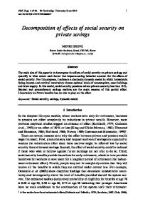
Condition A: Brain regions generically activated by the contrast of cued CRT vs baseline. Condition B: Brain regions generically activated by contrast of uncued CRT vs baseline. Condition C: Brain regions demonstrating enhanced activation after MPH on average over both versions of the CRT task. Condition D: Brain regions demonstrating significant positive PK/PD association between cued CRT task activation and AUC. Condition E: Brain regions demonstrating significant positive PK/PD association between uncued CRT task activation and AUC. Talairach coordinates are reported for the centroid of 3-D clusters of significantly activated voxels; negative x coordinates denote left-hemisphere regions and positive coordinates denote right-hemisphere regions The main effect of drug treatment estimated by two-way ANOVA demonstrated significantly enhanced activation (on average, over both versions of the task) after MPH in the lateral and medial premotor cortex and in the medial posterior parietal cortex (Fig. 3 and Table 2). Pharmacokinetic/pharmacodynamic (PK/PD) analysis Regression of task-related activation on pharmacokinetic variability identified several regions where the sum of standardised response statistics (or mass, m) of each activated cluster was significantly associated with the area under the plasma concentration–time curve, AUC0–scan, in both the cued and uncued versions of the task. Almost all brain regions where the mass of activated voxels (PD marker) was significantly associated with the AUC (PK marker) showed a positive pharmacokinetic/pharmacodynamic (PK/PD) association, meaning that subjects with greater AUC tended to have a larger mass of activation. For the relatively easy spatially cued condition of the CRT task, there were two areas in the right precentral gyrus (post hoc r=0.79, P=0.002) and left hippocampus/parahippocampal gyrus (post hoc r=0.85, P=0.001) where brain activation correlated positively with MPH plasma levels (Fig. 4 and Table 2). For the
more difficult uncued task, clusters of strong PK/PD association were located in the temporoparietal cortex around the right precuneus (r=0.86, P<0.001) and around the anterior cingulate cortex (r=0.83, P=0.001) including the left caudate nucleus and medial frontal gyrus (Fig. 4 and Table 2). MPH-enhanced activations in the motor cortex were located bilaterally in regions also generically activated by task performance. The ranking of all subjects was similar for each cluster of activation and consistent across the two conditions of the tasks. The findings were similar, albeit less pronounced, when correlations were calculated excluding one subject with extreme brain activation (subject 4) and one with poor performance on the CRT task (subject 5).
Fig. 4 Scatterplots showing PK/PD associations between mass (or sum) of activation test statistics in voxel clusters activated by the CRT task (y axis) and MPH pharmacokinetics (AUC0–scan, x axis). Top row, PK/PD associations in the left hippocampus and right premotor cortex for cued version of the task. Bottom row, PK/PD associations in medial parietal and medial premotor cortex for uncued version of the task. Points are numbered to identify individual participants. Individuals with greater AUC consistently tend to demonstrate greater activation in task-specific brain regions
Discussion Here we have shown that MPH has activation-enhancing effects on brain systems for choice motor reaction task performance that are comparable to those previously reported in fMRI studies of MPH in younger subjects. In addition, we have found an association between a pharmacokinetic marker of the highly variable bioavailability of MPH and magnitude of task-related activation in brain regions such as anterior cingulate, medial posterior parietal and lateral premotor cortex, which are known to be involved in visuospatial attention and response preparation. The anterior cingulate and parietal areas more strongly activated by the uncued task in participants exposed to a higher effective dose of MPH were adjacent to or coincident with some of the areas more strongly activated by the uncued version of the task after placebo. The neurocognitive networks generically activated in our study, especially by the more difficult version of the task, were similar to brain regions identified by Mesulam s group in fMRI studies using tasks demanding visuospatially cued attention with blocked (Kim et al. 1999; Mesulam et al. 2001) and event-related designs (Small et al. 2003). The network of brain regions showing significant PK/PD association included subcortical regions (left caudate) and more extended parietal regions typically involved in visuospatial attention and orienting. So far, only one pharmacological fMRI study has investigated the effects of two alpha receptor agonists on performance and brain activation during a similar attentional task. In the study of Coull et al. (2001), the alerting cue primarily activated the left lateralized prefrontal, premotor and parietal regions, and clonidine 0.2 mg, but not guanfacine 1 mg, impaired behavioural measures while attenuating brain activity in the left temporoparietal junction. The most important finding of our study, however, is the positive correlation between individual MPH plasma levels and task-related brain activations in attentional and (pre)motor areas. Increased activation with higher plasma exposure is consistent with the activation-enhancing properties of MPH demonstrated in other pharmacological fMRI studies (Vaidya et al. 1998; Bullmore et al. 2003); reduced cortical rCBF after MPH was previously only observed in PET studies (Mehta et al. 2000; Schweitzer et al. 2004). Two clinical fMRI studies measured plasma levels after administration of single levodopa doses. In patients with Tourette s syndrome a positive correlation between plasma levels and brain activation during a working memory task was observed (Hershey et al. 2004), whereas in patients with Parkinson s disease there was no association between activation by a motor task and levodopa plasma levels (Buhmann et al. 2003). Differences of MPH-related increases between the cued and uncued conditions of task can be explained by differences in phasic dopamine release: the cued version is relatively easy and will not elicit additional endogenous dopamine; only in the more difficult (uncued) version can additional, task-induced dopamine release be assumed (Mattay et al. 2003; Volkow et al. 2004). Our baseline control condition (cross-hair fixation) did not include finger movements; this has to be taken into consideration when interpreting differences between baseline and attentional activation (Goerendt et al. 2003). The lack of a behavioural effect of MPH 20 mg in our fMRI study corresponds with the only behavioural study in elderly volunteers that found no significant effects of single doses of 20 or 40 mg of MPH in a battery of executive and working memory tasks (Turner et al. 2003). The absence of significant PK/PD associations involving behavioural parameters is consistent with the view that brain activation, as measured by pharmacological fMRI, can be a more sensitive surrogate marker of drug effects on neurocognitive function than accuracy or latency of task performance.
Density of dopamine transporters declines with aging, as demonstrated by PET and SPECT radioligand imaging (Volkow et al. 1994; Lavalaye et al. 2000). There were no indications for deleterious effects on task performance or decreases in activation induced by MPH as predicted by the inverted-U model (Robbins 2000); this may be due to the high age of the volunteers resulting in a relatively low functioning dopaminergic system (Kaasinen and Rinne 2002), with catecholamine-enhancing effects hypothetically moving subjects from left to right, but not sufficiently far to exceed the optimal point, on the inverted U-shaped dose-response function (Arnsten and Robbins 2002). Methodological issues In our study, a minimal blood-sampling scheme was used to avoid disturbance of fMRI scanning procedures. Whilst this study was not designed to accurately estimate pharmacokinetic parameters, the results obtained are in good accordance with the literature and valid for correlational analyses. There are several reasons for the high variability in plasma levels. MPH as used in this study is a racemic mixture of d- and l-enantiomers, of which dexmethylphenidate is the pharmacologically effective isomer that binds to the dopamine transporter (Keating and Figgitt 2002; Volkow et al. 2002). Our volunteers did not receive a standardized breakfast; however, food consumption delays the rate of absorption after administration of immediate-release MPH for about 30 min (Midha et al. 2001). There are functional polymorphisms in genes for enzymes involved in MPH metabolism (Masellis et al. 2002). The effects of MPH are dependent on the rate of dopamine release; subjects with low dopamine tone will be less sensitive to MPH than those with a high dopamine tone (Volkow et al. 2002). The COMT val/met polymorphism is an important candidate gene that explains differences of tonic dopamine levels (Bilder et al. 2004). A combination of some or all of these factors might explain the observed PK variability. fMRI measures of drug effects on neurons are potentially complicated by drug effects on cerebral vasculature, which might also be expected to modulate the BOLD response. MPH inhibits the reuptake of dopamine via dopamine transporters and enhances blood pressure and pulse (Volkow et al. 2002); its direct and indirect effects on BOLD responses are incompletely known at present. However, one prior study combining fMRI and EEG measurements of the effects of sulpiride (a dopamine D2 receptor antagonist) on somatosensory cortical response to peripheral stimulation showed that both modalities can provide consistent markers of dopaminergic drug effects, implying that fMRI measures are not necessarily confounded by major vascular effects (Arthurs et al. 2004). Further limitations of this study include the lack of a stimulus onset asynchrony (SOA) jitter. The simplification of the task for elderly volunteers after pilot testing may have reduced the sensitivity to detect behavioural drug effects. On the other hand, the absence of drug effects on performance facilitates the interpretation of drug-related differences of brain activation (Honey and Bullmore 2004). We did not evaluate subjective effects of MPH and were therefore not able to relate those effects to performance or neuroimaging data. Findings of this study should not be generalized to a normal population, because we tested a group of elderly volunteers with perhaps subtly different hemodynamic response functions (D Esposito et al. 2003). In future work, we plan to test Age×Drug interactive effects on task-related brain activation more decisively by studying younger and elderly groups matched for performance and scanned using an identical protocol. To our knowledge this is the first demonstration of plasma-level-dependent effects of MPH on task-related fMRI signal changes in healthy volunteers. Pharmacological fMRI in combination with traditional methods of human psychopharmacology is a promising research tool to investigate brain and behavioural effects of clinically relevant drugs. Acknowledgements This work was supported by GlaxoSmithKline and the Wellcome Trust, by a research fellowship of the Alexander von Humboldt Foundation
(UM) and was completed within the MRC Behavioural and Clinical Neuroscience Centre (BCNC). We gratefully thank the volunteers who participated in this study for their cooperation, and colleagues at the MRI Unit, Maudsley Hospital, London, UK, for technical assistance with fMRI data acquisition.
References Arnsten AFT, Robbins TW (2002) Neurochemical modulation of prefrontal function in humans and animals. In: Stuss DT, Knight RT (eds) Principles of frontal lobe function. Oxford University Press, New York, pp 51–84 Arthurs OJ, Stephenson CM, Rice K, Lupson VC, Spiegelhalter DJ, Boniface SJ, Bullmore ET (2004) Dopaminergic effects on electrophysiological and functional MRI measures of human cortical stimulus-response power laws. NeuroImage 21:540–546 Biederman J, Spencer T, Wilens T (2004) Evidence-based pharmacotherapy for attention-deficit hyperactivity disorder. Int J Neuropsychopharmacol 7:77–97 Bilder RM, Volavka J, Lachman HM, Grace AA (2004) The catechol-O-methyltransferase polymorphism: relations to the tonic–phasic dopamine hypothesis and neuropsychiatric phenotypes. Neuropsychopharmacology 29:1943–1961 Brammer MJ, Bullmore ET, Simmons A, Williams SC, Grasby PM, Howard RJ, Woodruff PW, Rabe-Hesketh S (1997) Generic brain activation mapping in functional magnetic resonance imaging: a nonparametic approach. Magn Reson Imag 15:763–770
Braus DF, Brassen S, Weimer E, Tost H (2003) Functional magnetic resonance imaging of psychopharmacological brain effects: an update. Fortschr Neurol Psychiatr 71:72–83
Buhmann C, Glauche V, Sturenburg HJ, Oechsner M, Weiller C, Büchel C (2003) Pharmacologically modulated fMRI: cortical responsiveness to levodopa in drug-naive hemiparkinsonian patients. Brain 126:451–461 Bullmore ET, Suckling J, Overmeyer S, Rabe-Hesketh S, Taylor E, Brammer MJ (1999) Global, voxel, and cluster tests, by theory and permutation, for a difference between two groups of structural MR images of the brain. IEEE Trans Med Imag 18:32–42
Bullmore E, Long C, Suckling J, Fadili J, Calvert G, Zelaya F, Carpenter TA, Brammer M (2001) Colored noise and computational inference in neurophysiological (fMRI) time series analysis: resampling methods in time and wavelet domains. Hum Brain Mapp 12:61–78
Bullmore E, Suckling J, Zelaya F, Long C, Honey G, Reed L, Routledge C, Ng V, Fletcher P, Brown J, Williams SC (2003) Practice and difficulty evoke anatomically and
pharmacologically dissociable brain activation dynamics. Cereb Cortex 13:144–154 Chou WL (1978) Critical evaluation of the potential error in pharmacokinetic studies using the linear trapezoidal rule method for the calculation of the area under the plasma level– time curve. J Pharmacokinet Biopharm 6:539–547 Cools R, Barker RA, Sahakian BJ, Robbins TW (2001) Enhanced or impaired cognitive function in Parkinson s disease as a function of dopaminergic medication and task demands. Cereb Cortex 11:1136–1143 Coull JT, Nobre AC, Frith CD (2001) The noradrenergic alpha2 agonist clonidine modulates behavioural and neuroanatomical correlates of human attentional orienting and alerting. Cereb Cortex 11:73–84
Cox RW (1994) Analysis of functional neuroimages, version 1.01. Medical College of Wisconsin, Milwaukee D Esposito M, Deouell LY, Gazzaley A (2003) Alterations in the BOLD fMRI signal with ageing and disease: a challenge for neuroimaging. Nat Rev Neurosci 4:863–872 Furey M, Pietrini P, Haxby JV (2000) Cholinergic enhancement and increased selectivity of perceptual processing during working memory. Science 290:2315–2319 Gitelman DR, Nobre AC, Parrish TB, LaBar KS, Kim Y-H, Meyer JR, Mesulam MM (1999) A large-scale distributed network for covert spatial attention: further anatomical delineation based on stringent behavioural and cognitive controls. Brain 122:1093–1106
Goerendt IK, Messa C, Lawrence AD, Grasby PM, Piccini P, Brooks DJ (2003) Dopamine release during sequential finger movements in health and Parkinson s disease: a PET study. Brain 126:312–325 Goldman-Rakic PS, Muly EC III, Williams GV (2000) D1 receptors in prefrontal cells and circuits. Brain Res Rev 31:295–301
Greenhill L, Beyer DH, Finkleson J, Shaffer D, Biederman J, Conners CK, Gillberg C, Huss M, Jensen P, Kennedy JL, Klein R, Rapoport J, Sagvolden T, Spencer T, Swanson JM, Volkow N (2002) Guidelines and algorithms for the use of methylphenidate in children with attention-deficit hyperactivity disorder. J Atten Disord 6(Suppl 1):89–100 Hershey T, Black KJ, Hartlein JM, Barch DM, Braver TS, Carl JL, Perlmutter JS (2004) Cognitive–pharmacologic functional magnetic resonance imaging in Tourette syndrome: a pilot study. Biol Psychiatry 55:916–925 Honey G, Bullmore E (2004) Human pharmacological MRI. Trends Pharmacol Sci 25:366–374
Honey GD, Suckling J, Zelaya F, Long C, Routledge C, Jackson S, Ng V, Fletcher PC, Williams SCR, Brown J, Bullmore ET (2003) Dopaminergic drug effects on physiological connectivity in a human cortico–striato–thalamic system. Brain 126:1767–1781 Jahanshahi M, Brown RG, Marsden CD (1992) Simple and choice reaction time and the use of advance information for motor preparation in Parkinson s disease. Brain 115:539– 564 Kaasinen V, Rinne JO (2002) Functional imaging studies of dopamine system and cognition in normal aging and Parkinson s disease. Neurosci Biobehav Rev 26:785–793
Keating GM, Figgitt DP (2002) Dexmethylphenidate. Drugs 62:1899–1904 Kim YH, Gitelman DR, Nobre AC, Parrish TB, LaBar KS, Mesulam MM (1999) The largescale neural network for spatial attention displays multifunctional overlap but differential asymmetry. NeuroImage 9:269–277 Kimberg DY, D Esposito M (2003) Cognitive effects of the dopamine receptor agonist pergolide. Neuropsychologica 41:1020–1027 Knutson B, Bjork JM, Fong GW, Hommer D, Mattay VS, Weinberger DR (2004) Amphetamine modulates human incentive processing. Neuron 43:261–269 Laurienti PJ, Field AS, Burdette JH, Maldjian JA, Yen YF, Moody DM (2003) Relationship between caffeine-induced changes in resting cerebral perfusion and blood oxygenation level-dependent signal. Am J Neuroradiol 24:1607–1611 Lavalaye J, Booij J, Reneman L, Habraken JB, van Royen EA (2000) Effect of age and gender on dopamine transporter imaging with [123I]FP-CIT SPET in healthy volunteers. Eur J Nucl Med 27:867–869 Leonard BE, McCartan D, White J, King DJ (2004) Methylphenidate: a review of its neuropharmacological, neuropsychological and adverse clinical effects. Hum Psychopharmacol 19:151–180
Liu TT, Behzadi Y, Reston K, Uludag K, Lu K, Buracas GT, Dubowitz DJ, Buxton RB (2004) Caffeine alters the temporal dynamics of the visual BOLD response. NeuroImage 23:1402–1413 Masellis M, Basile VS, Muglia P, Ozdemir V, Macciardi FM, Kennedy JL (2002) Psychiatric pharmacogenetics: personalizing psychostimulant therapy in attentiondeficit/hyperactivity disorder. Behav Brain Res 130:85–90 Mattay VS, Goldberg TE, Fera F, Hariri AR, Tessitore A, Egan MF, Kolachana B, Callicott JH, Weinberger DR (2003) Catechol O-methyltransferase val158-met genotype and individual variation in the brain response to amphetamine. Proc Natl Acad Sci U S A 100:6186–6191
Mehta MA, Owen AM, Sahakian BJ, Mavaddat N, Pickard JD, Robbins TW (2000) Methylphenidate enhances working memory by modulating discrete frontal and parietal lobe regions in the human brain. J Neurosci 20:RC65 Mesulam MM (2000) Principles of cognitive and behavioural neurology. Oxford University Press, New York Mesulam MM, Nobre AC, Kim YH, Parrish TB, Gitelman DR (2001) Heterogeneity of cingulate contributions to spatial attention. NeuroImage 13:1065–1072 Midha KK, McKay G, Rawson MJ, Korchinski ED, Hubbard JW (2001) Effects of food on the pharmacokinetics of methylphenidate. Pharm Res 18:1185–1189 Mulderink TA, Gitelman DR, Mesulam MM, Parrish TB (2002) On the use of caffeine as a contrast booster for BOLD fMRI studies. NeuroImage 15:37–44 Murray GB, Cassem E (1998) Use of stimulants in depressed patients with medical illness. In: Nelson JG (ed) Geriatric psychopharmacology. Dekker, New York, pp 245–257 Posner MI (1980) Orienting of attention. Q J Exp Psychol 32:3025 Rao SM, Salmeron BJ, Durgerian S, Janowiak JA, Fischer M, Risinger RC, Conant LL, Stein EA (2000) Effects of methylphenidate on functional MRI blood-oxygen-leveldependent contrast. Am J Psychiatry 157:1697–1699 Robbins TW (2000) Chemical neuromodulation of frontal-executive functions in humans and other animals. Exp Brain Res 133:130–138
Schweitzer JB, Lee DO, Hanford RB, Zink CF, Ely TD, Tagamets MA, Hoffman JM, Grafton ST, Kilts CD (2004) Effect of methylphenidate on executive functioning in adults with attention-deficit/hyperactivity disorder: normalization of behavior but not related brain activity. Biol Psychiatry 56:597–606 Shafritz KM, Marchione KE, Gore JC, Shaywitz SE, Shaywitz BA (2004) The effects of methylphenidate on neural systems of attention in attention deficit hyperactivity disorder. Am J Psychiatry 161:1990–1997 Silveri MM, Anderson CM, McNeil JF, Diaz CI, Lukas SE, Mendelson JH, Renshaw PF, Kaufman MJ (2004) Oral methylphenidate challenge selectively decreases putaminal T2 in healthy subjects. Drug Alcohol Depend 76:173–180
Small DM, Gitelman DR, Gregory MD, Nobre AC, Parrish TB, Mesulam MM (2003) The posterior cingulate and medial prefrontal cortex mediate the anticipatory allocation of spatial attention. NeuroImage 18:633–641 Solanto MV (1998) Neuropsychopharmacological mechanisms of stimulant drug action in
attention-deficit hyperactivity disorder: a review and integration. Behav Brain Res 94:127– 152
Suckling J, Bullmore E (2004) Permutation tests for factorially designed neuroimaging experiments. Hum Brain Mapp 22:193–205 Talairach J, Tournoux P (1998) Coplanar stereotaxic atlas of the human brain. Thieme, Stuttgart Turner DC, Robbins TW, Clark L, Aron AR, Dowson J, Sahakian BJ (2003) Relative lack of cognitive effects of methylphenidate in elderly male volunteers. Psychopharmacology 168:455–464
Vaidya CJ, Austin G, Kirkorian G, Ridlehuber HW, Desmond JE, Glover GH, Gabrieli JD (1998) Selective effects of methylphenidate in attention deficit hyperactivity disorder: a functional magnetic resonance study. Proc Natl Acad Sci U S A 95:14494–14499 Volkow ND, Fowler JS, Wang GJ, Logan J, Schlyer D, MacGregor R, Hitzemann R, Wolf AP (1994) Decreased dopamine transporters with age in health human subjects. Ann Neurol 36:237–239 Volkow ND, Fowler JS, Wang GJ, Ding YS, Gatley SJ (2002) Role of dopamine in the therapeutic and reinforcing effects of methylphenidate in humans: results from imaging studies. Eur Neuropsychopharmacol 12:557–566 Volkow ND, Wang GJ, Fowler JS, Telang F, Maynard L, Logan J, Gatley SJ, Pappas N, Wong C, Vaska P, Zhu W, Swanson JM (2004) Evidence that methylphenidate enhances the saliency of a mathematical task by increasing dopamine in the human brain. Am J Psychiatry 161:1173–1180 Völlm BA, De Araujo IE, Cowen PJ, Rolls ET, Kringelbach ML, Smith KA, Jezzard P, Heal RJ, Matthews PM (2004) Methamphetamine activates reward circuitry in drug naive human subjects. Neuropsychopharmacology 29:1715–1722